MYCL1 is a member of the MYC family that was isolated from small cell lung carcinoma. Like the other members of the MYC family, MYCL1 is a proto-oncogene transcription factor belonging to the helix-loop-helix basic leucine zipper (HLH bzip) family. With its heterodimeric partners, MYCL1 binds to the DNA consensus site 5’ CACGTG 3’. MYCL1 is implicated in controlling a wide range of cellular processes from cellular proliferation to apoptosis.

Discontinued Product
AF4050 has been discontinued.
View all MYCL1/L-Myc products.
Key Product Details
Species Reactivity
Validated:
Human, Mouse
Cited:
Human
Applications
Validated:
Western Blot, Immunocytochemistry
Cited:
Western Blot
Label
Unconjugated
Antibody Source
Polyclonal Goat IgG
Loading...
Product Specifications
Immunogen
E. coli-derived recombinant human MYCL1/L‑Myc
Gly16-Asn139
Accession # P12524
Gly16-Asn139
Accession # P12524
Specificity
Detects human and mouse MYCL1/L‑Myc in Western blots.
Clonality
Polyclonal
Host
Goat
Isotype
IgG
Scientific Data Images for MYCL1/L-Myc Antibody
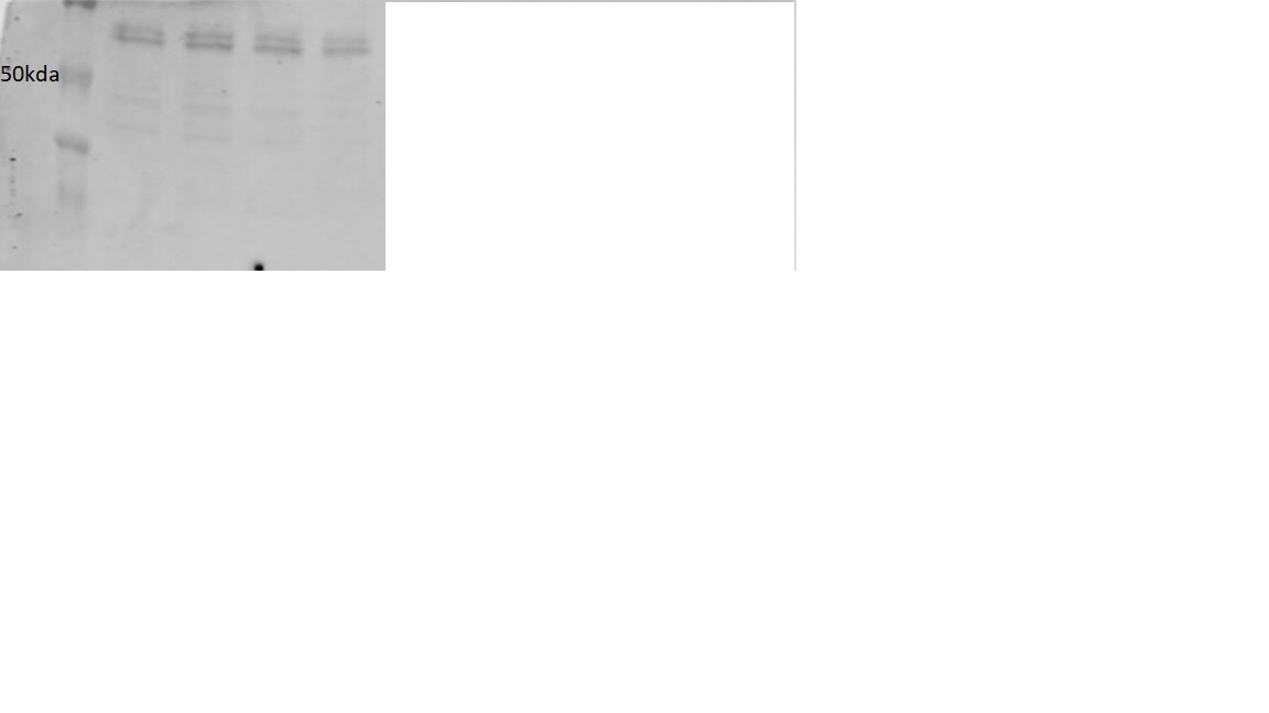
Detection of Human/Mouse MYCL1/ L‑Myc by Western Blot.
Western blot shows nuclear extracts of HeLa human cervical epithelial carcinoma cell line, A549 human lung carcinoma cell line, JEG-3 human epithelial choriocarcinoma cell line, and NIH-3T3 mouse embryonic fibroblast cell line. PVDF membrane was probed with 1 µg/mL of Human/Mouse MYCL1/L-Myc Antigen Affinity-purified Polyclonal Antibody (Catalog # AF4050) followed by HRP-conjugated Anti-Goat IgG Secondary Antibody (Catalog # HAF109). A specific band was detected for MYCL1/L-Myc at approximately 40 kDa (as indicated). This experiment was conducted under reducing conditions and using Immunoblot Buffer Group 2.
MYCL1/L‑Myc in HeLa Human Cell Line.
MYCL1/L-Myc was detected in immersion fixed HeLa human cervical epithelial carcinoma cell line using Goat Anti-Human/Mouse MYCL1/L-Myc Antigen Affinity-purified Polyclonal Antibody (Catalog # AF4050) at 10 µg/mL for 3 hours at room temperature. Cells were stained using the NorthernLights™ 557-conjugated Anti-Goat IgG Secondary Antibody (red, upper panel; Catalog # NL001) and counterstained with DAPI (blue, blue panel). Specific staining was localized to cytoplasm and nuclei. View our protocol for Fluorescent ICC Staining of Cells on Coverslips.
MYCL1/L‑Myc in NIH3T3 Mouse Cell Line.
MYCL1/L-Myc was detected in immersion fixed NIH3T3 mouse embryonic fibroblast cell line using Goat Anti-Human/Mouse MYCL1/L-Myc Antigen Affinity-purified Polyclonal Antibody (Catalog # AF4050) at 10 µg/mL for 3 hours at room temperature. Cells were stained using the Northern-Lights™ 557-conjugated Anti-Goat IgG Secondary Antibody (red, upper panel; Catalog # NL001) and counterstained with DAPI (blue, lower panel). Specific staining was localized to cytoplasm and nuclei. View our protocol for Fluorescent ICC Staining of Cells on Coverslips.
Detection of Human MYCL1/L‑Myc by Western Blot
Differential effects of MYC and NF-kappa B on glycolytic gene and MCT1 levels.A) Promoter region of MCT1 includes MYC and NF-kappa B binding sites. Shading reflects four independent IMR90 DNase I hypersensitivity datasets. B) IMR90 cells stably expressing ST, GFP or p53DD + hTERT (PH) and MCPyV tumor-derived early-region (PHE) with inducible expression of MYC, MYCN or MYCL were treated with dox (+) for 48 hours. Lysates were immunoblotted with the indicated antibodies. C) IMR90 PH and PHE cells inducibly expressing MYC or MYCL were transfected with RelA-specific pooled siRNA (siRelA) or non-targeting siRNA (siCtrl). After 24 hours, cells were refed with dox containing media and lysed after an additional 48 hours. Image collected and cropped by CiteAb from the following publication (https://dx.plos.org/10.1371/journal.ppat.1006020), licensed under a CC-BY license. Not internally tested by R&D Systems.
Detection of Human MYCL1/L‑Myc by Western Blot
MYC isoforms differentially regulate glycolysis gene expression and ECAR of MCC cells.A) MKL-1 and WaGa cells containing inducible vectors for MYC, MYCN or MYCL were treated with (+) or without (-) dox for 72 hours and lysates were immunoblotted with the indicated antibodies. B) ECAR (mpH/min) of MKL-1 cells inducibly expressing GFP, MYC, MYCN or MYCL after 72 hours of dox addition (minutes). Cells were treated with oligomycin (1 μM) at the indicated time point. *P < 0.05 calculated using unpaired student’s T test between MYC and MYCL samples. Image collected and cropped by CiteAb from the following publication (https://dx.plos.org/10.1371/journal.ppat.1006020), licensed under a CC-BY license. Not internally tested by R&D Systems.Applications for MYCL1/L-Myc Antibody
Application
Recommended Usage
Immunocytochemistry
5-15 µg/mL
Sample: Immersion fixed HeLa human cervical epithelial carcinoma cell line, and immersion fixed NIH3T3 mouse embryonic fibroblast cell line
Sample: Immersion fixed HeLa human cervical epithelial carcinoma cell line, and immersion fixed NIH3T3 mouse embryonic fibroblast cell line
Western Blot
1 µg/mL
Sample: HeLa human cervical epithelial carcinoma cell line, A549 human lung carcinoma cell line, JEG-3 human epithelial choriocarcinoma cell line, and NIH-3T3 mouse embryonic fibroblast cell line
Sample: HeLa human cervical epithelial carcinoma cell line, A549 human lung carcinoma cell line, JEG-3 human epithelial choriocarcinoma cell line, and NIH-3T3 mouse embryonic fibroblast cell line
Reviewed Applications
Read 1 review rated 3 using AF4050 in the following applications:
Formulation, Preparation, and Storage
Purification
Antigen Affinity-purified
Reconstitution
Reconstitute at 0.2 mg/mL in sterile PBS. For liquid material, refer to CoA for concentration.
Formulation
Lyophilized from a 0.2 μm filtered solution in PBS with Trehalose. *Small pack size (SP) is supplied either lyophilized or as a 0.2 µm filtered solution in PBS.
Shipping
Lyophilized product is shipped at ambient temperature. Liquid small pack size (-SP) is shipped with polar packs. Upon receipt, store immediately at the temperature recommended below.
Stability & Storage
Use a manual defrost freezer and avoid repeated freeze-thaw cycles.
- 12 months from date of receipt, -20 to -70 °C as supplied.
- 1 month, 2 to 8 °C under sterile conditions after reconstitution.
- 6 months, -20 to -70 °C under sterile conditions after reconstitution.
Calculators
Background: MYCL1/L-Myc
Long Name
v-Myc Myelocytomatosis Viral Oncogene Homolog 1, Lung Carcinoma Derived
Alternate Names
L-Myc, LMyc
Gene Symbol
MYCL
UniProt
Additional MYCL1/L-Myc Products
Product Documents for MYCL1/L-Myc Antibody
Certificate of Analysis
To download a Certificate of Analysis, please enter a lot or batch number in the search box below.
Note: Certificate of Analysis not available for kit components.
Product Specific Notices for MYCL1/L-Myc Antibody
For research use only
Citations for MYCL1/L-Myc Antibody
 Powered by Bioz
Customer Reviews for MYCL1/L-Myc Antibody (1)
3 out of 5
1 Customer Rating
Have you used MYCL1/L-Myc Antibody?
Submit a review and receive an Amazon gift card!
$25/€18/£15/$25CAN/¥2500 Yen for a review with an image
$10/€7/£6/$10CAN/¥1110 Yen for a review without an image
Submit a review
Customer Images
Showing
1
-
1 of
1 review
Showing All
Filter By:
-
Application: Western BlotSample Tested: small cell lung cancer cellsSpecies: HumanVerified Customer | Posted 12/18/2018ripa lysis 30ug protein loaded. Not sure which band or is it the right band

There are no reviews that match your criteria.
Protocols
Find general support by application which include: protocols, troubleshooting, illustrated assays, videos and webinars.
- Appropriate Fixation of IHC/ICC Samples
- Cellular Response to Hypoxia Protocols
- ClariTSA™ Fluorophore Kits
- Detection & Visualization of Antibody Binding
- ICC Cell Smear Protocol for Suspension Cells
- ICC Immunocytochemistry Protocol Videos
- ICC for Adherent Cells
- Immunocytochemistry (ICC) Protocol
- Immunocytochemistry Troubleshooting
- Immunofluorescence of Organoids Embedded in Cultrex Basement Membrane Extract
- Immunohistochemistry (IHC) and Immunocytochemistry (ICC) Protocols
- Preparing Samples for IHC/ICC Experiments
- Preventing Non-Specific Staining (Non-Specific Binding)
- Primary Antibody Selection & Optimization
- Protocol for VisUCyte™ HRP Polymer Detection Reagent
- Protocol for the Fluorescent ICC Staining of Cell Smears - Graphic
- Protocol for the Fluorescent ICC Staining of Cultured Cells on Coverslips - Graphic
- Protocol for the Preparation and Fluorescent ICC Staining of Cells on Coverslips
- Protocol for the Preparation and Fluorescent ICC Staining of Non-adherent Cells
- Protocol for the Preparation and Fluorescent ICC Staining of Stem Cells on Coverslips
- Protocol for the Preparation of a Cell Smear for Non-adherent Cell ICC - Graphic
- R&D Systems Quality Control Western Blot Protocol
- TUNEL and Active Caspase-3 Detection by IHC/ICC Protocol
- The Importance of IHC/ICC Controls
- Troubleshooting Guide: Western Blot Figures
- Western Blot Conditions
- Western Blot Protocol
- Western Blot Protocol for Cell Lysates
- Western Blot Troubleshooting
- Western Blot Troubleshooting Guide
- View all Protocols, Troubleshooting, Illustrated assays and Webinars
Loading...