Axin-1 is a negative regulator of the Wnt signaling pathway, which is involved in regulation of embryonic axis formation in vertebrates. Axin-1 binds to beta -catenin and glycogen synthase kinase-3 (GSK-3) to promote beta -catenin phosphorylation and subsequent degradation. Axin-1 also binds to the adenomatous polyposis coli (APC) gene, a known tumor suppressor that also regulates beta -catenin.

Key Product Details
Species Reactivity
Validated:
Human, Mouse, Rat
Cited:
Human, Mouse, Drosophila
Applications
Validated:
Western Blot
Cited:
Western Blot, Immunocytochemistry, Immunoprecipitation
Label
Unconjugated
Antibody Source
Polyclonal Goat IgG
Loading...
Product Specifications
Immunogen
E. coli-derived recombinant human Axin-1
Tyr210-Glu410
Accession # O15169
Tyr210-Glu410
Accession # O15169
Specificity
Detects human, mouse and rat Axin-1 in Western blots.
Clonality
Polyclonal
Host
Goat
Isotype
IgG
Scientific Data Images for Axin-1 Antibody
Detection of Human/Mouse/Rat Axin‑1 by Western Blot.
Western blot shows lysates of Raji human Burkitt's lymphoma cell line, Balb/3T3 mouse embryonic fibroblast cell line, and PC-12 rat adrenal pheochromocytoma cell line. PVDF membrane was probed with 0.5 µg/mL of Human/Mouse/Rat Axin-1 Antigen Affinity-purified Polyclonal Antibody (Catalog # AF3287) followed by HRP-conjugated Anti-Goat IgG Secondary Antibody (Catalog # HAF017). A specific band was detected for Axin-1 at approximately 120 kDa (as indicated). This experiment was conducted using Immunoblot Buffer Group 1.
Detection of Axin-1 by Immunocytochemistry/ Immunofluorescence
TNKSi treatment can induce axin expression and axin puncta associated with beta -catenin degradation. A. Western blot analysis of different cell lines treated +/- for 24 h with 2.5 μM of the TNKSi XAV939 revealed an increase in axin expression in different cell lines, and rescue of the degradation of beta -catenin in SW480 CRC cells where the wnt pathway is disrupted by APC mutation. beta -actin levels are shown as loading controls. Quantification of band intensities from two separate blots was performed and normalized to actin. The values shown are mean ± SD. B. SW480 cells were treated with 5 μM of the TNKSi G007-LK for 24 h, then immunostained with specific antibodies to detect different components of the beta -catenin degradation complex located at axin puncta. The following combinations of proteins were co-stained: axin and total beta -catenin, APC and Axin, GSK3 beta and Axin and phosphorylated and non-phosphorylated beta -catenin. Cells were imaged using a DeltaVision microscope system. C. SW480 control cells or cells treated with 5 μM of the TNKSi G007-LK for 24 h were stained for fluorescence. In control cells a disperse staining pattern is observed whereas in TNKSi treated cells the TNKS-1 and -2 (red) specifically colocalize at TNKSi induced axin puncta (green) as shown by immunofluorescence microscopy. Image collected and cropped by CiteAb from the following open publication (https://pubmed.ncbi.nlm.nih.gov/26930278), licensed under a CC-BY license. Not internally tested by R&D Systems.
Detection of Axin-1 by Western Blot
Tankyrase inhibitors promote inclusion of axin and TNKS into insoluble complexes. A. NIH 3T3 cells were left untreated (- CSK) or washed for 5 min with 0.2% Triton X-100 containing MT-buffer (+CSK) to permeabilize the plasma membrane and remove soluble proteins, before immunolabeling with fluorescent antibodies for axin and tubulin. Axin puncta in untreated cells and after induction by TNKSi were still visible after the detergent extraction, despite a decrease in background staining. This implies that the axin puncta are part of an insoluble pool resistant to extraction. B. To determine if axin and TNKS accrued in an insoluble fraction in vitro after TNKSi treatment in SW480 cells, cells were extracted either with RIPA buffer (left panel) or enriched for the insoluble fraction by extracting cells on the plate with SDS containing sample buffer (right panel). The addition of TNKSi (G007-LK) increased the accumulation of the TNKS-1 and -2 in the insoluble fractions and this was at least partly reduced for TNKS2 and axin following MG132 treatment, indicating that TNKSi might reduce solubility and mobility of axin and TNKS. Quantification of band intensity was measured and values were corrected to the endogenous control beta -actin (a second experiment is shown in S6 Fig). Image collected and cropped by CiteAb from the following open publication (https://pubmed.ncbi.nlm.nih.gov/26930278), licensed under a CC-BY license. Not internally tested by R&D Systems.
Detection of Axin-1 by Western Blot
Proteasome inhibitor induces PARylation of TNKSs and TNKSi reduces basal PARylation levels. A. SW480 and HEK293 cells were exposed to combinations of TNKSi (G007-LK) and MG132 treatments similar to that shown in legend to Fig 3. The cells were then lysed and extracts analysed by IP assay using a specific antibody against PAR. PARylated forms of TNKS1/2 were then detected by western blot. The IgG control was negative. Pull-down of PARylated proteins revealed an increase in PARylation of TNKS1 by MG132 treatment, and a decrease of PARylation induced by TNKSi. When combined, the MG132 dominated and caused some induction of PARylated TNKS1 in both SW480 and HEK293 cells. Levels of PARylated TNKS2 (and also axin, not shown) were too low for detection. Quantification of band intensities (mean ± SD) was from two separate experiments. B. Total extract western blot shows similar TNKS levels after different treatments. Image collected and cropped by CiteAb from the following open publication (https://pubmed.ncbi.nlm.nih.gov/26930278), licensed under a CC-BY license. Not internally tested by R&D Systems.
Detection of Axin-1 by Immunocytochemistry/ Immunofluorescence
Induction of axin puncta by TNKSi is proteasome-dependent. A. SW480 cells were treated with single or combined doses of tankyrase (5 μM G007LK) and proteasome (20 μM MG132) inhibitors. The MG132 was added for 6 h, either simultaneously with G007-LK for a 6 h treatment, or during the last 6 h of a 24 h G007-LK treatment. As shown in the immunofluorescence images, the addition of MG132 caused a reduction in the % of cells with visible TNKSi-induced axin puncta after 6 h treatment. The 6 h MG132 treatment also caused a modest increase in nuclear staining of axin. The later addition of MG132 (at the end of a 24 h G007-LK treatment) caused the induced axin puncta to relocate to the perinuclear region. B. The number of axin puncta per cell were scored by microscopy and categorized (<5, 5–15 or >15 puncta per cell). As shown, TNKSi induced a high number of puncta per cell (more than 80% of cells scored >15 puncta per cell) and this was blocked at 6 h or reduced after 24 h by MG132 treatment. C. A diagram summarizing the effect of G007-LK +/- MG132 on axin pattern and cellular distribution (shown in green). Image collected and cropped by CiteAb from the following open publication (https://pubmed.ncbi.nlm.nih.gov/26930278), licensed under a CC-BY license. Not internally tested by R&D Systems.
Detection of Axin-1 by Western Blot
Endocytosis inhibitors cause a decrease in the level of Armadillo/ beta -catenin both in stimulated and unstimulated cells. (A) The level of Armadillo increased in S2R+ cells that had been treated with Wingless-conditioned medium (lane 2) or SB-216763 (lane 5). This was prevented by treatment with Dyngo-4a (lanes 3 and 6) or Dynasore (lanes 4 and 7). Lamin, Actin and Syntaxin levels were unaffected. (B) Dyngo-4a reversibly reduced the level of Armadillo in uninduced cells. (C) Dyngo-4a reduced signalling-induced accumulation of beta -catenin in RKO cells. Cells were pre-incubated with SB-216763 to activate signalling and then exposed to a mixture of SB-216763 and Dyngo-4a. The total time of treatment with either drug is indicated. As in Drosophila cells, Dyngo-4a caused a decrease in beta -catenin levels in unstimulated cells. A progressive decrease can be seen after 0.5, 1 and 2 hours of treatment with Dyngo-4a (no SB-216763) in lanes 9–11. (D) The effect of Dyngo-4a and SB-216763 on the level of various components of the Wnt pathway. LRP6, GSK3 beta and CK1 alpha were largely unaffected, whereas the levels of APC and Axin1 dropped markedly 30–60 minutes after treatment with Dyngo-4a. SB-216763 caused an increase in the amount of beta -catenin. This correlated with a decrease in phosphorylated beta -catenin (pT42/S37/S33 beta -catenin; lane 3), as expected because phosphorylated beta -catenin reflects the activity of the degradation complex (Hernández et al., 2012). By contrast, the (mild) decrease in beta -catenin caused by Dyngo-4a (lanes 7 and 8) is paralleled by a similar decrease in phosphorylated beta -catenin, suggesting that Dyngo-4a impacts on the level of beta -catenin through a mechanism that is independent of the destruction complex. Image collected and cropped by CiteAb from the following open publication (https://pubmed.ncbi.nlm.nih.gov/25236598), licensed under a CC-BY license. Not internally tested by R&D Systems.Applications for Axin-1 Antibody
Application
Recommended Usage
Western Blot
0.5 µg/mL
Sample: Raji human Burkitt's lymphoma cell line, Balb/3T3 mouse embryonic fibroblast cell line, and PC-12 rat adrenal pheochromocytoma cell line
Sample: Raji human Burkitt's lymphoma cell line, Balb/3T3 mouse embryonic fibroblast cell line, and PC-12 rat adrenal pheochromocytoma cell line
Reviewed Applications
Read 1 review rated 5 using AF3287 in the following applications:
Formulation, Preparation, and Storage
Purification
Antigen Affinity-purified
Reconstitution
Reconstitute at 0.2 mg/mL in sterile PBS. For liquid material, refer to CoA for concentration.
Loading...
Formulation
Lyophilized from a 0.2 μm filtered solution in PBS with Trehalose. *Small pack size (SP) is supplied either lyophilized or as a 0.2 µm filtered solution in PBS.
Shipping
Lyophilized product is shipped at ambient temperature. Liquid small pack size (-SP) is shipped with polar packs. Upon receipt, store immediately at the temperature recommended below.
Stability & Storage
Use a manual defrost freezer and avoid repeated freeze-thaw cycles.
- 12 months from date of receipt, -20 to -70 °C as supplied.
- 1 month, 2 to 8 °C under sterile conditions after reconstitution.
- 6 months, -20 to -70 °C under sterile conditions after reconstitution.
Calculators
Background: Axin-1
Long Name
Axis Inhibitor 1
Alternate Names
Axin1, MGC52315
Gene Symbol
AXIN1
UniProt
Additional Axin-1 Products
Product Documents for Axin-1 Antibody
Certificate of Analysis
To download a Certificate of Analysis, please enter a lot or batch number in the search box below.
Note: Certificate of Analysis not available for kit components.
Product Specific Notices for Axin-1 Antibody
For research use only
Related Research Areas
Citations for Axin-1 Antibody
 Powered by Bioz
Customer Reviews for Axin-1 Antibody (1)
5 out of 5
1 Customer Rating
Have you used Axin-1 Antibody?
Submit a review and receive an Amazon gift card!
$25/€18/£15/$25CAN/¥2500 Yen for a review with an image
$10/€7/£6/$10CAN/¥1110 Yen for a review without an image
Submit a review
Customer Images
Showing
1
-
1 of
1 review
Showing All
Filter By:
-
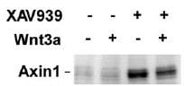
Application: Western BlotSample Tested: EMT6 cellsSpecies: MouseVerified Customer | Posted 10/26/2015From left to right are lanes: negative control without or with Wnt3a treatment, and Axin1 stabilizer XAV939 without or with Wnt3a treatment. <br />Specificity: Specific<br />Sensitivity: Sensitive<br />Buffer: TBS+Tween 0.1%<br />Dilution: 1/1000

There are no reviews that match your criteria.
Protocols
Find general support by application which include: protocols, troubleshooting, illustrated assays, videos and webinars.
- Cellular Response to Hypoxia Protocols
- R&D Systems Quality Control Western Blot Protocol
- Troubleshooting Guide: Western Blot Figures
- Western Blot Conditions
- Western Blot Protocol
- Western Blot Protocol for Cell Lysates
- Western Blot Troubleshooting
- Western Blot Troubleshooting Guide
- View all Protocols, Troubleshooting, Illustrated assays and Webinars
Loading...
Associated Pathways


