Dopamine- and cAMP-Regulated Phosphoprotein, Mr 32 kDa (DARPP-32), also known as PPP1R1B, is a 23 kDa protein that anomalously migrates at about 32‑35 kDa on SDS-PAGE. When phosphorylated at T34 by protein kinase A (PKA), DARPP-32 is a potent inhibitor of protein phosphatase 1 (PP1). Dephosphorylation of DARPP‑32 at T34 is achieved primarily by the calcium-dependent activation of the phosphatase calcineurin. DARPP-32 is expressed almost exclusively in neuronal tissues, with highest levels in dopamine-innervated neurons.

Discontinued Product
AF6259 has been discontinued.
View all DARPP-32 products.
Key Product Details
Species Reactivity
Validated:
Human, Mouse, Rat
Cited:
Mouse, Transgenic Mouse
Applications
Validated:
Immunohistochemistry, Western Blot
Cited:
Immunohistochemistry, Immunohistochemistry-Frozen, Western Blot
Label
Unconjugated
Antibody Source
Polyclonal Goat IgG
Loading...
Product Specifications
Immunogen
E. coli-derived recombinant human DARPP‑32
Arg51-Ala204
Accession # Q9UD71
Arg51-Ala204
Accession # Q9UD71
Specificity
Detects human, mouse, and rat DARPP‑32 in Western blots.
Clonality
Polyclonal
Host
Goat
Isotype
IgG
Scientific Data Images for DARPP-32 Antibody
Detection of Human, Mouse, and Rat DARPP‑32 by Western Blot.
Western blot shows lysates of human brain tissue, mouse brain tissue, and rat brain tissue. PVDF Membrane was probed with 1 µg/mL of Goat Anti-Human/Mouse/Rat DARPP-32 Antigen Affinity-purified Polyclonal Antibody (Catalog # AF6259) followed by HRP-conjugated Anti-Goat IgG Secondary Antibody (Catalog # HAF109). A specific band was detected for DARPP-32 at approximately 32 - 35 kDa (as indicated). This experiment was conducted under reducing conditions and using Immunoblot Buffer Group 1.
DARPP‑32 in Human Brain.
DARPP-32 was detected in immersion fixed paraffin-embedded sections of human brain (hippocampus) using Goat Anti-Human/Mouse/Rat DARPP-32 Antigen Affinity-purified Polyclonal Antibody (Catalog # AF6259) at 15 µg/mL overnight at 4 °C. Tissue was stained using the Anti-Goat HRP-DAB Cell & Tissue Staining Kit (brown; Catalog # CTS008) and counterstained with hematoxylin (blue). Specific staining was localized to neurons and glial cells. View our protocol for Chromogenic IHC Staining of Paraffin-embedded Tissue Sections.
DARPP‑32 in Mouse Brain.
DARPP-32 was detected in perfusion fixed frozen sections of mouse brain (caudate putamen) using Goat Anti-Human/Mouse/Rat DARPP-32 Antigen Affinity-purified Polyclonal Antibody (Catalog # AF6259) at 15 µg/mL overnight at 4 °C. Tissue was stained using the Anti-Goat HRP-DAB Cell & Tissue Staining Kit (brown; Catalog # CTS008) and counterstained with hematoxylin (blue). Specific staining was localized to neuronal processes. View our protocol for Chromogenic IHC Staining of Frozen Tissue Sections.
DARPP‑32 in Rat Brain.
DARPP-32 was detected in perfusion fixed frozen sections of rat brain (globus pallidus) using Goat Anti-Human/Mouse/Rat DARPP-32 Antigen Affinity-purified Polyclonal Antibody (Catalog # AF6259) at 15 µg/mL overnight at 4 °C. Tissue was stained using the Anti-Goat HRP-DAB Cell & Tissue Staining Kit (brown; Catalog # CTS008) and counterstained with hematoxylin (blue). Specific staining was localized to neuronal processes. View our protocol for Chromogenic IHC Staining of Frozen Tissue Sections.Applications for DARPP-32 Antibody
Application
Recommended Usage
Immunohistochemistry
5-15 µg/mL
Sample: Immersion fixed paraffin-embedded sections of human brain (hippocampus), and perfusion fixed frozen sections of mouse brain (caudate putamen) and rat brain (globus pallidus)
Sample: Immersion fixed paraffin-embedded sections of human brain (hippocampus), and perfusion fixed frozen sections of mouse brain (caudate putamen) and rat brain (globus pallidus)
Western Blot
1 µg/mL
Sample: Human brain tissue, mouse brain tissue, and rat brain tissue
Sample: Human brain tissue, mouse brain tissue, and rat brain tissue
Reviewed Applications
Read 1 review rated 5 using AF6259 in the following applications:
Formulation, Preparation, and Storage
Purification
Antigen Affinity-purified
Reconstitution
Reconstitute at 0.2 mg/mL in sterile PBS. For liquid material, refer to CoA for concentration.
Formulation
Lyophilized from a 0.2 μm filtered solution in PBS with Trehalose. *Small pack size (SP) is supplied either lyophilized or as a 0.2 µm filtered solution in PBS.
Shipping
Lyophilized product is shipped at ambient temperature. Liquid small pack size (-SP) is shipped with polar packs. Upon receipt, store immediately at the temperature recommended below.
Stability & Storage
Use a manual defrost freezer and avoid repeated freeze-thaw cycles.
- 12 months from date of receipt, -20 to -70 °C as supplied.
- 1 month, 2 to 8 °C under sterile conditions after reconstitution.
- 6 months, -20 to -70 °C under sterile conditions after reconstitution.
Calculators
Background: DARPP-32
Long Name
Dopamine and cAMP-regulated Phosphoprotein
Alternate Names
DARPP32, PPP1R1B
Gene Symbol
PPP1R1B
UniProt
Additional DARPP-32 Products
Product Documents for DARPP-32 Antibody
Certificate of Analysis
To download a Certificate of Analysis, please enter a lot or batch number in the search box below.
Note: Certificate of Analysis not available for kit components.
Product Specific Notices for DARPP-32 Antibody
For research use only
Related Research Areas
Citations for DARPP-32 Antibody
 Powered by Bioz
Customer Reviews for DARPP-32 Antibody (1)
5 out of 5
1 Customer Rating
Have you used DARPP-32 Antibody?
Submit a review and receive an Amazon gift card!
$25/€18/£15/$25CAN/¥2500 Yen for a review with an image
$10/€7/£6/$10CAN/¥1110 Yen for a review without an image
Submit a review
Customer Images
Showing
1
-
1 of
1 review
Showing All
Filter By:
-
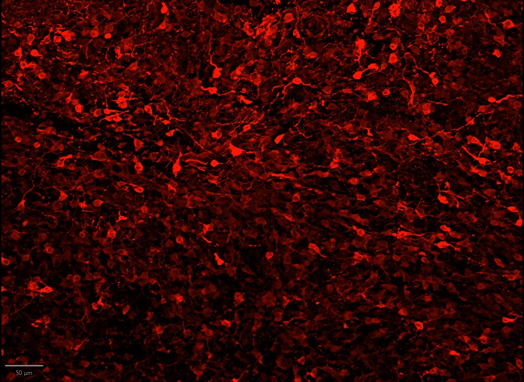
Application: ImmunofluorescenceSample Tested: Striatal organoidSpecies: HumanVerified Customer | Posted 04/17/2026Darpp32 staining of d85 striatal orgnoid showing MSNfixation: 4% PFA 4° overnight blocking + permeablization: 10% BSA + 1% TX + 0.05% Tween20 in PBS Primary antibody: DARPP‑32 (1:200) diluted in 4% BSA + 0.1% TX in PBS 4° overnight Secondary antibody: anti-goat 568 antibody diluted in 4% BSA + 0.1% TX RT 1h

There are no reviews that match your criteria.
Protocols
Find general support by application which include: protocols, troubleshooting, illustrated assays, videos and webinars.
- Antigen Retrieval Protocol (PIER)
- Antigen Retrieval for Frozen Sections Protocol
- Appropriate Fixation of IHC/ICC Samples
- Cellular Response to Hypoxia Protocols
- Chromogenic IHC Staining of Formalin-Fixed Paraffin-Embedded (FFPE) Tissue Protocol
- Chromogenic Immunohistochemistry Staining of Frozen Tissue
- ClariTSA™ Fluorophore Kits
- Detection & Visualization of Antibody Binding
- Fluorescent IHC Staining of Frozen Tissue Protocol
- Graphic Protocol for Heat-induced Epitope Retrieval
- Graphic Protocol for the Preparation and Fluorescent IHC Staining of Frozen Tissue Sections
- Graphic Protocol for the Preparation and Fluorescent IHC Staining of Paraffin-embedded Tissue Sections
- Graphic Protocol for the Preparation of Gelatin-coated Slides for Histological Tissue Sections
- IHC Sample Preparation (Frozen sections vs Paraffin)
- Immunofluorescent IHC Staining of Formalin-Fixed Paraffin-Embedded (FFPE) Tissue Protocol
- Immunohistochemistry (IHC) and Immunocytochemistry (ICC) Protocols
- Immunohistochemistry Frozen Troubleshooting
- Immunohistochemistry Paraffin Troubleshooting
- Preparing Samples for IHC/ICC Experiments
- Preventing Non-Specific Staining (Non-Specific Binding)
- Primary Antibody Selection & Optimization
- Protocol for Heat-Induced Epitope Retrieval (HIER)
- Protocol for Making a 4% Formaldehyde Solution in PBS
- Protocol for VisUCyte™ HRP Polymer Detection Reagent
- Protocol for the Preparation & Fixation of Cells on Coverslips
- Protocol for the Preparation and Chromogenic IHC Staining of Frozen Tissue Sections
- Protocol for the Preparation and Chromogenic IHC Staining of Frozen Tissue Sections - Graphic
- Protocol for the Preparation and Chromogenic IHC Staining of Paraffin-embedded Tissue Sections
- Protocol for the Preparation and Chromogenic IHC Staining of Paraffin-embedded Tissue Sections - Graphic
- Protocol for the Preparation and Fluorescent IHC Staining of Frozen Tissue Sections
- Protocol for the Preparation and Fluorescent IHC Staining of Paraffin-embedded Tissue Sections
- Protocol for the Preparation of Gelatin-coated Slides for Histological Tissue Sections
- R&D Systems Quality Control Western Blot Protocol
- TUNEL and Active Caspase-3 Detection by IHC/ICC Protocol
- The Importance of IHC/ICC Controls
- Troubleshooting Guide: Immunohistochemistry
- Troubleshooting Guide: Western Blot Figures
- Western Blot Conditions
- Western Blot Protocol
- Western Blot Protocol for Cell Lysates
- Western Blot Troubleshooting
- Western Blot Troubleshooting Guide
- View all Protocols, Troubleshooting, Illustrated assays and Webinars
Loading...
Associated Pathways
