The heat shock proteins are a highly conserved family of stress response proteins. HSPs function primarily as molecular chaperones, facilitating the folding of other cellular proteins, preventing protein aggregation, or targeting improperly folded proteins to specific degradative pathways. Some HSPs are expressed at low levels under normal physiological conditions but show dramatically increased expression in response to cellular stress, others are constitutively expressed. Specific HSPs play a role in regulating apoptosis by interacting directly with key components of the apoptotic pathway.

Key Product Details
Validated by
Biological Validation
Species Reactivity
Validated:
Human, Mouse, Rat
Cited:
Human, Mouse, Rat, Primate - Chlorocebus aethiops (African Green Monkey)
Applications
Validated:
Immunohistochemistry, Western Blot, Simple Western
Cited:
Immunohistochemistry, Western Blot, Flow Cytometry, Immunocytochemistry, Immunoprecipitation, Microarray
Label
Unconjugated
Antibody Source
Monoclonal Mouse IgG2A Clone # 242707
Loading...
Product Specifications
Immunogen
E. coli-derived recombinant human HSP70/HSPA1A
Met1-Asp641
Accession # NP_005336
Met1-Asp641
Accession # NP_005336
Specificity
Detects the induced form of human and mouse HSP70/HSPA1A in Western blots. In Western blots, no cross-reactivity with the constitutively expressed HSC70 (HSP73) is detected.
Clonality
Monoclonal
Host
Mouse
Isotype
IgG2A
Scientific Data Images for HSP70/HSPA1A Antibody (242707)
Detection of Human and Mouse HSP70/ HSPA1A by Western Blot.
Western blot shows lysates of Jurkat human acute T cell leukemia cell line and NIH-3T3 mouse embryonic fibroblast cell line untreated (-) or treated (+) with a 42 °C heat shock for 30 minutes with a 3 hour recovery. PVDF membrane was probed with 0.1 µg/mL of Mouse Anti-Human/Mouse/Rat HSP70/HSPA1A Monoclonal Antibody (Catalog # MAB1663), followed by HRP-conjugated Anti-Mouse IgG Secondary Antibody (Catalog # HAF007). A specific band was detected for HSP70/HSPA1A at approximately 70 kDa (as indicated). This experiment was conducted under reducing conditions and using Immunoblot Buffer Group 2.
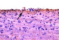
HSP70/HSPA1A in Human Liver Cancer Tissue.
HSP70/HSPA1A was detected in immersion fixed paraffin-embedded sections of human liver cancer tissue using Mouse Anti-Human/Mouse/Rat HSP70/HSPA1A Monoclonal Antibody (Catalog # MAB1663) at 25 µg/mL overnight at 4 °C. Tissue was stained using the Anti-Mouse HRP-DAB Cell & Tissue Staining Kit (brown; Catalog # CTS002) and counterstained with hematoxylin (blue). View our protocol for Chromogenic IHC Staining of Paraffin-embedded Tissue Sections.
Detection of Human HSP70/HSPA1A by Simple WesternTM.
Simple Western lane view shows lysates of Jurkat human acute T cell leukemia cell line untreated (-) or treated (+) by heat shocked (HS), loaded at 0.2 mg/mL. A specific band was detected for HSP70/HSPA1A at approximately 67 kDa (as indicated) using 0.5 µg/mL of Mouse Anti-Human/Mouse/Rat HSP70/HSPA1A Monoclonal Antibody (Catalog # MAB1663). This experiment was conducted under reducing conditions and using the 12-230 kDa separation system.
Detection of Mouse HSP70/HSPA1A by Western Blot
KNK437 inhibition assay of cell cultures. (A) Viability test results of KNK437 (10, 50 and 100 μM) inhibition assay of mononuclear peripheral blood burst formation unit-erythroid (BFU-E) cultures, with and without EPO. (B) Viability test results of a KNK437 (50 and 100 μM) inhibition assay of CD34+ bone marrow BFU-E cultures, with and without EPO. (C) Viability test results of KNK437 treatment (50 μM) on HEL and Ba/F3 JAK2 V617F (BAF3MUT) cell lines. X axis: concentration (μM); Y axis: % of viability cells (viability test) or BFU-E (BFU-E cultures) (D) Molecular effects of KNK437 in HEL cell line Western blot of HSP90, HSP70, p-JAK2 (decreased with KNK437 treatment), p-ERK and p-P38. Actin was used as the housekeeping control. (E) Molecular effects of KNK437 in Ba/F3 cell line Western blot of JAK2, p-ERK, ERK, p-STAT5, STAT5 AND Actin, (F) Western blot of HSP90, HSP70, JAK2, STAT5 and p-STAT5. Expression levels of HSP70, JAK2 and p-STAT5 decreased after HSP70 siRNA interference on HEL cell line. Actin was used as the housekeeping control. Image collected and cropped by CiteAb from the following publication (https://pubmed.ncbi.nlm.nih.gov/24252366), licensed under a CC-BY license. Not internally tested by R&D Systems.
Detection of Human HSP70/HSPA1A by Western Blot
KNK437 inhibition assay of cell cultures. (A) Viability test results of KNK437 (10, 50 and 100 μM) inhibition assay of mononuclear peripheral blood burst formation unit-erythroid (BFU-E) cultures, with and without EPO. (B) Viability test results of a KNK437 (50 and 100 μM) inhibition assay of CD34+ bone marrow BFU-E cultures, with and without EPO. (C) Viability test results of KNK437 treatment (50 μM) on HEL and Ba/F3 JAK2 V617F (BAF3MUT) cell lines. X axis: concentration (μM); Y axis: % of viability cells (viability test) or BFU-E (BFU-E cultures) (D) Molecular effects of KNK437 in HEL cell line Western blot of HSP90, HSP70, p-JAK2 (decreased with KNK437 treatment), p-ERK and p-P38. Actin was used as the housekeeping control. (E) Molecular effects of KNK437 in Ba/F3 cell line Western blot of JAK2, p-ERK, ERK, p-STAT5, STAT5 AND Actin, (F) Western blot of HSP90, HSP70, JAK2, STAT5 and p-STAT5. Expression levels of HSP70, JAK2 and p-STAT5 decreased after HSP70 siRNA interference on HEL cell line. Actin was used as the housekeeping control. Image collected and cropped by CiteAb from the following publication (https://pubmed.ncbi.nlm.nih.gov/24252366), licensed under a CC-BY license. Not internally tested by R&D Systems.Applications for HSP70/HSPA1A Antibody (242707)
Application
Recommended Usage
Immunohistochemistry
8-25 µg/mL
Sample: Immersion fixed paraffin-embedded sections of human liver cancer tissue
Sample: Immersion fixed paraffin-embedded sections of human liver cancer tissue
Simple Western
0.5 µg/mL
Sample: Heat shock treated Jurkat human acute T cell leukemia cell line
Sample: Heat shock treated Jurkat human acute T cell leukemia cell line
Western Blot
0.1 µg/mL
Sample: 42° C heat shock-treated Jurkat human acute T cell leukemia cell line and DA3 mouse myeloma cell line
Sample: 42° C heat shock-treated Jurkat human acute T cell leukemia cell line and DA3 mouse myeloma cell line
Reviewed Applications
Read 1 review rated 5 using MAB1663 in the following applications:
Formulation, Preparation, and Storage
Purification
Protein A or G purified from hybridoma culture supernatant
Reconstitution
Reconstitute at 0.5 mg/mL in sterile PBS. For liquid material, refer to CoA for concentration.
Loading...
Formulation
Lyophilized from a 0.2 μm filtered solution in PBS with Trehalose. *Small pack size (SP) is supplied either lyophilized or as a 0.2 µm filtered solution in PBS.
Shipping
Lyophilized product is shipped at ambient temperature. Liquid small pack size (-SP) is shipped with polar packs. Upon receipt, store immediately at the temperature recommended below.
Stability & Storage
Use a manual defrost freezer and avoid repeated freeze-thaw cycles.
- 12 months from date of receipt, -20 to -70 °C as supplied.
- 1 month, 2 to 8 °C under sterile conditions after reconstitution.
- 6 months, -20 to -70 °C under sterile conditions after reconstitution.
Calculators
Background: HSP70/HSPA1A
Long Name
Heat Shock Protein 70
Alternate Names
HSP70-1A, HSP72, HSPA1A
Gene Symbol
HSPA1A
UniProt
Additional HSP70/HSPA1A Products
Product Documents for HSP70/HSPA1A Antibody (242707)
Certificate of Analysis
To download a Certificate of Analysis, please enter a lot or batch number in the search box below.
Note: Certificate of Analysis not available for kit components.
Product Specific Notices for HSP70/HSPA1A Antibody (242707)
For research use only
Related Research Areas
Citations for HSP70/HSPA1A Antibody (242707)
 Powered by Bioz
Customer Reviews for HSP70/HSPA1A Antibody (242707) (1)
5 out of 5
1 Customer Rating
Have you used HSP70/HSPA1A Antibody (242707)?
Submit a review and receive an Amazon gift card!
$25/€18/£15/$25CAN/¥2500 Yen for a review with an image
$10/€7/£6/$10CAN/¥1110 Yen for a review without an image
Submit a review
Customer Images
Showing
1
-
1 of
1 review
Showing All
Filter By:
-
Application: ImmunohistochemistrySample Tested: Spinal cordSpecies: MouseVerified Customer | Posted 08/19/2021

There are no reviews that match your criteria.
Protocols
Find general support by application which include: protocols, troubleshooting, illustrated assays, videos and webinars.
- Antigen Retrieval Protocol (PIER)
- Antigen Retrieval for Frozen Sections Protocol
- Appropriate Fixation of IHC/ICC Samples
- Cellular Response to Hypoxia Protocols
- Chromogenic IHC Staining of Formalin-Fixed Paraffin-Embedded (FFPE) Tissue Protocol
- Chromogenic Immunohistochemistry Staining of Frozen Tissue
- ClariTSA™ Fluorophore Kits
- Detection & Visualization of Antibody Binding
- Fluorescent IHC Staining of Frozen Tissue Protocol
- Graphic Protocol for Heat-induced Epitope Retrieval
- Graphic Protocol for the Preparation and Fluorescent IHC Staining of Frozen Tissue Sections
- Graphic Protocol for the Preparation and Fluorescent IHC Staining of Paraffin-embedded Tissue Sections
- Graphic Protocol for the Preparation of Gelatin-coated Slides for Histological Tissue Sections
- IHC Sample Preparation (Frozen sections vs Paraffin)
- Immunofluorescent IHC Staining of Formalin-Fixed Paraffin-Embedded (FFPE) Tissue Protocol
- Immunohistochemistry (IHC) and Immunocytochemistry (ICC) Protocols
- Immunohistochemistry Frozen Troubleshooting
- Immunohistochemistry Paraffin Troubleshooting
- Preparing Samples for IHC/ICC Experiments
- Preventing Non-Specific Staining (Non-Specific Binding)
- Primary Antibody Selection & Optimization
- Protocol for Heat-Induced Epitope Retrieval (HIER)
- Protocol for Making a 4% Formaldehyde Solution in PBS
- Protocol for VisUCyte™ HRP Polymer Detection Reagent
- Protocol for the Preparation & Fixation of Cells on Coverslips
- Protocol for the Preparation and Chromogenic IHC Staining of Frozen Tissue Sections
- Protocol for the Preparation and Chromogenic IHC Staining of Frozen Tissue Sections - Graphic
- Protocol for the Preparation and Chromogenic IHC Staining of Paraffin-embedded Tissue Sections
- Protocol for the Preparation and Chromogenic IHC Staining of Paraffin-embedded Tissue Sections - Graphic
- Protocol for the Preparation and Fluorescent IHC Staining of Frozen Tissue Sections
- Protocol for the Preparation and Fluorescent IHC Staining of Paraffin-embedded Tissue Sections
- Protocol for the Preparation of Gelatin-coated Slides for Histological Tissue Sections
- R&D Systems Quality Control Western Blot Protocol
- TUNEL and Active Caspase-3 Detection by IHC/ICC Protocol
- The Importance of IHC/ICC Controls
- Troubleshooting Guide: Immunohistochemistry
- Troubleshooting Guide: Western Blot Figures
- Western Blot Conditions
- Western Blot Protocol
- Western Blot Protocol for Cell Lysates
- Western Blot Troubleshooting
- Western Blot Troubleshooting Guide
- View all Protocols, Troubleshooting, Illustrated assays and Webinars
Loading...
Associated Pathways
