
Netrin-1 (from Sanskrit netr meaning "to guide"; also known as Epididymis tissue protein Li 131P) is a 66-76 kDa secreted member of the laminin superfamily of molecules. It is expressed by multiple cell types, including embryonic neural tube ventral midline cells plus postnatal renal tubule epithelium and oligodendroglia. Netrin-1 has multiple receptors, including beta 4-integrins, DCC, DSCAM and UNC5A-thru-D. Its effects are context specific. For example, on neurons, binding to either DCC or DSCAM alone promotes chemoattraction, while a DCC:UNC5 complex promotes chemorepulsion. In addition, the ligation of UNC5B on leukocytes suppresses cytokine production, while the binding of netrin to DCC expressed on select cell types inhibits apoptosis. Mature human netrin-1 is 580 amino acids (aa) in length (aa 25-604). It contains an N-terminal laminin domain (aa 47-284), three EGF-like domains (aa 285-453) and a C-terminal cell-interaction netrin-like domain (aa 472-601). Over aa 22-604, human netrin-1 shares 99% aa sequence identity with mouse netrin-1.

Scientific images may be resized, cropped, or adjusted for brightness or contrast for presentation purposes. These updates are limited to presentation and do not affect the underlying scientific interpretation of the data. Where available, additional source data may be provided upon request.
Key Product Details
Species Reactivity
Validated:
Cited:
Applications
Validated:
Cited:
Label
Antibody Source
Product Specifications
Immunogen
Val22-Ala604
Accession # O95631
Specificity
Clonality
Host
Isotype
Scientific Data Images for Netrin-1 Antibody
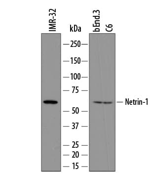
Detection of Human, Mouse, and Rat Netrin‑1 by Western Blot.
Western blot shows lysates of IMR-32 human neuroblastoma cell line, bEnd.3 mouse endothelioma cell line, and C6 rat glioma cell line. PVDF membrane was probed with 0.5 µg/mL of Sheep Anti-Human/Mouse/Rat Netrin-1 Antigen Affinity-purified Polyclonal Antibody (Catalog # AF6419) followed by HRP-conjugated Anti-Sheep IgG Secondary Antibody (Catalog # HAF016). A specific band was detected for Netrin-1 at approximately 66 kDa (as indicated). This experiment was conducted under reducing conditions and using Immunoblot Buffer Group 8.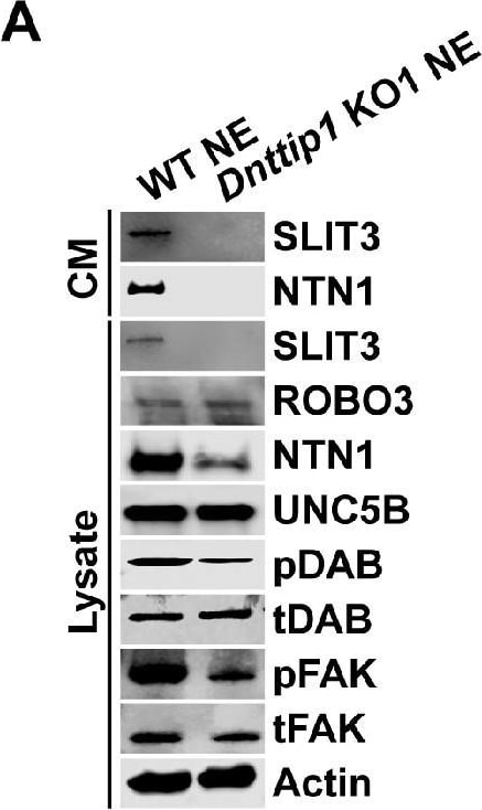
Detection of Mouse Netrin-1 by Western Blot
MiDAC regulates neurite outgrowth via the SLIT3/ROBO3 and NTN1/UNC5B signaling pathways.(A) WB for signaling components of the SLIT3/ROBO3 and NTN1/DCC/UNC5B signaling axes from CM and total cell lysates of WT and Dnttip1 KO1 NE after 12 days of differentiation. To enrich SLIT3 and NTN1 from CM, IPs were performed with SLIT3 and NTN1 antibodies from CM of WT and Dnttip1 KO1 NE. Actin is the loading control for the total cell lysates. (B) Assay to rescue the neurite outgrowth defects in Dnttip1 KO1 NE. CM of Dnttip1 KO1 NE was supplemented with the recombinant signaling ligands SLIT3 and/or NTN1 from day 7–12 without or with preblocking of Dnttip1 KO1 NE with IgG or signaling receptor antibodies against ROBO3 and/or UNC5B. MAP2 IF staining was performed after 12 days of differentiation. Nuclei were stained with DAPI. To facilitate analysis the neuronal cell body (blue) and its neurites were manually traced with ImageJ software and for each sample one traced neuron is displayed in the inlet. The white scale bar represents 50 µm. (C, D) Quantification of (C) neurite length and (D) the total number of neurites per neuron from the MAP2 IF staining in (B) using ImageJ. (C) Neurite length was divided into two categories of short neurites <50 µm (green box plots) and longer neurites ≥50 µm (white box plots). (C, D) The neurites of 200 neurons were assessed per sample. One-way ANOVA was performed throughout where ***, p≤0.001; and ns, p>0.05 is not significant.MiDAC regulates neurite outgrowth via the SLIT3/ROBO3 and NTN1/UNC5B signaling pathways.(A) Chamber assay to rescue the network formation defects of GNP-derived neurons that were supplemented with CM of Dnttip1 KO1 NE. CM of Dnttip1 KO1 NE was supplemented with the recombinant signaling ligands SLIT3 and/or NTN1 from day 7–12 without or with pre-blocking of GNP-derived neurons with control IgG or signaling receptor antibodies against ROBO3 and/or UNC5B. TUBB3 IF staining was performed after 12 days of differentiation.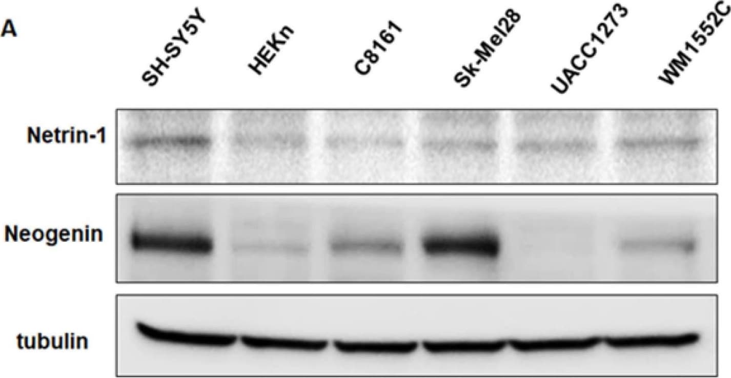
Detection of Netrin-1 by Western Blot
Expression of Netrin-1 and Neogenin in human melanoma cells. (A) Western blot results show different levels of Netrin-1 and Neogenin expression in human melanoma cell lines. The human neuroblastoma cell line SH-SY5Y and the normal human epidermal keratinocyte cell line HEKn were used as positive-expressing and poorly expressing controls for cellular expression of Netrin-1, respectively. (B) Results from immunofluorescence (IF) staining shows more intense expression of Neogenin in the aggressive melanoma cell lines C8161 and Sk-Mel28 compared to the poorly aggressive melanoma cell line UACC1273. SH-SY5Y was used as positive control for the IF staining for Neogenin. (Original magnification 20X). (C) The histogram shows the ratio of Neogenin to Netrin-1 expression determined from densitometric analysis of the respective WB bands and represented as a fold difference relative to the Neogenin-to-Netrin-1 ratio calculated for SH-SY5Y, which was used as control. These results clearly show that Neogenin expression relative to Netrin-1 is significantly greater in aggressive compared to poorly aggressive melanoma cells (* p < 0.05, n = 3). Image collected and cropped by CiteAb from the following open publication (https://pubmed.ncbi.nlm.nih.gov/36361539), licensed under a CC-BY license. Not internally tested by R&D Systems.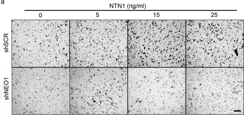
Detection of Netrin-1 by Immunohistochemistry
NEO1 promotes chemotactic NTN1-mediated cell migration. a: Representative transwell assay images performed with shSCR and shNEO1 SK-N-SH cells which migrated for 4 hours in increasing concentrations of NTN1 indicated in Figure. Bar = 100 µm. b: Quantification of the photographs taken for each condition. Values are expressed as induction times of migration relative to the condition without chemotactic stimulus (0 ng/ml NTN1) for shSCR and shNeo1 cells. N = 3, n = 5 fields per condition were counted, * p < 0.05 0 v/s 25 ng/ml NTN1. c: Representative images of confocal microscopy of spheroid-based migration assay on fibronectin for 1 h, comparing shSCR versus NEO1 knock-down cells. The images reveal F-actin labeling. d: Quantification of cells that migrated away from the spheroid for each condition tested. N = 3, n = 15. *** p < 0.01 shSCR versus shNEO1 Image collected and cropped by CiteAb from the following open publication (https://pubmed.ncbi.nlm.nih.gov/33724150), licensed under a CC-BY license. Not internally tested by R&D Systems.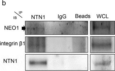
Detection of Netrin-1 by Western Blot
NEO1/NTN1 form a complex with integrin beta 1 in SK-N-SH cells. a: Representative western blots (WB) of protein co-immunoprecipitation assays used to evaluate interaction between NEO1 with NTN1 and integrin beta 1. b: Representative WB of protein co-immunoprecipitation assays used to evaluate interaction between NTN1 with NEO1 and integrin beta 1, c: Representative WB of protein co-immunoprecipitation assays used to evaluate interaction between integrin beta 1 with NTN1 and NEO1. N = 2. WCL: whole cell lysate. N = 2, n = 3 Image collected and cropped by CiteAb from the following open publication (https://pubmed.ncbi.nlm.nih.gov/33724150), licensed under a CC-BY license. Not internally tested by R&D Systems.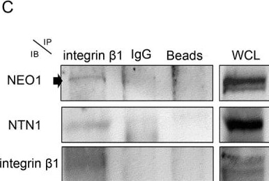
Detection of Netrin-1 by Western Blot
NEO1/NTN1 form a complex with integrin beta 1 in SK-N-SH cells. a: Representative western blots (WB) of protein co-immunoprecipitation assays used to evaluate interaction between NEO1 with NTN1 and integrin beta 1. b: Representative WB of protein co-immunoprecipitation assays used to evaluate interaction between NTN1 with NEO1 and integrin beta 1, c: Representative WB of protein co-immunoprecipitation assays used to evaluate interaction between integrin beta 1 with NTN1 and NEO1. N = 2. WCL: whole cell lysate. N = 2, n = 3 Image collected and cropped by CiteAb from the following open publication (https://pubmed.ncbi.nlm.nih.gov/33724150), licensed under a CC-BY license. Not internally tested by R&D Systems.Applications for Netrin-1 Antibody
Western Blot
Sample: IMR‑32 human neuroblastoma cell line, bEnd.3 mouse endothelioma cell line, and C6 rat glioma cell line
Formulation, Preparation, and Storage
Purification
Reconstitution
Sterile PBS to a final concentration of 0.2 mg/mL. For liquid material, refer to CoA for concentration.
Formulation
Shipping
Stability & Storage
- 12 months from date of receipt, -20 to -70 °C as supplied.
- 1 month, 2 to 8 °C under sterile conditions after reconstitution.
- 6 months, -20 to -70 °C under sterile conditions after reconstitution.
Calculators
Background: Netrin-1
Alternate Names
Gene Symbol
UniProt
Additional Netrin-1 Products
Product Documents for Netrin-1 Antibody
Certificate of Analysis
To download a Certificate of Analysis, please enter a lot or batch number in the search box below.
Note: Certificate of Analysis not available for kit components.
Product Specific Notices for Netrin-1 Antibody
For research use only
Related Research Areas
Citations for Netrin-1 Antibody
 Powered by Bioz
Customer Reviews for Netrin-1 Antibody
There are currently no reviews for this product. Be the first to review Netrin-1 Antibody and earn rewards!
Have you used Netrin-1 Antibody?
Submit a review and receive an Amazon gift card!
$25/€18/£15/$25CAN/¥2500 for a review with an image
$10/€7/£6/$10CAN/¥1110 for a review without an image
Submit a review
Protocols
Find general support by application which include: protocols, troubleshooting, illustrated assays, videos and webinars.
- Cellular Response to Hypoxia Protocols
- R&D Systems Quality Control Western Blot Protocol
- Troubleshooting Guide: Western Blot Figures
- Western Blot Conditions
- Western Blot Protocol
- Western Blot Protocol for Cell Lysates
- Western Blot Troubleshooting
- Western Blot Troubleshooting Guide
- View all Protocols, Troubleshooting, Illustrated assays and Webinars
Associated Pathways
