
RGM-C, also known as hemojuvelin, is a member of the repulsive guidance molecule (RGM) family of GPI-linked neuronal and muscle membrane glycoproteins (1, 2). RGM-C is expressed in striated muscle and periportal hepatocytes (3-5). The protein undergoes partial cleavage intracellularly, resulting in a disulfide-linked dimer of the 14 kDa N-terminal and 33 kDa C-terminal portions (4, 6, 7). The N-terminal fragment contains an RGD motif, while the C-terminal fragment carries the GPI attachment site (4, 7). Two alternatively spliced isoforms lack either approximately half or the entire N-terminal fragment. Full length RGM-C can also be released from the cell and circulates in the blood (6, 8). RGM-C is disrupted in type 2A juvenile hemochromatosis, a hereditary iron homeostasis disorder characterized by excessive iron accumulation (5). In mouse, loss of RGM-C function results in decreased expression of the iron regulatory hormone hepicidin and increased iron deposition in liver, pancreas, and heart (5, 9). Membrane associated RGM-C upregulates hepicidin while soluble RGM-C downregulates hepicidin expression (8). This appears to be an iron-responsive regulatory system, as high blood iron levels reduce the amount of soluble RGM-C produced (8). RGM-C, similar to RGM-A, associates with neogenin (7). Disease-related point mutations can prevent internal RGM-C cleavage or its ability to interact with neogenin (6, 7). Experimental inflammatory conditions result in decreased RGM-C expression and increased hepicidin expression, although the two effects occur independently (5, 10). RGM-C also functions as a BMP coreceptor and enhances BMP-2 and BMP-4 signaling (11). In this context, RGM-C enhances the BMP-2 upregulation of hepatic hepicidin (11). Mature human RGM-C shares 89% amino acid (aa) sequence identity with mouse and rat RGM-C. It shares 49% and 44% aa sequence identity with human RGM-A and RGM-B, respectively.
RGM-C/Hemojuvelin Antibody
R&D Systems | Catalog # AF3720

Scientific images may be resized, cropped, or adjusted for brightness or contrast for presentation purposes. These updates are limited to presentation and do not affect the underlying scientific interpretation of the data. Where available, additional source data may be provided upon request.
Key Product Details
Species Reactivity
Validated:
Cited:
Applications
Validated:
Cited:
Label
Antibody Source
Product Specifications
Immunogen
Gln36-Asp400
Accession # Q6ZVN8
Specificity
Clonality
Host
Isotype
Scientific Data Images for RGM-C/Hemojuvelin Antibody
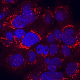
RGM‑C/Hemojuvelin in HepG2 Human Cell Line.
RGM-C/Hemojuvelin was detected in immersion fixed HepG2 human hepatocellular carcinoma cell line using Goat Anti-Human/Mouse RGM-C/Hemojuvelin Antigen Affinity-purified Polyclonal Antibody (Catalog # AF3720) at 10 µg/mL for 3 hours at room temperature. Cells were stained using the NorthernLights™ 557-conjugated Anti-Goat IgG Secondary Antibody (red; Catalog # NL001) and counterstained with DAPI (blue). Specific staining was localized to cytoplasm. View our protocol for Fluorescent ICC Staining of Cells on Coverslips.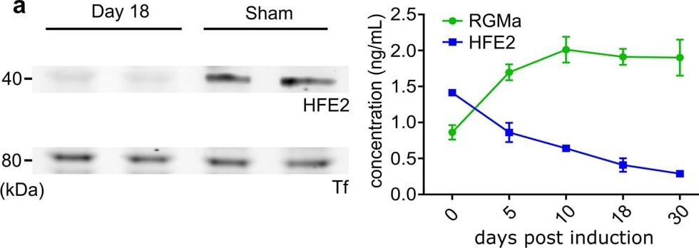
Detection of RGM-C/Hemojuvelin by Western Blot
HFE2 administration significantly improves EAE disease outcome.a Western Blot analysis shows a strong HFE2 reduction in EAE animal serum when compared to control. ELISA quantification showing HFE2 decrease and RGMa increase in EAE animal serum (mean ± s.d.; EAE n = 3 and RGMa n = 3). b Body conditioning assessment (‘0’ no paralysis to ‘4’ forelimb weakness) of EAE-induced mice with HFE2 + /- RGMa treatment every 3 days (d3, 6, 9, 12 and 15; mean ± s.e.m.; unpaired two-tail t-tests; PBS n = 8, HFE2 n = 6, RGMa+ HFE2 n = 8). c Body conditioning assessment of EAE-induced Hfe2fl/fl and Hfe2 delta Alb-cre mice (mean ± s.e.m.; unpaired two-tail t-tests; Hfe2fl/fln = 8 and Hfe2 delta Alb-cren = 8). d Representative confocal images of CD3 +, B220+ and CD11b+ immune staining in spinal cord of EAE-induced mice 3 weeks after induction and quantification (mean ± s.e.m.; unpaired two-tail t-test; PBS n = 7, HFE2 n = 6 mice; technical replicates shown). e Schematic of activated immune cell adoptive transfer. Representative confocal images of CD4-positive labelled transferred cell (CellVue) in PBS-treated or HFE2-treated recipient WT mice and quantification (mean ± s.e.m.; unpaired two-tail t-test; PBS n = 6, HFE2 n = 6). Ptx; Pertussis Toxin. f Representative confocal images of fibrinogen in the spinal cord of HFE2-treated EAE-mice and quantification (mean ± s.e.m.; unpaired two-tail t-test; PBS-treated n = 6 and HFE2 n = 6). *P < 0.05, **P < 0.01, ***P < 0.001. Source data are provided as a Source Data file (includes exact p-values). Image collected and cropped by CiteAb from the following open publication (https://pubmed.ncbi.nlm.nih.gov/38310100), licensed under a CC-BY license. Not internally tested by R&D Systems.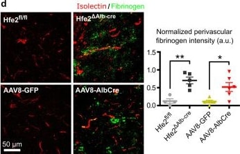
Detection of RGM-C/Hemojuvelin by Immunohistochemistry
Hfe2-deficient mice displays severe BCB disruption.a Schematic of the BCB assessment method using light-sheet microscopy. b 3D rendered representative light-sheet microscopic images of TR-dextran deposition in CUBIC-cleared brains (scale bars, 400 µm) and time-series quantification (mean ± s.e.m.; unpaired two-tail t-test; Hfe2fl/fl (control) n = 5, Hfe2 delta AlbCren = 6, AAV8-GFP n = 5, AAV8-AlbCre n = 7; n representing data generated from one brain). c Representative in-vivo multiphoton images of TR-dextran at 40 mins time-point (scale bar, 50 µm) from the parietal lobes of the cerebral cortex. The normalized extravascular fluorescence ( delta F/F) intensity was plotted over time (mean ± s.e.m.; unpaired two-tail t-tests) and the groups differences were assessed with spatial leakage log-likelihood (repeated measures factorial ANOVA rendered as approximate unpaired two-tail t-test; Hfe2fl/fln = 5, Hfe2 delta AlbCren = 10, AAV8-GFP n = 6, AAV8-AlbCre n = 5). d Representative confocal images of fibrinogen disposition around the vessels of cerebral cortex (isolectin) of Hfe2-deficient mice (scale bar, 50 µm) and quantification (mean ± s.e.m.; unpaired two-tail t-test; Hfe2fl/fln = 4, Hfe2 delta AlbCren = 5, AAV8-GFP n = 5, AAV8-AlbCre n = 6). e Confocal imaging of TR-dextran deposition in brain slices from the cerebral cortex with endothelium counterstain. The same observation was made in sections from 3 AAV8-GFP and 3 AAV8-AlbCre brains. f 3D rendered representative light-sheet microscopic images of TR-dextran deposition in CUBIC-cleared brains (scale bars, 400 µm; mean ± s.e.m.; unpaired two-tail t-test; Hfe2fl/fln = 5, Hfe2 delta ActaCren = 6). *P < 0.05, **P < 0.01, ***P < 0.001. Source data are provided as a Source Data file (includes exact p-values). Image collected and cropped by CiteAb from the following open publication (https://pubmed.ncbi.nlm.nih.gov/38310100), licensed under a CC-BY license. Not internally tested by R&D Systems.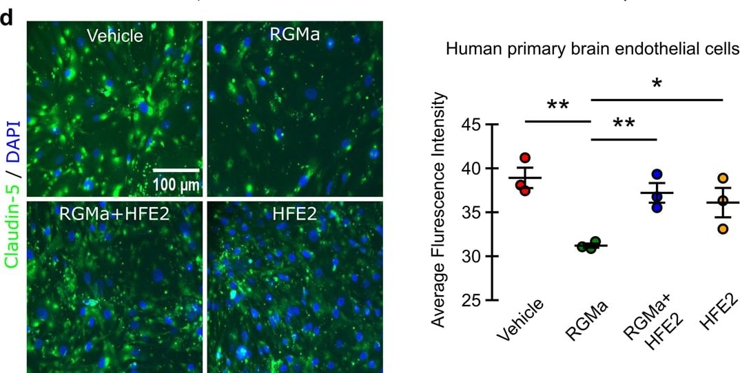
Detection of RGM-C/Hemojuvelin by Immunohistochemistry
Soluble HFE2 and RGMa effects on endothelial cells.a Knockout of liver Hfe2 reduces pericyte coverage in section from the cerebral cortex (mean ± s.e.m.; unpaired two-tail t-test; AAV8-GFP n = 3, Hfe2 delta Alb-cren = 4, and AAV8-AlbCre n = 3). Scale bar, 50μm. b Quantification through quantitative RT-PCR shows a reduction in PDGF-B mRNA levels in liver knock out animals (mean ± s.e.m.; paired two-tail t-test; Hfe2fl/fln = 3 and Hfe2 delta Alb-cren = 3). c Quantification of PDGF-B mRNA in cultured bEnd.3 cells following indicated treatments. Treatment with RGMa leads to a significant reduction of PDGF-B mRNA levels that is rescued with HFE2 addition (mean ± s.e.m.; paired two-tail t-test; HFE2 n = 3, RGMa n = 4, and RGMa+ HFE2 n = 5; n representing an independent experiment). d Immunocytochemistry of claudin-5 in human primary endothelial cell monolayer after PBS, HFE2, RGMa, and RGMa+HFE2 treatments and quantification (mean ± s.e.m.; unpaired two-tail t-test; replicates n = 3). Scale bar, 100 µm. e Western blotting of Claudin-5 expression in bEnd3 cell lysates after indicated treatment (mean ± s.e.m.; unpaired two-tailed t-test; replicates n = 3). f Transwell permeability leakage assay performed on a monolayer of bEnd3 cells using HRP (mean ± s.e.m.; paired one-tail t-test; replicates n = 3). g Transwell permeability leakage assay performed on a monolayer of human primary endothelial cells using 70 kDa FITC-dextran (mean ± s.e.m.; paired two-tail t-test; replicates n = 3). h In vitro TEER analysis shows that RGMa alters the integrity of a monolayer of bEnd3 cells, this is rescued by HFE2 (mean ± s.e.m.; unpaired two-tail t-test; replicates n = 3). *P < 0.05, **P < 0.01, ***P < 0.001. Source data are provided as a Source Data file (includes exact p-values). Image collected and cropped by CiteAb from the following open publication (https://pubmed.ncbi.nlm.nih.gov/38310100), licensed under a CC-BY license. Not internally tested by R&D Systems.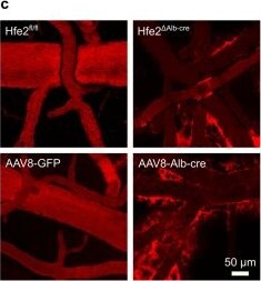
Detection of RGM-C/Hemojuvelin by Immunohistochemistry
Hfe2-deficient mice displays severe BCB disruption.a Schematic of the BCB assessment method using light-sheet microscopy. b 3D rendered representative light-sheet microscopic images of TR-dextran deposition in CUBIC-cleared brains (scale bars, 400 µm) and time-series quantification (mean ± s.e.m.; unpaired two-tail t-test; Hfe2fl/fl (control) n = 5, Hfe2 delta AlbCren = 6, AAV8-GFP n = 5, AAV8-AlbCre n = 7; n representing data generated from one brain). c Representative in-vivo multiphoton images of TR-dextran at 40 mins time-point (scale bar, 50 µm) from the parietal lobes of the cerebral cortex. The normalized extravascular fluorescence ( delta F/F) intensity was plotted over time (mean ± s.e.m.; unpaired two-tail t-tests) and the groups differences were assessed with spatial leakage log-likelihood (repeated measures factorial ANOVA rendered as approximate unpaired two-tail t-test; Hfe2fl/fln = 5, Hfe2 delta AlbCren = 10, AAV8-GFP n = 6, AAV8-AlbCre n = 5). d Representative confocal images of fibrinogen disposition around the vessels of cerebral cortex (isolectin) of Hfe2-deficient mice (scale bar, 50 µm) and quantification (mean ± s.e.m.; unpaired two-tail t-test; Hfe2fl/fln = 4, Hfe2 delta AlbCren = 5, AAV8-GFP n = 5, AAV8-AlbCre n = 6). e Confocal imaging of TR-dextran deposition in brain slices from the cerebral cortex with endothelium counterstain. The same observation was made in sections from 3 AAV8-GFP and 3 AAV8-AlbCre brains. f 3D rendered representative light-sheet microscopic images of TR-dextran deposition in CUBIC-cleared brains (scale bars, 400 µm; mean ± s.e.m.; unpaired two-tail t-test; Hfe2fl/fln = 5, Hfe2 delta ActaCren = 6). *P < 0.05, **P < 0.01, ***P < 0.001. Source data are provided as a Source Data file (includes exact p-values). Image collected and cropped by CiteAb from the following open publication (https://pubmed.ncbi.nlm.nih.gov/38310100), licensed under a CC-BY license. Not internally tested by R&D Systems.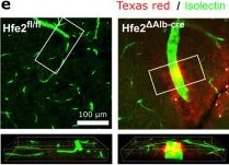
Detection of RGM-C/Hemojuvelin by Immunohistochemistry
Hfe2-deficient mice displays severe BCB disruption.a Schematic of the BCB assessment method using light-sheet microscopy. b 3D rendered representative light-sheet microscopic images of TR-dextran deposition in CUBIC-cleared brains (scale bars, 400 µm) and time-series quantification (mean ± s.e.m.; unpaired two-tail t-test; Hfe2fl/fl (control) n = 5, Hfe2 delta AlbCren = 6, AAV8-GFP n = 5, AAV8-AlbCre n = 7; n representing data generated from one brain). c Representative in-vivo multiphoton images of TR-dextran at 40 mins time-point (scale bar, 50 µm) from the parietal lobes of the cerebral cortex. The normalized extravascular fluorescence ( delta F/F) intensity was plotted over time (mean ± s.e.m.; unpaired two-tail t-tests) and the groups differences were assessed with spatial leakage log-likelihood (repeated measures factorial ANOVA rendered as approximate unpaired two-tail t-test; Hfe2fl/fln = 5, Hfe2 delta AlbCren = 10, AAV8-GFP n = 6, AAV8-AlbCre n = 5). d Representative confocal images of fibrinogen disposition around the vessels of cerebral cortex (isolectin) of Hfe2-deficient mice (scale bar, 50 µm) and quantification (mean ± s.e.m.; unpaired two-tail t-test; Hfe2fl/fln = 4, Hfe2 delta AlbCren = 5, AAV8-GFP n = 5, AAV8-AlbCre n = 6). e Confocal imaging of TR-dextran deposition in brain slices from the cerebral cortex with endothelium counterstain. The same observation was made in sections from 3 AAV8-GFP and 3 AAV8-AlbCre brains. f 3D rendered representative light-sheet microscopic images of TR-dextran deposition in CUBIC-cleared brains (scale bars, 400 µm; mean ± s.e.m.; unpaired two-tail t-test; Hfe2fl/fln = 5, Hfe2 delta ActaCren = 6). *P < 0.05, **P < 0.01, ***P < 0.001. Source data are provided as a Source Data file (includes exact p-values). Image collected and cropped by CiteAb from the following open publication (https://pubmed.ncbi.nlm.nih.gov/38310100), licensed under a CC-BY license. Not internally tested by R&D Systems.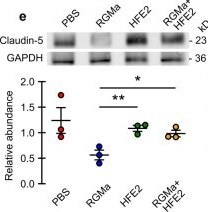
Detection of RGM-C/Hemojuvelin by Western Blot
Soluble HFE2 and RGMa effects on endothelial cells.a Knockout of liver Hfe2 reduces pericyte coverage in section from the cerebral cortex (mean ± s.e.m.; unpaired two-tail t-test; AAV8-GFP n = 3, Hfe2 delta Alb-cren = 4, and AAV8-AlbCre n = 3). Scale bar, 50μm. b Quantification through quantitative RT-PCR shows a reduction in PDGF-B mRNA levels in liver knock out animals (mean ± s.e.m.; paired two-tail t-test; Hfe2fl/fln = 3 and Hfe2 delta Alb-cren = 3). c Quantification of PDGF-B mRNA in cultured bEnd.3 cells following indicated treatments. Treatment with RGMa leads to a significant reduction of PDGF-B mRNA levels that is rescued with HFE2 addition (mean ± s.e.m.; paired two-tail t-test; HFE2 n = 3, RGMa n = 4, and RGMa+ HFE2 n = 5; n representing an independent experiment). d Immunocytochemistry of claudin-5 in human primary endothelial cell monolayer after PBS, HFE2, RGMa, and RGMa+HFE2 treatments and quantification (mean ± s.e.m.; unpaired two-tail t-test; replicates n = 3). Scale bar, 100 µm. e Western blotting of Claudin-5 expression in bEnd3 cell lysates after indicated treatment (mean ± s.e.m.; unpaired two-tailed t-test; replicates n = 3). f Transwell permeability leakage assay performed on a monolayer of bEnd3 cells using HRP (mean ± s.e.m.; paired one-tail t-test; replicates n = 3). g Transwell permeability leakage assay performed on a monolayer of human primary endothelial cells using 70 kDa FITC-dextran (mean ± s.e.m.; paired two-tail t-test; replicates n = 3). h In vitro TEER analysis shows that RGMa alters the integrity of a monolayer of bEnd3 cells, this is rescued by HFE2 (mean ± s.e.m.; unpaired two-tail t-test; replicates n = 3). *P < 0.05, **P < 0.01, ***P < 0.001. Source data are provided as a Source Data file (includes exact p-values). Image collected and cropped by CiteAb from the following open publication (https://pubmed.ncbi.nlm.nih.gov/38310100), licensed under a CC-BY license. Not internally tested by R&D Systems.Applications for RGM-C/Hemojuvelin Antibody
Immunocytochemistry
Sample: Immersion fixed HepG2 human hepatocellular carcinoma cell line
Western Blot
Sample:
Recombinant Human RGM-C (Catalog # 3720-RG)
Recombinant Mouse RGM-C (Catalog # 3634-RG)
Formulation, Preparation, and Storage
Purification
Reconstitution
Reconstitute at 0.2 mg/mL in sterile PBS. For liquid material, refer to CoA for concentration.
Formulation
Shipping
Stability & Storage
- 12 months from date of receipt, -20 to -70 °C as supplied.
- 1 month, 2 to 8 °C under sterile conditions after reconstitution.
- 6 months, -20 to -70 °C under sterile conditions after reconstitution.
Calculators
Background: RGM-C/Hemojuvelin
References
- Papanikolaou, G. et al. (2004) Nat. Genet. 36:77.
- Schmidtmer, J. and D. Engelkamp (2004) Gene Exp. Patterns 4:105.
- Oldekamp, J. et al. (2004) Gene Exp. Patterns 4:283.
- Niederkofler, V. et al. (2004) J. Neurosci. 24:808.
- Niederkofler, V. et al. (2005) J. Clin. Invest. 115:2180.
- Kuninger, D. et al. (2006) J. Cell Sci. 119:3273.
- Zhang, A.S. et al. (2005) J. Biol. Chem. 280:33885.
- Lin, L. et al. (2005) Blood 106:2884.
- Huang, F.W. et al. (2005) J. Clin. Invest. 115:2187.
- Krijt, J. et al. (2004) Blood 104:4308.
- Babitt, J.L. et al. (2006) Nat. Genet. 38:531.
Long Name
Alternate Names
Gene Symbol
UniProt
Additional RGM-C/Hemojuvelin Products
Product Documents for RGM-C/Hemojuvelin Antibody
Certificate of Analysis
To download a Certificate of Analysis, please enter a lot or batch number in the search box below.
Note: Certificate of Analysis not available for kit components.
Product Specific Notices for RGM-C/Hemojuvelin Antibody
For research use only
Related Research Areas
Citations for RGM-C/Hemojuvelin Antibody
 Powered by Bioz
Customer Reviews for RGM-C/Hemojuvelin Antibody
There are currently no reviews for this product. Be the first to review RGM-C/Hemojuvelin Antibody and earn rewards!
Have you used RGM-C/Hemojuvelin Antibody?
Submit a review and receive an Amazon gift card!
$25/€18/£15/$25CAN/¥2500 for a review with an image
$10/€7/£6/$10CAN/¥1110 for a review without an image
Submit a review
Protocols
Find general support by application which include: protocols, troubleshooting, illustrated assays, videos and webinars.
- Appropriate Fixation of IHC/ICC Samples
- Cellular Response to Hypoxia Protocols
- ClariTSA™ Fluorophore Kits
- Detection & Visualization of Antibody Binding
- ICC Cell Smear Protocol for Suspension Cells
- ICC Immunocytochemistry Protocol Videos
- ICC for Adherent Cells
- Immunocytochemistry (ICC) Protocol
- Immunocytochemistry Troubleshooting
- Immunofluorescence of Organoids Embedded in Cultrex Basement Membrane Extract
- Immunohistochemistry (IHC) and Immunocytochemistry (ICC) Protocols
- Preparing Samples for IHC/ICC Experiments
- Preventing Non-Specific Staining (Non-Specific Binding)
- Primary Antibody Selection & Optimization
- Protocol for VisUCyte™ HRP Polymer Detection Reagent
- Protocol for the Fluorescent ICC Staining of Cell Smears - Graphic
- Protocol for the Fluorescent ICC Staining of Cultured Cells on Coverslips - Graphic
- Protocol for the Preparation and Fluorescent ICC Staining of Cells on Coverslips
- Protocol for the Preparation and Fluorescent ICC Staining of Non-adherent Cells
- Protocol for the Preparation and Fluorescent ICC Staining of Stem Cells on Coverslips
- Protocol for the Preparation of a Cell Smear for Non-adherent Cell ICC - Graphic
- R&D Systems Quality Control Western Blot Protocol
- TUNEL and Active Caspase-3 Detection by IHC/ICC Protocol
- The Importance of IHC/ICC Controls
- Troubleshooting Guide: Western Blot Figures
- Western Blot Conditions
- Western Blot Protocol
- Western Blot Protocol for Cell Lysates
- Western Blot Troubleshooting
- Western Blot Troubleshooting Guide
- View all Protocols, Troubleshooting, Illustrated assays and Webinars