
Key Product Details
Species Reactivity
Validated:
Human, Mouse
Cited:
Human, Mouse
Applications
Validated:
Immunocytochemistry
Cited:
Immunohistochemistry, Western Blot, Immunocytochemistry, Infared Microscopy
Label
Unconjugated
Antibody Source
Monoclonal Rat IgG2A Clone # 303728
Loading...
Product Specifications
Immunogen
E. coli-derived recombinant human TOR
Phe1720-Ala2020
Accession # P42345
Phe1720-Ala2020
Accession # P42345
Specificity
Detects human TOR in direct ELISAs.
Clonality
Monoclonal
Host
Rat
Isotype
IgG2A
Scientific Data Images for TOR Antibody (303728)
TOR in MCF-7 Human Cell Line.
TOR was detected in immersion fixed MCF-7 human breast cancer cell line using 10 µg/mL Human/Mouse TOR Monoclonal Antibody (Catalog # MAB1537) for 3 hours at room temperature. Cells were stained with the NorthernLights™ 557-conjugated Anti-Rat IgG Secondary Antibody (red; Catalog # NL013) and counterstained with DAPI (blue). View our protocol for Fluorescent ICC Staining of Cells on Coverslips.Applications for TOR Antibody (303728)
Application
Recommended Usage
Immunocytochemistry
8-25 µg/mL
Sample: Immersion fixed MCF-7 human breast cancer cell line
Sample: Immersion fixed MCF-7 human breast cancer cell line
Reviewed Applications
Read 1 review rated 5 using MAB1537 in the following applications:
Formulation, Preparation, and Storage
Purification
Protein A or G purified from hybridoma culture supernatant
Reconstitution
Reconstitute at 0.5 mg/mL in sterile PBS. For liquid material, refer to CoA for concentration.
Loading...
Formulation
Lyophilized from a 0.2 μm filtered solution in PBS with Trehalose. *Small pack size (SP) is supplied either lyophilized or as a 0.2 µm filtered solution in PBS.
Shipping
Lyophilized product is shipped at ambient temperature. Liquid small pack size (-SP) is shipped with polar packs. Upon receipt, store immediately at the temperature recommended below.
Stability & Storage
Use a manual defrost freezer and avoid repeated freeze-thaw cycles.
- 12 months from date of receipt, -20 to -70 °C as supplied.
- 1 month, 2 to 8 °C under sterile conditions after reconstitution.
- 6 months, -20 to -70 °C under sterile conditions after reconstitution.
Calculators
Background: TOR
Long Name
Mammalian Target of Rapamycin
Alternate Names
FRAP1, FRAP2, MTOR, RAFT1
Gene Symbol
MTOR
UniProt
Additional TOR Products
Product Documents for TOR Antibody (303728)
Certificate of Analysis
To download a Certificate of Analysis, please enter a lot or batch number in the search box below.
Note: Certificate of Analysis not available for kit components.
Product Specific Notices for TOR Antibody (303728)
For research use only
Related Research Areas
Citations for TOR Antibody (303728)
 Powered by Bioz
Customer Reviews for TOR Antibody (303728) (1)
5 out of 5
1 Customer Rating
Have you used TOR Antibody (303728)?
Submit a review and receive an Amazon gift card!
$25/€18/£15/$25CAN/¥2500 Yen for a review with an image
$10/€7/£6/$10CAN/¥1110 Yen for a review without an image
Submit a review
Customer Images
Showing
1
-
1 of
1 review
Showing All
Filter By:
-
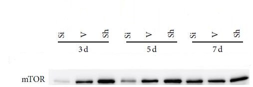
Application: Western BlotSample Tested: rat tissuesSpecies: RatVerified Customer | Posted 07/29/2017The western blot was utilized to detect the expression of mTORThe anti-mTOR antibody was used primary antibodies (1 : 1000; Novus Biologicals, USA).Our data showed it worked well.Antibody is good.

There are no reviews that match your criteria.
Protocols
Find general support by application which include: protocols, troubleshooting, illustrated assays, videos and webinars.
- Appropriate Fixation of IHC/ICC Samples
- Cellular Response to Hypoxia Protocols
- ClariTSA™ Fluorophore Kits
- Detection & Visualization of Antibody Binding
- ICC Cell Smear Protocol for Suspension Cells
- ICC Immunocytochemistry Protocol Videos
- ICC for Adherent Cells
- Immunocytochemistry (ICC) Protocol
- Immunocytochemistry Troubleshooting
- Immunofluorescence of Organoids Embedded in Cultrex Basement Membrane Extract
- Immunohistochemistry (IHC) and Immunocytochemistry (ICC) Protocols
- Preparing Samples for IHC/ICC Experiments
- Preventing Non-Specific Staining (Non-Specific Binding)
- Primary Antibody Selection & Optimization
- Protocol for VisUCyte™ HRP Polymer Detection Reagent
- Protocol for the Fluorescent ICC Staining of Cell Smears - Graphic
- Protocol for the Fluorescent ICC Staining of Cultured Cells on Coverslips - Graphic
- Protocol for the Preparation and Fluorescent ICC Staining of Cells on Coverslips
- Protocol for the Preparation and Fluorescent ICC Staining of Non-adherent Cells
- Protocol for the Preparation and Fluorescent ICC Staining of Stem Cells on Coverslips
- Protocol for the Preparation of a Cell Smear for Non-adherent Cell ICC - Graphic
- TUNEL and Active Caspase-3 Detection by IHC/ICC Protocol
- The Importance of IHC/ICC Controls
- View all Protocols, Troubleshooting, Illustrated assays and Webinars


