
N-Acetylglucosaminyltransferase V (GnT-V), also known as mannosylglycoprotein N-acetyl-glucosaminyltransferase 5 (MGAT5), adds an N-acetylglucosamine to the alpha 1-6-linked core mannose of an N-linked oligosaccharide in the Golgi apparatus (1). This reaction is the committing step for the biosynthesis of beta 1-6GlcNAc-branched arm in N-glycans. The degree of N-glycan branching has been shown to regulate cell proliferation and differentiation (2). An increase in the GnT-V activity and its glycan products is also known to positively correlate with the progression of invasive malignancies (3, 4). For example, ectopic expression of GnT-V in epithelial cells results in morphological transformation and tumor growth in mice and overexpression in carcinoma cells has been shown to induce metastatic spread (3-5).
Human N-Acetylglucosaminyltransferase V/MGAT5 Antibody (706824)
R&D Systems | Catalog # MAB5469

Scientific images may be resized, cropped, or adjusted for brightness or contrast for presentation purposes. These updates are limited to presentation and do not affect the underlying scientific interpretation of the data. Where available, additional source data may be provided upon request.
Key Product Details
Species Reactivity
Validated:
Cited:
Applications
Validated:
Cited:
Label
Antibody Source
Product Specifications
Immunogen
Leu189-Leu741
Accession # Q09328
Specificity
Clonality
Host
Isotype
Scientific Data Images for Human N-Acetylglucosaminyltransferase V/MGAT5 Antibody (706824)
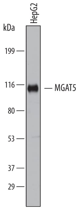
Detection of Human N‑Acetylglucosaminyltransferase V/MGAT5 by Western Blot.
Western blot shows lysates of HepG2 human hepatocellular carcinoma cell line. PVDF membrane was probed with 2 µg/mL of Mouse Anti-Human N-Acetylglucosaminyltransferase V/MGAT5 Monoclonal Antibody (Catalog # MAB5469) followed by HRP-conjugated Anti-Mouse IgG Secondary Antibody (Catalog # HAF007). A specific band was detected for N-Acetylglucosaminyltransferase V/MGAT5 at approximately 100 kDa (as indicated). This experiment was conducted under reducing conditions and using Immunoblot Buffer Group 1.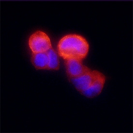
N‑Acetylglucosaminyltransferase V/MGAT5 in MCF‑7 Human Cell Line.
N-Acetylglucosaminyltransferase V/MGAT5 was detected in immersion fixed MCF-7 human breast cancer cell line using Mouse Anti-Human N-Acetylglucosaminyltransferase V/MGAT5 Monoclonal Antibody (Catalog # MAB5469) at 25 µg/mL for 3 hours at room temperature. Cells were stained using the NorthernLights™ 557-conjugated Anti-Mouse IgG Secondary Antibody (red; Catalog # NL007) and counterstained with DAPI (blue). Specific staining was localized to plasma membranes and cytoplasm. View our protocol for Fluorescent ICC Staining of Cells on Coverslips.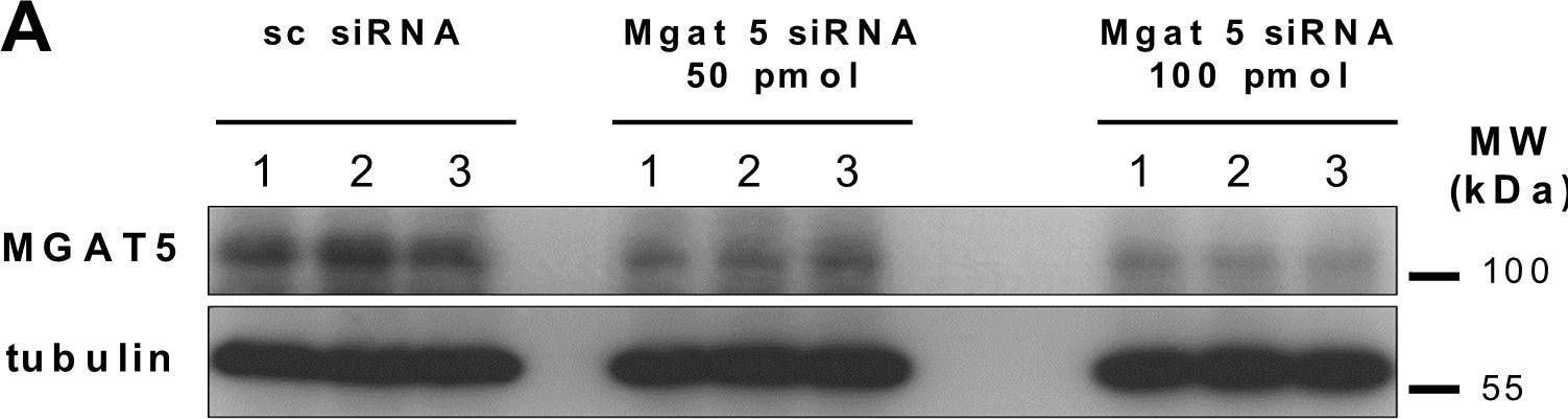
Detection of N-Acetylglucosaminyltransferase V/MGAT5 by Western Blot
Knockdown of Mgat5 expression in cultured human RPE cells attenuates binding of Gal-3 to RPE cells.(A) Western blot analysis of Mgat5 expression in RPE cells transfected with siRNA containing the same nucleotides as Mgat5 siRNA in random order (sc siRNA), and the same cell line transfected with 50 pmol and 100 pmol of double stranded siRNA complementary to Mgat5 (Mgat5 siRNA), respectively. Lysates containing approximately equal amounts of protein were separated by SDS-PAGE and blotted for immunochemical detection of Mgat5 content. Experiments were repeated at least three times. MW; molecular weight. Image collected and cropped by CiteAb from the following open publication (https://pubmed.ncbi.nlm.nih.gov/26760037), licensed under a CC-BY license. Not internally tested by R&D Systems.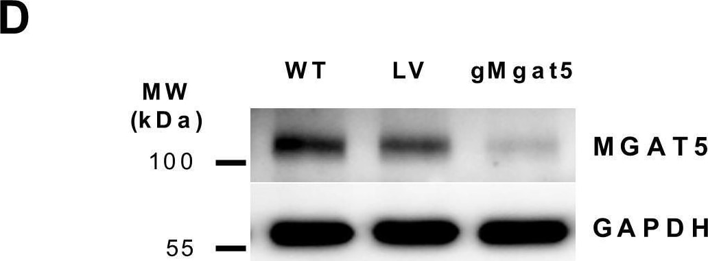
Detection of N-Acetylglucosaminyltransferase V/MGAT5 by Western Blot
Knockdown of Mgat5 expression in cultured human RPE cells attenuates binding of Gal-3 to RPE cells. The target sequence derived from the genomic sequence of Mgat5 was inserted into a CRISPR-Cas9 nuclease expressing lentiviral vector and ARPE19 cells were transfected using Lipfectamine 2000 Plus reagent. (D) Western blot analysis of Mgat5 expression in ARPE19 cells transduced with guide RNA leading to specific knockdown of the Mgat5 (gMgat5), or cells tranduced with a CRISPR-Cas9 lentiviral vector encoding for an none-coding filler RNA (LV), or wild-type ARPE-19 cells. Lysates containing approximately equal amounts of protein were separated by SDS-PAGE and blotted for immunochemical detection of Mgat5 content. Image collected and cropped by CiteAb from the following open publication (https://pubmed.ncbi.nlm.nih.gov/26760037), licensed under a CC-BY license. Not internally tested by R&D Systems.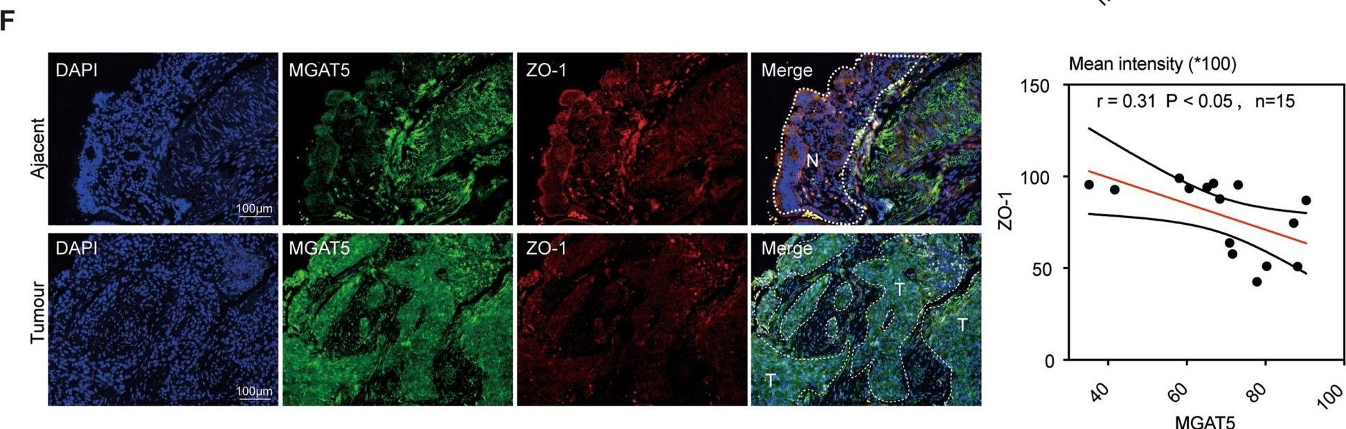
Detection of Human N-Acetylglucosaminyltransferase V/MGAT5 by Immunohistochemistry
Clinical significance of MGAT5 as a potential therapeutic target in tumour EMT. A Representative IHC staining of MGAT5 in CRC patient tissue from the TMA (ZL-Cocsur1241). B MGAT5 protein expression in malignant colorectal cancer (T) and adjacent nonmalignant tissues (N). The data are presented as a box plot. Whiskers show the min-to-max values, n = 69 per group. C Kaplan‒Meier analysis of OS in patients with varying MGAT5 expression levels based on data from selected CRC tissues. D Kaplan‒Meier analyses were performed to assess the associations between MGAT5 expression and RFS. E MGAT5 protein levels in nonmetastatic and metastatic colorectal cancer (T) tissues. The data are presented as a box plot. The whiskers show the min-to-max values. F Immunostaining of the CRC tissue TMA for MGAT5 or ZO-1. Correlations between the mean MGAT5 intensity and the mean ZO-1 intensity in CRC tissue samples (n = 15). Pearson's correlation analysis yielded the R and p values. Scale bar: 100 µm Image collected and cropped by CiteAb from the following open publication (https://pubmed.ncbi.nlm.nih.gov/39511539), licensed under a CC-BY license. Not internally tested by R&D Systems.Applications for Human N-Acetylglucosaminyltransferase V/MGAT5 Antibody (706824)
Immunocytochemistry
Sample: Immersion fixed MCF‑7 human breast cancer cell line
Western Blot
Sample: HepG2 human hepatocellular carcinoma cell line
Reviewed Applications
Read 1 review rated 4 using MAB5469 in the following applications:
Formulation, Preparation, and Storage
Purification
Reconstitution
Sterile PBS to a final concentration of 0.5 mg/mL. For liquid material, refer to CoA for concentration.
Formulation
Shipping
Stability & Storage
- 12 months from date of receipt, -20 to -70 °C as supplied.
- 1 month, 2 to 8 °C under sterile conditions after reconstitution.
- 6 months, -20 to -70 °C under sterile conditions after reconstitution.
Calculators
Background: N-Acetylglucosaminyltransferase V/MGAT5
References
- Saito, H. et al. (1994) Biochem. Biophys. Res. Commun. 198:318.
- Lau, K.S. et al. (2007) Cell 198:123.
- Dennis, J.W. et al. (2002) Biochim. Biophys. Acta 1573:414.
- Granovsky, M. et al. (2008) Nat. Med. 7:1.
- Kim, Y.S. et al. (2008) Mol. Cell. Proteomics 7:1.
Long Name
Alternate Names
Gene Symbol
UniProt
Additional N-Acetylglucosaminyltransferase V/MGAT5 Products
Product Documents for Human N-Acetylglucosaminyltransferase V/MGAT5 Antibody (706824)
Certificate of Analysis
To download a Certificate of Analysis, please enter a lot or batch number in the search box below.
Note: Certificate of Analysis not available for kit components.
Product Specific Notices for Human N-Acetylglucosaminyltransferase V/MGAT5 Antibody (706824)
For research use only
Related Research Areas
Citations for Human N-Acetylglucosaminyltransferase V/MGAT5 Antibody (706824)
 Powered by Bioz
Customer Reviews for Human N-Acetylglucosaminyltransferase V/MGAT5 Antibody (706824) (1)
Have you used Human N-Acetylglucosaminyltransferase V/MGAT5 Antibody (706824)?
Submit a review and receive an Amazon gift card!
$25/€18/£15/$25CAN/¥2500 for a review with an image
$10/€7/£6/$10CAN/¥1110 for a review without an image
Submit a review
Customer Images
-
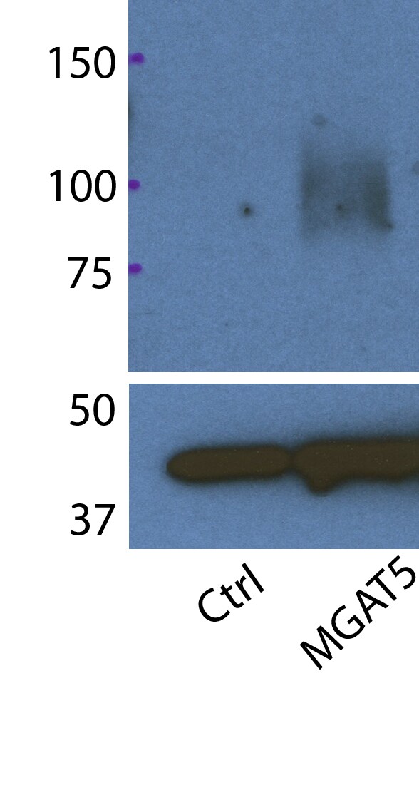
Application: Western BlotSample Tested: Cell LysatesSpecies: HumanVerified Customer | Posted 03/23/2018MGAT5 western blotting after MGAT5A transfection to 293 cells (Figure Legend) Ctrl: Control vector was transfected for 48 hrs. MGAT5: MGAT5A vector was transfected for 48 hrs. Above blots: human MGAT5 (80-100 kDa) Lower blots: beta-actin (42-kDa) Western blot condition: Tris buffer (0.5% Triton X)-lysed cell extract (15 ug each) was loaded with non-reducing condition (:No beta-MercaptoEthanol on loading buffer). Primary Ab: anti-MGAT5 antibody (1 ug/ml) overnight at 4C Secondary Ab: anti-mouse HRP antibody (1:10000 dilution) 90 minute at room temperature. Development: SuperSignal West-Pico PLUS for 15 sec.

There are no reviews that match your criteria.
Protocols
Find general support by application which include: protocols, troubleshooting, illustrated assays, videos and webinars.
- Appropriate Fixation of IHC/ICC Samples
- Cellular Response to Hypoxia Protocols
- ClariTSA™ Fluorophore Kits
- Detection & Visualization of Antibody Binding
- ICC Cell Smear Protocol for Suspension Cells
- ICC Immunocytochemistry Protocol Videos
- ICC for Adherent Cells
- Immunocytochemistry (ICC) Protocol
- Immunocytochemistry Troubleshooting
- Immunofluorescence of Organoids Embedded in Cultrex Basement Membrane Extract
- Immunohistochemistry (IHC) and Immunocytochemistry (ICC) Protocols
- Preparing Samples for IHC/ICC Experiments
- Preventing Non-Specific Staining (Non-Specific Binding)
- Primary Antibody Selection & Optimization
- Protocol for VisUCyte™ HRP Polymer Detection Reagent
- Protocol for the Fluorescent ICC Staining of Cell Smears - Graphic
- Protocol for the Fluorescent ICC Staining of Cultured Cells on Coverslips - Graphic
- Protocol for the Preparation and Fluorescent ICC Staining of Cells on Coverslips
- Protocol for the Preparation and Fluorescent ICC Staining of Non-adherent Cells
- Protocol for the Preparation and Fluorescent ICC Staining of Stem Cells on Coverslips
- Protocol for the Preparation of a Cell Smear for Non-adherent Cell ICC - Graphic
- R&D Systems Quality Control Western Blot Protocol
- TUNEL and Active Caspase-3 Detection by IHC/ICC Protocol
- The Importance of IHC/ICC Controls
- Troubleshooting Guide: Western Blot Figures
- Western Blot Conditions
- Western Blot Protocol
- Western Blot Protocol for Cell Lysates
- Western Blot Troubleshooting
- Western Blot Troubleshooting Guide
- View all Protocols, Troubleshooting, Illustrated assays and Webinars