Human Osteopontin/OPN Antibody (190312)
R&D Systems | Catalog # MAB1433

Key Product Details
Species Reactivity
Validated:
Human
Cited:
Human, Rat
Applications
Validated:
Immunohistochemistry
Cited:
Immunohistochemistry, Immunohistochemistry-Paraffin, Immunocytochemistry, ELISA Capture
Label
Unconjugated
Antibody Source
Monoclonal Mouse IgG2B Clone # 190312
Loading...
Product Specifications
Immunogen
Mouse myeloma cell line NS0-derived recombinant human Osteopontin
Ile17-Asn300
Accession # NP_000573.1
Ile17-Asn300
Accession # NP_000573.1
Specificity
Detects human Osteopontin in direct ELISAs.
Clonality
Monoclonal
Host
Mouse
Isotype
IgG2B
Scientific Data Images for Human Osteopontin/OPN Antibody (190312)
Osteopontin/OPN in Human Breast Cancer Tissue.
Osteopontin/OPN was detected in immersion fixed paraffin-embedded sections of human breast cancer tissue using Mouse Anti-Human Osteopontin/OPN Monoclonal Antibody (Catalog # MAB1433) at 25 µg/mL overnight at 4 °C. Tissue was stained using the Anti-Mouse HRP-DAB Cell & Tissue Staining Kit (brown; Catalog # CTS002) and counterstained with hematoxylin (blue). Specific staining was localized to cytoplasm in epithelial cells. View our protocol for Chromogenic IHC Staining of Paraffin-embedded Tissue Sections.
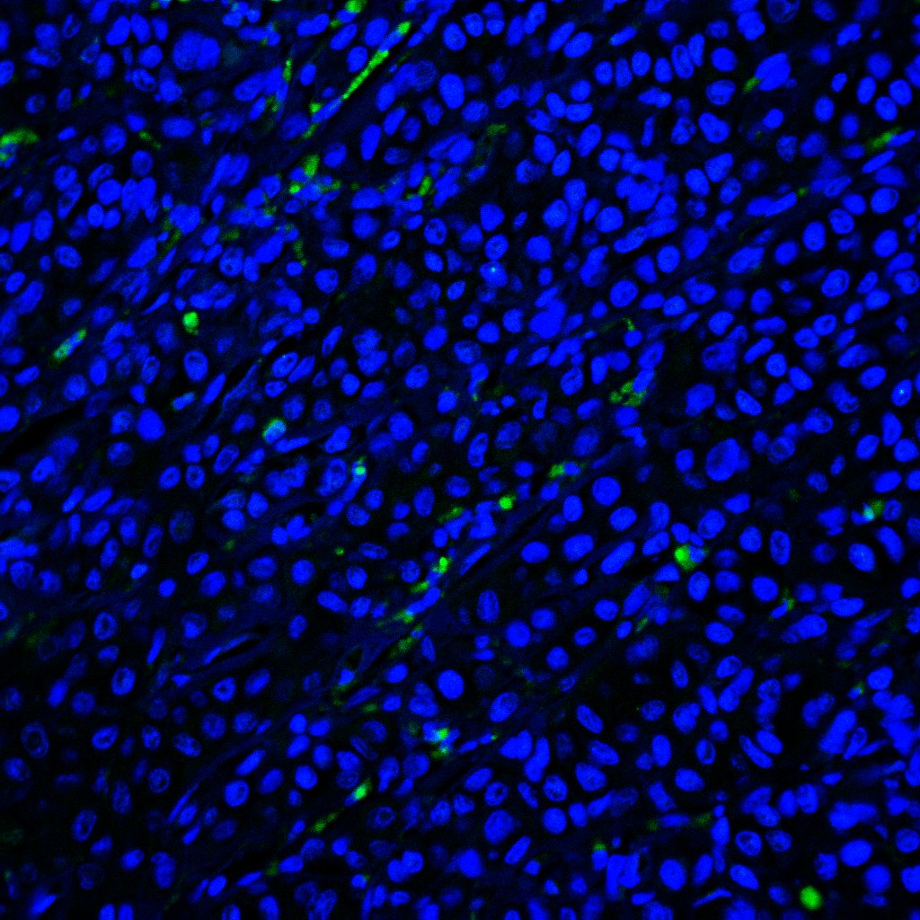
Detection of Osteopontin/OPN by Immunocytochemistry/ Immunofluorescence
SPP1 and OPN expression in BAL cells. (A) SSP1 relative mRNA levels in IPF and N-IPF relative to healthy (Brown-Forsythe Anova, with individual comparisons), (B) Violin plots of mean OPN expression/cell per disease group (Kruskal-Wallis test and individual comparisons) and (C) Typical Images of BAL cytospins stained with anti-human OPN antibody and ToPro-633 nuclear stain in (i) healthy, (ii) IPF and (iii) N-IPF samples (*p < 0.05, **P < 0.01, ****p < 0.0001). Image collected and cropped by CiteAb from the following open publication (https://pubmed.ncbi.nlm.nih.gov/34867934), licensed under a CC-BY license. Not internally tested by R&D Systems.Applications for Human Osteopontin/OPN Antibody (190312)
Application
Recommended Usage
Immunohistochemistry
8-25 µg/mL
Sample: Immersion fixed paraffin-embedded sections of human breast cancer tissue
Sample: Immersion fixed paraffin-embedded sections of human breast cancer tissue
Reviewed Applications
Read 2 reviews rated 4 using MAB1433 in the following applications:
Formulation, Preparation, and Storage
Purification
Protein A or G purified from hybridoma culture supernatant
Reconstitution
Reconstitute at 0.5 mg/mL in sterile PBS. For liquid material, refer to CoA for concentration.
Loading...
Formulation
Lyophilized from a 0.2 μm filtered solution in PBS with Trehalose. *Small pack size (SP) is supplied either lyophilized or as a 0.2 µm filtered solution in PBS.
Shipping
Lyophilized product is shipped at ambient temperature. Liquid small pack size (-SP) is shipped with polar packs. Upon receipt, store immediately at the temperature recommended below.
Stability & Storage
Use a manual defrost freezer and avoid repeated freeze-thaw cycles.
- 12 months from date of receipt, -20 to -70 °C as supplied.
- 1 month, 2 to 8 °C under sterile conditions after reconstitution.
- 6 months, -20 to -70 °C under sterile conditions after reconstitution.
Calculators
Background: Osteopontin/OPN
(6‑8). OPN can also be cleaved by MMP3, 7, 9, and 12 within the SVVYGLR motif and at sites closer to the C‑terminus (8, 9). OPN is widely expressed and is prominent in mineralized tissues. It inhibits bone mineralization and kidney stone formation, and promotes inflammation and cell adhesion and migration (1, 2, 4, 6). Its expression is up‑regulated during inflammation, obesity, atherosclerosis, cancer, and tissue damage, and contributes to the pathophysiology of these conditions (1, 2, 6, 9, 10).
References
- Scatena, M. et al. (2007) Arterioscler. Thromb. Vasc. Biol. 27:2302.
- Rangaswami, H. et al. (2006) Trends Cell Biol. 16:79.
- Young, M.F. et al. (1990) Genomics 7:491.
- Weber, G.F. et al. (2002) J. Leukoc. Biol. 72:752.
- Keykhosravani, M. et al. (2005) Biochemistry 44:6990.
- Kazanecki, C.C. et al. (2007) J. Cell. Biochem. 102:912.
- Senger, D.R. et al. (1994) Mol. Biol. Cell 5:565.
- Yokosaki, Y. et al. (2005) Matrix Biol. 24:418.
- Takafuji, V. et al. (2007) Oncogene 26:6361.
- Kiefer, F.W. et al. (2010) Diabetes 59:935.
Long Name
Secreted Phosphoprotein 1 [BNSP]
Alternate Names
Eta-1, OPN, Spp1
Gene Symbol
SPP1
UniProt
Additional Osteopontin/OPN Products
Product Documents for Human Osteopontin/OPN Antibody (190312)
Certificate of Analysis
To download a Certificate of Analysis, please enter a lot or batch number in the search box below.
Note: Certificate of Analysis not available for kit components.
Product Specific Notices for Human Osteopontin/OPN Antibody (190312)
For research use only
Citations for Human Osteopontin/OPN Antibody (190312)
 Powered by Bioz
Customer Reviews for Human Osteopontin/OPN Antibody (190312) (2)
4 out of 5
2 Customer Ratings
Have you used Human Osteopontin/OPN Antibody (190312)?
Submit a review and receive an Amazon gift card!
$25/€18/£15/$25CAN/¥2500 Yen for a review with an image
$10/€7/£6/$10CAN/¥1110 Yen for a review without an image
Submit a review
Customer Images
Showing
1
-
2 of
2 reviews
Showing All
Filter By:
-
Application: Immunocytochemistry/ImmunofluorescenceSample Tested: Melanoma tissueSpecies: HumanVerified Customer | Posted 09/27/2023

-
Application: Immunohistochemistry-ParaffinSample Tested: See PMID 20562127Species: HumanVerified Customer | Posted 02/10/2015
There are no reviews that match your criteria.
Protocols
Find general support by application which include: protocols, troubleshooting, illustrated assays, videos and webinars.
- Antigen Retrieval Protocol (PIER)
- Antigen Retrieval for Frozen Sections Protocol
- Appropriate Fixation of IHC/ICC Samples
- Cellular Response to Hypoxia Protocols
- Chromogenic IHC Staining of Formalin-Fixed Paraffin-Embedded (FFPE) Tissue Protocol
- Chromogenic Immunohistochemistry Staining of Frozen Tissue
- ClariTSA™ Fluorophore Kits
- Detection & Visualization of Antibody Binding
- Fluorescent IHC Staining of Frozen Tissue Protocol
- Graphic Protocol for Heat-induced Epitope Retrieval
- Graphic Protocol for the Preparation and Fluorescent IHC Staining of Frozen Tissue Sections
- Graphic Protocol for the Preparation and Fluorescent IHC Staining of Paraffin-embedded Tissue Sections
- Graphic Protocol for the Preparation of Gelatin-coated Slides for Histological Tissue Sections
- IHC Sample Preparation (Frozen sections vs Paraffin)
- Immunofluorescent IHC Staining of Formalin-Fixed Paraffin-Embedded (FFPE) Tissue Protocol
- Immunohistochemistry (IHC) and Immunocytochemistry (ICC) Protocols
- Immunohistochemistry Frozen Troubleshooting
- Immunohistochemistry Paraffin Troubleshooting
- Preparing Samples for IHC/ICC Experiments
- Preventing Non-Specific Staining (Non-Specific Binding)
- Primary Antibody Selection & Optimization
- Protocol for Heat-Induced Epitope Retrieval (HIER)
- Protocol for Making a 4% Formaldehyde Solution in PBS
- Protocol for VisUCyte™ HRP Polymer Detection Reagent
- Protocol for the Preparation & Fixation of Cells on Coverslips
- Protocol for the Preparation and Chromogenic IHC Staining of Frozen Tissue Sections
- Protocol for the Preparation and Chromogenic IHC Staining of Frozen Tissue Sections - Graphic
- Protocol for the Preparation and Chromogenic IHC Staining of Paraffin-embedded Tissue Sections
- Protocol for the Preparation and Chromogenic IHC Staining of Paraffin-embedded Tissue Sections - Graphic
- Protocol for the Preparation and Fluorescent IHC Staining of Frozen Tissue Sections
- Protocol for the Preparation and Fluorescent IHC Staining of Paraffin-embedded Tissue Sections
- Protocol for the Preparation of Gelatin-coated Slides for Histological Tissue Sections
- TUNEL and Active Caspase-3 Detection by IHC/ICC Protocol
- The Importance of IHC/ICC Controls
- Troubleshooting Guide: Immunohistochemistry
- View all Protocols, Troubleshooting, Illustrated assays and Webinars
Loading...
Associated Pathways
