PRELP (Proline aRginine-rich End Leucine-rich repeat Protein; also Prolargin) is a 55‑62 kDa secreted glycoprotein that belongs to the small leucine-rich proteoglycan (SLRP) superfamily of extracellular matrix (ECM) molecules (1‑4). Within this family, it is considered a class II member, implying that it is unlikely to form dimeric structures (3). PRELP is synthesized as a 382 amino acid (aa) precursor that contains a 20 aa signal sequence plus a 362 aa mature region (1, 5). Like other SLRPs, PRELP contains an N-terminal extension (aa 72‑107) coupled to multiple Leu-rich repeats (LRRs) (aa 95‑382) (6). Unlike other SLRPs, PRELP does not contain any proteoglycan chains, and its N‑terminal extension is highly basic in charge. The N-terminus reportedly binds to negatively-charged heparin/heparin-sulfate, chondroitin sulfate, and Gram- bacterial cell walls, while the LRR region participates in protein-protein interactions (7‑9). Although PRELP is known to be synthesized by only a few cell types, including osteoblasts, skeletal muscle and chondrocytes, its expression is likely to be more widespread, given its presence in the basement membrane (BM) of Bowman’s capsule, epididymal epithelium and the stratified squamous epithelium of the skin (1, 10, 11). The dual binding profile of PRELP is key to its function. In cartilage, PRELP likely links chondrocyte cell membrane heparin sulfate (HS) chains to endogenous type II collagen. Within the context of the BM, PRELP likely plays an anchoring role. The BM is composed of type IV collagen and laminin, linked together by nidogen. BM Perlecan reinforces this linkage by binding to all three components. PRELP, on the edge of the BM, can bind to free perlecan HS chains (via its N-terminus), and to underlying type I collagen (via its LRRs), thus forming an anchor for the BM (11). Notably, the N-terminus appears to do more than simply provide part of a linkage mechanism. In bone, osteoblast secreted PRELP is hypothesized to undergo proteolysis by enzymes such as LysC and glutamyl endopeptidase. This will generate 40‑75 aa N‑terminal fragments that can bind to chondroitin sulfate adducts that exist on the surface of prefusion osteoclast precursors. Following binding, PRELP is internalized, complexed to annexin-II, and translocated to the nucleus, where it interacts with NF kappa Bp65 to block osteoclast maturation (8). In tissue, PRELP may also undergo proteolytic processing during inflammation to release an N‑terminal fragment containing aa 21‑42 of the precursor (7). This sequence has been shown to possess potent antimicrobial activity by creating pores in bacterial cell walls. Mature human PRELP shares 91% aa identity with mouse PRELP (10).

Key Product Details
Species Reactivity
Validated:
Human
Cited:
Human, Mouse, Xenograft
Applications
Validated:
Western Blot, Immunocytochemistry
Cited:
Immunohistochemistry
Label
Unconjugated
Antibody Source
Polyclonal Sheep IgG
Loading...
Product Specifications
Immunogen
Chinese hamster ovary cell line CHO-derived recombinant human PRELP
Gln21-Ile382
Accession # P51888
Gln21-Ile382
Accession # P51888
Specificity
Detects human PRELP in direct ELISAs and Western blots.
Clonality
Polyclonal
Host
Sheep
Isotype
IgG
Scientific Data Images for Human PRELP Antibody
Detection of Human PRELP by Western Blot.
Western blot shows lysates of human liver tissue and human pancreas tissue. PVDF membrane was probed with 1 µg/mL of Sheep Anti-Human PRELP Antigen Affinity-purified Polyclonal Antibody (Catalog # AF6447) followed by HRP-conjugated Anti-Sheep IgG Secondary Antibody (Catalog # HAF016). A specific band was detected for PRELP at approximately 65 kDa (as indicated). This experiment was conducted under reducing conditions and using Immunoblot Buffer Group 1.
PRELP in Human Mesenchymal Stem Cells.
PRELP was detected in immersion fixed human mesenchymal stem cells differentiated into chondrocytes using Sheep Anti-Human PRELP Antigen Affinity-purified Polyclonal Antibody (Catalog # AF6447) at 10 µg/mL overnight at 4 °C. Cells were stained using the NorthernLights™ 557-conjugated Anti-Sheep IgG Secondary Antibody (red; Catalog # NL010) and counterstained with DAPI (blue). Specific staining was localized to cytoplasm. View our protocol for Fluorescent ICC Staining of Cells on Coverslips.
Detection of Zebrafish PRELP by Immunohistochemistry
SLRPs are enriched in human spinal cord lesions.Anti-LUM, anti-PRELP, anti-CHAD, and anti-FMOD immunoreactivity is increased (arrowheads; see calibration bar of lookup table (LUT)) in the epicenter of the injured human spinal cord at nine days-post injury, as compared to rostral control segments of the same patient. Note that immunoreactivity is mainly observed in proximity to the hemorrhage (black; see calibration bar of LUT). Also note that with the exception of blood-derived autofluorescence, individual channels show distinct pattern of immunofluorescence (IF) signal (insets). Shown are transversal sections (dorsal is up). Similar results were obtained for anti-LUM in five out of six cases, for anti-PRELP and anti-FMOD in four out of six cases, and anti-CHAD in two out of six cases (Supplementary Table 3). Scale bars: 2 mm, 200 µm (insets). The human icon in the panel was created using BioRender. Image collected and cropped by CiteAb from the following open publication (https://pubmed.ncbi.nlm.nih.gov/37884489), licensed under a CC-BY license. Not internally tested by R&D Systems.
Detection of Zebrafish PRELP by Immunohistochemistry
SLRPs are enriched in human brain lesions.Anti-CHAD, anti-FMOD, anti-LUM, and anti-PRELP immunoreactivity is locally increased in areas of scarring caused by contusion, local hemorrhage, or previous surgery (asterisks indicate lesion center, arrowheads mark scar boundaries) in the human brain, as compared to regions distant to the primary lesion site (hash sign), as well as human brain controls with no signs of fibrotic scarring (see Supplementary Fig. 3). Shown are coronal sections of brain tissue from patients with traumatic brain injury (TBI) or previous surgery (re-OP; bottom panel). Six (n = 6) cases with scars following TBI or previous surgery were analyzed and showed similar results (Supplementary Table 1). Scale bars: 500 µm, 50 µm (insets). The human icon in the panel was created using BioRender. Image collected and cropped by CiteAb from the following open publication (https://pubmed.ncbi.nlm.nih.gov/37884489), licensed under a CC-BY license. Not internally tested by R&D Systems.Applications for Human PRELP Antibody
Application
Recommended Usage
Immunocytochemistry
5-15 µg/mL
Sample: Immersion fixed human mesenchymal stem cells differentiated into chondrocytes
Sample: Immersion fixed human mesenchymal stem cells differentiated into chondrocytes
Western Blot
1 µg/mL
Sample: Human liver tissue and human pancreas tissue
Sample: Human liver tissue and human pancreas tissue
Reviewed Applications
Read 1 review rated 5 using AF6447 in the following applications:
Formulation, Preparation, and Storage
Purification
Antigen Affinity-purified
Reconstitution
Sterile PBS to a final concentration of 0.2 mg/mL. For liquid material, refer to CoA for concentration.
Loading...
Formulation
Lyophilized from a 0.2 μm filtered solution in PBS with Trehalose. *Small pack size (SP) is supplied either lyophilized or as a 0.2 µm filtered solution in PBS.
Shipping
Lyophilized product is shipped at ambient temperature. Liquid small pack size (-SP) is shipped with polar packs. Upon receipt, store immediately at the temperature recommended below.
Stability & Storage
Use a manual defrost freezer and avoid repeated freeze-thaw cycles.
- 12 months from date of receipt, -20 to -70 °C as supplied.
- 1 month, 2 to 8 °C under sterile conditions after reconstitution.
- 6 months, -20 to -70 °C under sterile conditions after reconstitution.
Calculators
Background: PRELP
References
- Bengtsson, E. et al. (1995) J. Biol. Chem. 270:25639.
- Merline, R. et al. (2009) J. Cell Commun. Signal. 3:323.
- McEwan, P.A. et al. (2006) J. Struct. Biol. 155:294.
- Neame, P.J. et al. (1999) Cell. Mol. Life Sci. 55:1327.
- Grover, J. et al. (1996) Genomics 38:109.
- SwissProt # P51888.
- Bengtsson, E. et al. (2000) J. Biol. Chem. 275:40695.
- Rucci, N. et. al. (2009) J. Cell Biol. 187:669.
- Malmsten, M. et al. (2006) Matrix Biol. 25:294.
- Grover, J. & P.J. Roughley (2001) Matrix Biol. 20:555.
- Bengtsson, E. et al. (2002) J. Biol. Chem. 277:15061.
Long Name
Proline-arginine-Rich End Leucine-rich repeat Protein
Alternate Names
MST161, MSTP161, Prolargin, SLRR2A
Entrez Gene IDs
5549 (Human)
Gene Symbol
PRELP
UniProt
Additional PRELP Products
Product Documents for Human PRELP Antibody
Certificate of Analysis
To download a Certificate of Analysis, please enter a lot or batch number in the search box below.
Note: Certificate of Analysis not available for kit components.
Product Specific Notices for Human PRELP Antibody
For research use only
Related Research Areas
Citations for Human PRELP Antibody
 Powered by Bioz
Customer Reviews for Human PRELP Antibody (1)
5 out of 5
1 Customer Rating
Have you used Human PRELP Antibody?
Submit a review and receive an Amazon gift card!
$25/€18/£15/$25CAN/¥2500 Yen for a review with an image
$10/€7/£6/$10CAN/¥1110 Yen for a review without an image
Submit a review
Customer Images
Showing
1
-
1 of
1 review
Showing All
Filter By:
-
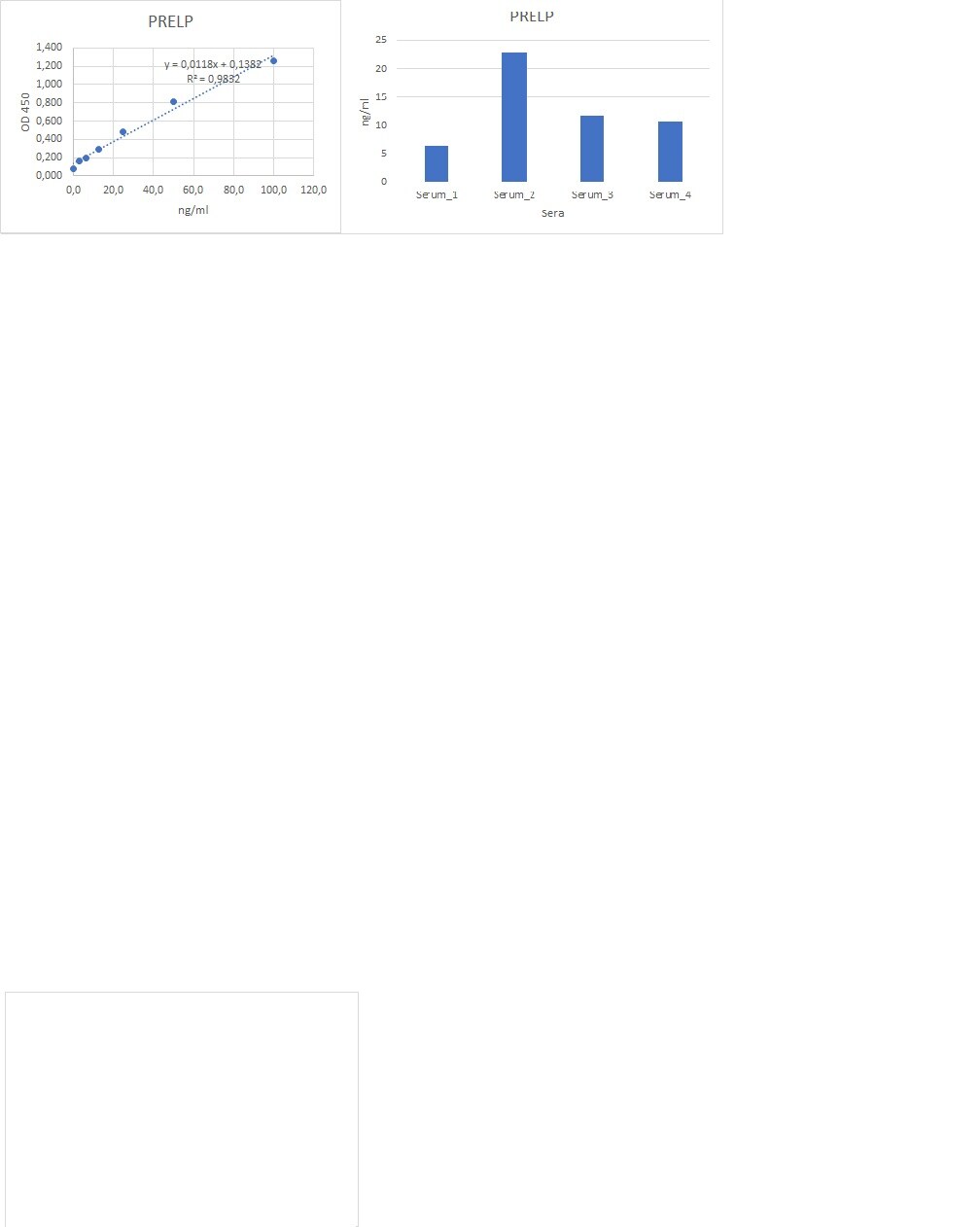
Application: ELISASample Tested: Serum and PlasmaSpecies: HumanVerified Customer | Posted 07/05/2022works very well for ELISA in combination with mAb

There are no reviews that match your criteria.
Protocols
Find general support by application which include: protocols, troubleshooting, illustrated assays, videos and webinars.
- Appropriate Fixation of IHC/ICC Samples
- Cellular Response to Hypoxia Protocols
- ClariTSA™ Fluorophore Kits
- Detection & Visualization of Antibody Binding
- ICC Cell Smear Protocol for Suspension Cells
- ICC Immunocytochemistry Protocol Videos
- ICC for Adherent Cells
- Immunocytochemistry (ICC) Protocol
- Immunocytochemistry Troubleshooting
- Immunofluorescence of Organoids Embedded in Cultrex Basement Membrane Extract
- Immunohistochemistry (IHC) and Immunocytochemistry (ICC) Protocols
- Preparing Samples for IHC/ICC Experiments
- Preventing Non-Specific Staining (Non-Specific Binding)
- Primary Antibody Selection & Optimization
- Protocol for VisUCyte™ HRP Polymer Detection Reagent
- Protocol for the Fluorescent ICC Staining of Cell Smears - Graphic
- Protocol for the Fluorescent ICC Staining of Cultured Cells on Coverslips - Graphic
- Protocol for the Preparation and Fluorescent ICC Staining of Cells on Coverslips
- Protocol for the Preparation and Fluorescent ICC Staining of Non-adherent Cells
- Protocol for the Preparation and Fluorescent ICC Staining of Stem Cells on Coverslips
- Protocol for the Preparation of a Cell Smear for Non-adherent Cell ICC - Graphic
- R&D Systems Quality Control Western Blot Protocol
- TUNEL and Active Caspase-3 Detection by IHC/ICC Protocol
- The Importance of IHC/ICC Controls
- Troubleshooting Guide: Western Blot Figures
- Western Blot Conditions
- Western Blot Protocol
- Western Blot Protocol for Cell Lysates
- Western Blot Troubleshooting
- Western Blot Troubleshooting Guide
- View all Protocols, Troubleshooting, Illustrated assays and Webinars
Loading...
Associated Pathways
