
Human Repulsive Guidance molecule (RGM) is a 33 kDa GPI-linked member of an expanding RGM-related family of neuronal and muscle-expressed membrane proteins (1). It is synthesized as a 450 amino acid (aa) preproprotein that contains a 47 aa signal sequence, a 121 aa N-terminal prosegment, a 256 mature region and a 26 aa C-terminal prosegment (2). The N-terminal prosegment contains an RGD tripeptide and the molecule’s only two potential N-linked glycosylation sites. The mature segment shows an abbreviated von Willebrand factor domain. Proteolytic processing occurs at an aspartic acid-proline bond, creating a predicted 32 kDa mature region (2). The mature region of human RGM-A has 88% and 93% aa identity to the chick and mouse mature region of RGM-A, respectively. When compared to human RGMb and c, the mature region of human RGM-A shows 58% and 54% aa identity, respectively. Recombinant chick RGM-A has been reported to induce collapse of temporal but not nasal growth cones, and to repel temporal retinal axons in vitro. This suggests a role in the development of the retina-superior colliculus connection. In mammals, however, this activity is not so evident, and thus its function in this system is uncertain (3). Alternatively, in mice, RGM-A is said to be needed for neural tube closure, and may play a role in entorhinal-hippocampal connections (3, 4). The receptor for RGM-A is reported to be neogenin (5, 6). RGM-A has also been shown to be a bone morphogenic protein co-receptor, able to bind both BMP-2 and BMP-4 (7).

Scientific images may be resized, cropped, or adjusted for brightness or contrast for presentation purposes. These updates are limited to presentation and do not affect the underlying scientific interpretation of the data. Where available, additional source data may be provided upon request.
Key Product Details
Species Reactivity
Validated:
Cited:
Applications
Validated:
Cited:
Label
Antibody Source
Product Specifications
Immunogen
Cys48-Gly422
Accession # Q96B86
Specificity
Clonality
Host
Isotype
Scientific Data Images for Human RGM-A Antibody
RGM‑A in Human Brain.
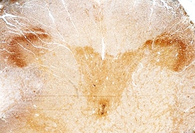
RGM-A was detected in immersion fixed paraffin-embedded sections of human brain (spinal cord) using Goat Anti-Human RGM-A Antigen Affinity-purified Polyclonal Antibody (Catalog # AF2459) at 15 µg/mL overnight at 4 °C. Before incubation with the primary antibody, tissue was subjected to heat-induced epitope retrieval using Antigen Retrieval Reagent-Basic (Catalog # CTS013). Tissue was stained using the Anti-Goat HRP-DAB Cell & Tissue Staining Kit (brown; Catalog # CTS008) and counterstained with hematoxylin (blue). Specific staining was localized to the dorsal horn. View our protocol for Chromogenic IHC Staining of Paraffin-embedded Tissue Sections.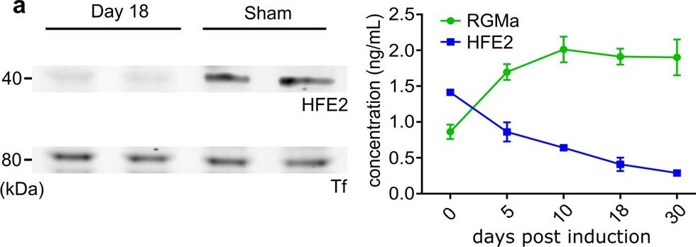
Detection of RGM-A by Western Blot
HFE2 administration significantly improves EAE disease outcome. A Western Blot analysis shows a strong HFE2 reduction in EAE animal serum when compared to control. ELISA quantification showing HFE2 decrease and RGMa increase in EAE animal serum (mean ± s.d.; EAE n = 3 and RGMa n = 3). b Body conditioning assessment (‘0’ no paralysis to ‘4’ forelimb weakness) of EAE-induced mice with HFE2 + /- RGMa treatment every 3 days (d3, 6, 9, 12 and 15; mean ± s.e.m.; unpaired two-tail t-tests; PBS n = 8, HFE2 n = 6, RGMa+ HFE2 n = 8). c Body conditioning assessment of EAE-induced Hfe2fl/fl and Hfe2 delta Alb-cre mice (mean ± s.e.m.; unpaired two-tail t-tests; Hfe2fl/fln = 8 and Hfe2 delta Alb-cren = 8). d Representative confocal images of CD3 +, B220+ and CD11b+ immune staining in spinal cord of EAE-induced mice 3 weeks after induction and quantification (mean ± s.e.m.; unpaired two-tail t-test; PBS n = 7, HFE2 n = 6 mice; technical replicates shown). e Schematic of activated immune cell adoptive transfer. Representative confocal images of CD4-positive labelled transferred cell (CellVue) in PBS-treated or HFE2-treated recipient WT mice and quantification (mean ± s.e.m.; unpaired two-tail t-test; PBS n = 6, HFE2 n = 6). Ptx; Pertussis Toxin. f Representative confocal images of fibrinogen in the spinal cord of HFE2-treated EAE-mice and quantification (mean ± s.e.m.; unpaired two-tail t-test; PBS-treated n = 6 and HFE2 n = 6). *P < 0.05, **P < 0.01, ***P < 0.001. Source data are provided as a Source Data file (includes exact p-values). Image collected and cropped by CiteAb from the following open publication (https://www.nature.com/articles/s41467-024-45303-1), licensed under a CC-BY license. Not internally tested by R&D Systems.Applications for Human RGM-A Antibody
Immunohistochemistry
Sample: Formalin fixed paraffin-embedded sections of human spinal cord subjected to Antigen Retrieval Reagent-Basic (Catalog # CTS013)
Western Blot
Sample: Recombinant Human Repulsive Guidance Molecule A/RGM-A (Catalog # 2459-RM)
Reviewed Applications
Read 1 review rated 5 using AF2459 in the following applications:
Formulation, Preparation, and Storage
Purification
Reconstitution
Reconstitute at 0.2 mg/mL in sterile PBS. For liquid material, refer to CoA for concentration.
Formulation
Shipping
Stability & Storage
- 12 months from date of receipt, -20 to -70 °C as supplied.
- 1 month, 2 to 8 °C under sterile conditions after reconstitution.
- 6 months, -20 to -70 °C under sterile conditions after reconstitution.
Calculators
Background: RGM-A
References
- Samad, T.A. et al. (2004) J. Neurosci. 24:2027.
- Monnier P. et al. (2002) Nature 419:392.
- Niederkofler V. et al. (2004) J. Neurosci. 24:808.
- Brinks, H. et al. (2004) J. Neurosci. 24:3862.
- Rajagopalan S. et al. (2004) Nat. Cell Biol. 6:756.
- Matsunaga E. et al. (2004) Nat. Cell Biol. 6:749.
- Babitt J.L. et al. (2005) J. Biol. Chem. 280(33):29820.
Long Name
Alternate Names
Gene Symbol
UniProt
Additional RGM-A Products
Product Documents for Human RGM-A Antibody
Certificate of Analysis
To download a Certificate of Analysis, please enter a lot or batch number in the search box below.
Note: Certificate of Analysis not available for kit components.
Product Specific Notices for Human RGM-A Antibody
For research use only
Related Research Areas
Citations for Human RGM-A Antibody
 Powered by Bioz
Customer Reviews for Human RGM-A Antibody (1)
Have you used Human RGM-A Antibody?
Submit a review and receive an Amazon gift card!
$25/€18/£15/$25CAN/¥2500 for a review with an image
$10/€7/£6/$10CAN/¥1110 for a review without an image
Submit a review
Customer Images
-
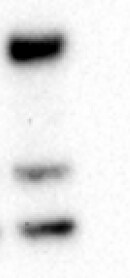
Application: Functional AssaySample Tested: Recombinant proteinSpecies: HumanVerified Customer | Posted 02/04/2025Worked in detecting 2459-RM in in vitro assay

There are no reviews that match your criteria.
Protocols
Find general support by application which include: protocols, troubleshooting, illustrated assays, videos and webinars.
- Antigen Retrieval Protocol (PIER)
- Antigen Retrieval for Frozen Sections Protocol
- Appropriate Fixation of IHC/ICC Samples
- Cellular Response to Hypoxia Protocols
- Chromogenic IHC Staining of Formalin-Fixed Paraffin-Embedded (FFPE) Tissue Protocol
- Chromogenic Immunohistochemistry Staining of Frozen Tissue
- ClariTSA™ Fluorophore Kits
- Detection & Visualization of Antibody Binding
- Fluorescent IHC Staining of Frozen Tissue Protocol
- Graphic Protocol for Heat-induced Epitope Retrieval
- Graphic Protocol for the Preparation and Fluorescent IHC Staining of Frozen Tissue Sections
- Graphic Protocol for the Preparation and Fluorescent IHC Staining of Paraffin-embedded Tissue Sections
- Graphic Protocol for the Preparation of Gelatin-coated Slides for Histological Tissue Sections
- IHC Sample Preparation (Frozen sections vs Paraffin)
- Immunofluorescent IHC Staining of Formalin-Fixed Paraffin-Embedded (FFPE) Tissue Protocol
- Immunohistochemistry (IHC) and Immunocytochemistry (ICC) Protocols
- Immunohistochemistry Frozen Troubleshooting
- Immunohistochemistry Paraffin Troubleshooting
- Preparing Samples for IHC/ICC Experiments
- Preventing Non-Specific Staining (Non-Specific Binding)
- Primary Antibody Selection & Optimization
- Protocol for Heat-Induced Epitope Retrieval (HIER)
- Protocol for Making a 4% Formaldehyde Solution in PBS
- Protocol for VisUCyte™ HRP Polymer Detection Reagent
- Protocol for the Preparation & Fixation of Cells on Coverslips
- Protocol for the Preparation and Chromogenic IHC Staining of Frozen Tissue Sections
- Protocol for the Preparation and Chromogenic IHC Staining of Frozen Tissue Sections - Graphic
- Protocol for the Preparation and Chromogenic IHC Staining of Paraffin-embedded Tissue Sections
- Protocol for the Preparation and Chromogenic IHC Staining of Paraffin-embedded Tissue Sections - Graphic
- Protocol for the Preparation and Fluorescent IHC Staining of Frozen Tissue Sections
- Protocol for the Preparation and Fluorescent IHC Staining of Paraffin-embedded Tissue Sections
- Protocol for the Preparation of Gelatin-coated Slides for Histological Tissue Sections
- R&D Systems Quality Control Western Blot Protocol
- TUNEL and Active Caspase-3 Detection by IHC/ICC Protocol
- The Importance of IHC/ICC Controls
- Troubleshooting Guide: Immunohistochemistry
- Troubleshooting Guide: Western Blot Figures
- Western Blot Conditions
- Western Blot Protocol
- Western Blot Protocol for Cell Lysates
- Western Blot Troubleshooting
- Western Blot Troubleshooting Guide
- View all Protocols, Troubleshooting, Illustrated assays and Webinars