SorCS1 is a type I transmembrane receptor of the mammalian Vps10p (vacuolar protein-sorting 10 protein) family (1, 2). These sorting receptors include sortilin, SorLA, and three SorCS proteins. Three splicing variants (SorCS1a, b and c) differ only in their cytoplasmic domains (3). All variants are predominantly expressed in the central nervous system, but SorCS1 can also be identified in heart, kidney and pancreatic islets (2‑5). SorCS1a mediates endocytosis, and only ~10% of it is expressed on the cell surface. SorCS1b shows higher surface expression (~45%) and is much less involved in endocytosis. SorCS1c is intermediate. Human SorCS1a is synthesized as a 1159 amino acid (aa) preproform with a 33 aa signal sequence and a 77 aa propeptide. After proteolytic processing at a furin-type consensus sequence, the mature SorCS1a is a 1049 aa, 130 kDa protein with a 989 aa extracellular/lumenal domain (ECD). Within the ECD, human SorCS1 shares 93%, 94%, 93% and 98% aa identity with mouse, rat, bovine and canine SorCS1, respectively. It also shares 70% and 46% aa identity with the ECD of human SorCS3 and SorCS2, respectively. The ECD contains an imperfect leucine-rich repeat (LRR) and a Vps10p domain and binds the growth factor PDGF-BB (1, 2, 6). Expression in the hippocampus indicates that SorCS1 may modulate PDGF-BB activity in this location (6). SorCS1 has also been identified as a susceptibility gene for type 2 diabetes in overweight females (4). Consequently, it has been proposed to affect insulin secretion by modifying PDGF-mediated growth of the islet vasculature (7). The 80 kDa ECD may be constitutively or inducibly shed, mainly via the metalloproteinase TACE/ADAM17 (6). The shed soluble form also binds PDGF. The cellular portion appears to undergo regulated intramembrane proteolysis (8).

Key Product Details
Species Reactivity
Validated:
Human
Cited:
Human, Mouse
Applications
Validated:
Western Blot, Immunocytochemistry
Cited:
Western Blot, Immunocytochemistry
Label
Unconjugated
Antibody Source
Polyclonal Goat IgG
Loading...
Product Specifications
Immunogen
Mouse myeloma cell line NS0-derived recombinant human SorCS1
Ser111-Ser1099 (Ser231Gly)
Accession # Q8WY21.3
Ser111-Ser1099 (Ser231Gly)
Accession # Q8WY21.3
Specificity
Detects human SorCS1 in direct ELISAs and Western blots. In direct ELISAs, approximately 20% cross-reactivity with recombinant mouse SorCS1 is observed and less than 1% cross-reactivity with recombinant human (rh) SorCS2 and rhSorCS3 is observed.
Clonality
Polyclonal
Host
Goat
Isotype
IgG
Scientific Data Images for Human SorCS1 Antibody
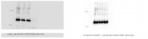
Detection of SorCS1 by Western Blot
MPEP decreases SORT1 expression and increases extracellular PGRN in mammalian cell lines. (A and B) M17 cells were treated with control siRNA (siR-Ctrl) or gene-specific SORT1 siRNA (siR-SORT1). (A) Intracellular levels of PGRN, SORT1 and GAPDH were evaluated by western blot at a 48 h time-point. (B) Suppression of SORT1 levels increased extracellular PGRN levels. (C) Chemical name and structure of MPEP. (D–I) Treatment of M17 cells (D and E), HeLa cells (F and G) or NIH3T3 cells (H and I) with MPEP for 24 h dose dependently reduced SORT1 levels (D, F and H) and increased exPGRN levels (E, G and I) at 10 and 20 μM. (J) Under the same conditions, MPEP did not affect levels of SORLA, SORCS1 and ubiquitinated proteins in M17 cells. ***P < 0.001 versus vehicle control, analysis performed by one-way ANOVA followed by Tukey's post-test. Image collected and cropped by CiteAb from the following open publication (https://pubmed.ncbi.nlm.nih.gov/24163244), licensed under a CC-BY license. Not internally tested by R&D Systems.Applications for Human SorCS1 Antibody
Application
Recommended Usage
Immunocytochemistry
5-15 µg/mL
Sample: Immersion fixed beta TC-6 mouse beta cell insulinoma cell line
Sample: Immersion fixed beta TC-6 mouse beta cell insulinoma cell line
Western Blot
0.1 µg/mL
Sample: Recombinant Human SorCS1 (Catalog # 3457-SR)
Sample: Recombinant Human SorCS1 (Catalog # 3457-SR)
Reviewed Applications
Read 3 reviews rated 5 using AF3457 in the following applications:
Formulation, Preparation, and Storage
Purification
Antigen Affinity-purified
Reconstitution
Reconstitute at 0.2 mg/mL in sterile PBS. For liquid material, refer to CoA for concentration.
Loading...
Formulation
Lyophilized from a 0.2 μm filtered solution in PBS with Trehalose. *Small pack size (SP) is supplied either lyophilized or as a 0.2 µm filtered solution in PBS.
Shipping
Lyophilized product is shipped at ambient temperature. Liquid small pack size (-SP) is shipped with polar packs. Upon receipt, store immediately at the temperature recommended below.
Stability & Storage
Use a manual defrost freezer and avoid repeated freeze-thaw cycles.
- 12 months from date of receipt, -20 to -70 °C as supplied.
- 1 month, 2 to 8 °C under sterile conditions after reconstitution.
- 6 months, -20 to -70 °C under sterile conditions after reconstitution.
Calculators
Background: SorCS1
References
- Hampe, W. et al. (2001) Hum. Genet. 108:529.
- Hermey, G. et al. (1999) Biochem. Biophys. Res. Commun. 266:347.
- Hermey, G. et al. (2003) J. Biol. Chem. 278:7390.
- Clee, S.M. et al. (2006) Nat. Genet. 38:688.
- Hermey, G. et al. (2001) Neurosci. Lett. 313:83.
- Hermey, G. et al. (2006) Biochem. J. 395:285.
- Goodarzi, M.O. et al. (2007) Diabetes Apr 10 [Epub ahead of print].
- Nyborg, A.C. et al. (2006) Mol. Neurodegen. 1:3.
Long Name
Sortilin-related VPS10 Domain Containing Receptor 1
Alternate Names
FLJ41758, FLJ43475, FLJ44957, hSorCS, SORCS, SORCS receptor 1, sorCS1, sortilin-related VPS10 domain containing receptor 1, VPS10 domain receptor protein SORCS 1, VPS10 domain receptor SorCS, VPS10 domain-containing receptor SorCS1
Gene Symbol
SORCS1
UniProt
Additional SorCS1 Products
Product Documents for Human SorCS1 Antibody
Certificate of Analysis
To download a Certificate of Analysis, please enter a lot or batch number in the search box below.
Note: Certificate of Analysis not available for kit components.
Product Specific Notices for Human SorCS1 Antibody
For research use only
Related Research Areas
Citations for Human SorCS1 Antibody
 Powered by Bioz
Customer Reviews for Human SorCS1 Antibody (3)
5 out of 5
3 Customer Ratings
Have you used Human SorCS1 Antibody?
Submit a review and receive an Amazon gift card!
$25/€18/£15/$25CAN/¥2500 Yen for a review with an image
$10/€7/£6/$10CAN/¥1110 Yen for a review without an image
Submit a review
Customer Images
Showing
1
-
3 of
3 reviews
Showing All
Filter By:
-
Application: ImmunoprecipitationSample Tested: IP of human SorCS1 transfected into HEK293 cellsSpecies: HumanVerified Customer | Posted 12/18/2014

-
Application: ELISASample Tested: N/ASpecies: HumanVerified Customer | Posted 12/18/2014
-
Application: ImmunofluorescenceSample Tested: CHO-cells stable expressing mouse SorCS1 variant cSpecies: OtherVerified Customer | Posted 12/18/2014
There are no reviews that match your criteria.
Protocols
Find general support by application which include: protocols, troubleshooting, illustrated assays, videos and webinars.
- Appropriate Fixation of IHC/ICC Samples
- Cellular Response to Hypoxia Protocols
- ClariTSA™ Fluorophore Kits
- Detection & Visualization of Antibody Binding
- ICC Cell Smear Protocol for Suspension Cells
- ICC Immunocytochemistry Protocol Videos
- ICC for Adherent Cells
- Immunocytochemistry (ICC) Protocol
- Immunocytochemistry Troubleshooting
- Immunofluorescence of Organoids Embedded in Cultrex Basement Membrane Extract
- Immunohistochemistry (IHC) and Immunocytochemistry (ICC) Protocols
- Preparing Samples for IHC/ICC Experiments
- Preventing Non-Specific Staining (Non-Specific Binding)
- Primary Antibody Selection & Optimization
- Protocol for VisUCyte™ HRP Polymer Detection Reagent
- Protocol for the Fluorescent ICC Staining of Cell Smears - Graphic
- Protocol for the Fluorescent ICC Staining of Cultured Cells on Coverslips - Graphic
- Protocol for the Preparation and Fluorescent ICC Staining of Cells on Coverslips
- Protocol for the Preparation and Fluorescent ICC Staining of Non-adherent Cells
- Protocol for the Preparation and Fluorescent ICC Staining of Stem Cells on Coverslips
- Protocol for the Preparation of a Cell Smear for Non-adherent Cell ICC - Graphic
- R&D Systems Quality Control Western Blot Protocol
- TUNEL and Active Caspase-3 Detection by IHC/ICC Protocol
- The Importance of IHC/ICC Controls
- Troubleshooting Guide: Western Blot Figures
- Western Blot Conditions
- Western Blot Protocol
- Western Blot Protocol for Cell Lysates
- Western Blot Troubleshooting
- Western Blot Troubleshooting Guide
- View all Protocols, Troubleshooting, Illustrated assays and Webinars
Loading...