Vascular endothelial (VE)-cadherin (VE-CAD), also called 7B4 and cadherin‑5 (CDH5), is a member of the cadherin family of cell adhesion molecules. Cadherins are calcium‑dependent transmembrane proteins which bind to one another in a homophilic manner. On their cytoplasmic side, they associate with the three catenins, alpha, beta, and gamma (plakoglobin). This association links the cadherin protein to the cytoskeleton. Without association with the catenins, the cadherins are non-adhesive. Cadherins play a role in development, specifically in tissue formation. They may also help to maintain tissue architecture in the adult. VE-cadherin has been shown to play important roles in vasculogenesis and angiogenesis. VE-cadherin is a classical cadherin molecule. Classical cadherins consist of a large extracellular domain which contains DXD and DXNDN repeats responsible for mediating calcium-dependent adhesion, a single-pass transmembrane domain, and a short carboxy-terminal cytoplasmic domain responsible for interacting with the catenins. Human VE-cadherin is a 784 amino acid (aa) residue protein with a 25 aa signal sequence and a 759 aa propeptide. The mature protein begins at amino acid 48 and has a 546 aa extracellular domain, a 27 aa transmembrane domain, and a 164 aa cytoplasmic domain. The human and mouse mature VE-cadherin proteins share approximately 74% homology.
Human VE-Cadherin Antibody (123413)
R&D Systems | Catalog # MAB9381

Key Product Details
Validated by
Species Reactivity
Validated:
Cited:
Applications
Validated:
Cited:
Label
Antibody Source
Product Specifications
Immunogen
Asp48-Gln593
Accession # P33151
Specificity
Clonality
Host
Isotype
Scientific Data Images for Human VE-Cadherin Antibody (123413)
Detection of Human VE‑Cadherin by Western Blot.
Western blot shows lysate of HUVEC human umbilical vein endothelial cells. PVDF membrane was probed with 1 µg/mL of Mouse Anti-Human VE-Cadherin Monoclonal Antibody (Catalog # MAB9381) followed by HRP-conjugated Anti-Mouse IgG Secondary Antibody (HAF018). A specific band was detected for VE-Cadherin at approximately 125 kDa (as indicated). This experiment was conducted under reducing conditions and using Immunoblot Buffer Group 1.
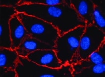
VE‑Cadherin in HUVEC Cells.
VE-Cadherin was detected in immersion fixed HUVEC cells using Mouse Anti-Human VE-Cadherin Monoclonal Antibody (Catalog # MAB9381) at 0.5 µg/mL for 3 hours at room temperature. Cells were stained using the NorthernLights™ 557-conjugated Anti-Mouse IgG Secondary Antibody (red; Catalog # NL007) and counterstained with DAPI (blue). Specific staining was localized to plasma membrane. View our protocol for Fluorescent ICC Staining of Cells on Coverslips.
Detection of VE‑Cadherin in HUVEC Human Cells by Flow Cytometry.
HUVEC human umbilical vein endothelial cells were stained with Mouse Anti-Human VE-Cadherin Monoclonal Antibody (Catalog # MAB9381, filled histogram) or isotype control antibody (MAB004, open histogram) followed by Allophycocyanin-conjugated Anti-Mouse IgG Secondary Antibody (F0101B). View our protocol for Staining Membrane-associated Proteins.
Detection of Human VE-Cadherin by Flow Cytometry
Immortalized HAECs retain phenotypic and functional characteristics of primary cells. (A) (Top) Immortalized HAEC confluent monolayers stained with CD31-AF488 antibody (green) and DAPI (blue). (Bottom) Flow cytometry histogram plots of primary or immortalized HAECs labeled with CD31-AF488 antibody. Secondary antibody only (dashed line) and nonstained cells (dotted line) were used as negative controls. (B) (Top) Immortalized HAEC confluent monolayers stained with VE-cadherin-AF488 (green) antibody, Acti-Stain 555 phalloidin (red), and DAPI (blue). (Bottom) Flow cytometry histogram plots of immortalized HAECs labeled with VE-cadherin-AF488 and ZO-1-AF647 antibodies. Secondary antibody only (dashed line) was used as a negative control. (C) Tube forming assay of primary (top) and immortalized (bottom) HAECs at 0 h and 8 h after seeding in Matrigel-coated wells. (D) Acetylated low-density lipoprotein (Ac-LDL) uptake assay of primary HAECs (top), immortalized HAECs (middle), and fibroblasts (negative control; bottom) treated with fluorescently labeled Ac-LDL (red) for 2.5 h and stained with DAPI (blue). (E) IL-8 detection in culture supernatants 24 h after treatment of primary or immortalized HAECs with increasing concentrations of TNF-alpha, IFN-gamma, or IL-6. (F) IL-8 detection in culture supernatants 24 h after treatment of immortalized HAECs with increasing concentrations of IL-2 or IL-1. Image collected and cropped by CiteAb from the following publication (https://pubmed.ncbi.nlm.nih.gov/29229737), licensed under a CC-BY license. Not internally tested by R&D Systems.Applications for Human VE-Cadherin Antibody (123413)
CyTOF-ready
Flow Cytometry
Sample: HUVEC human umbilical vein endothelial cells stained in buffer containing Ca2+ and Mg2+
Immunocytochemistry
Sample: Immersion fixed HUVEC human umbilical vein endothelial cells
Western Blot
Sample: HUVEC human umbilical vein endothelial cells
Reviewed Applications
Read 2 reviews rated 4.5 using MAB9381 in the following applications:
Flow Cytometry Panel Builder
Bio-Techne Knows Flow Cytometry
Save time and reduce costly mistakes by quickly finding compatible reagents using the Panel Builder Tool.
Advanced Features
- Spectra Viewer - Custom analysis of spectra from multiple fluorochromes
- Spillover Popups - Visualize the spectra of individual fluorochromes
- Antigen Density Selector - Match fluorochrome brightness with antigen density

Formulation, Preparation, and Storage
Purification
Reconstitution
Reconstitute at 0.5 mg/mL in sterile PBS. For liquid material, refer to CoA for concentration.
Formulation
*Small pack size (-SP) is supplied either lyophilized or as a 0.2 µm filtered solution in PBS.
Shipping
Stability & Storage
- 12 months from date of receipt, -20 to -70 °C as supplied.
- 1 month, 2 to 8 °C under sterile conditions after reconstitution.
- 6 months, -20 to -70 °C under sterile conditions after reconstitution.
Calculators
Background: VE-Cadherin
References
- Shimoyama, Y. et al. (1989) J. Cell Biol. 109:1787.
- Bussemakers, M.J.G. et al. (1993) Mol. Biol. Reports 17:123.
- Overduin, M. et al. (1995) Science 267:386.
- Takeichi, M. (1991) Science 251:1451.
- Nose, A. et al. (1987) EMBO J. 6:3655.
- Carmeliet, P. et al. (1999) Cell 98:147.
- Gory-Faure, S. et al. (1999) Development 126:2093.
Long Name
Alternate Names
Gene Symbol
UniProt
Additional VE-Cadherin Products
Product Documents for Human VE-Cadherin Antibody (123413)
Certificate of Analysis
To download a Certificate of Analysis, please enter a lot or batch number in the search box below.
Note: Certificate of Analysis not available for kit components.
Product Specific Notices for Human VE-Cadherin Antibody (123413)
For research use only
Citations for Human VE-Cadherin Antibody (123413)
 Powered by Bioz
Customer Reviews for Human VE-Cadherin Antibody (123413) (2)
Have you used Human VE-Cadherin Antibody (123413)?
Submit a review and receive an Amazon gift card!
$25/€18/£15/$25CAN/¥2500 Yen for a review with an image
$10/€7/£6/$10CAN/¥1110 Yen for a review without an image
Submit a review
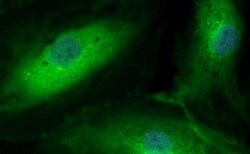
Customer Images
-
Application: Immunocytochemistry/ImmunofluorescenceSample Tested: HUVEC human umbilical vein endothelial cellsSpecies: HumanVerified Customer | Posted 04/05/2022

-
Application: Immunocytochemistry/ImmunofluorescenceSample Tested: Human Coronary Artery Endothelial CellsSpecies: HumanVerified Customer | Posted 09/10/2021
There are no reviews that match your criteria.
Protocols
Find general support by application which include: protocols, troubleshooting, illustrated assays, videos and webinars.
- 7-Amino Actinomycin D (7-AAD) Cell Viability Flow Cytometry Protocol
- Appropriate Fixation of IHC/ICC Samples
- Cellular Response to Hypoxia Protocols
- ClariTSA™ Fluorophore Kits
- Detection & Visualization of Antibody Binding
- Extracellular Membrane Flow Cytometry Protocol
- Flow Cytometry Protocol for Cell Surface Markers
- Flow Cytometry Protocol for Staining Membrane Associated Proteins
- Flow Cytometry Staining Protocols
- Flow Cytometry Troubleshooting Guide
- ICC Cell Smear Protocol for Suspension Cells
- ICC Immunocytochemistry Protocol Videos
- ICC for Adherent Cells
- Immunocytochemistry (ICC) Protocol
- Immunocytochemistry Troubleshooting
- Immunofluorescence of Organoids Embedded in Cultrex Basement Membrane Extract
- Immunohistochemistry (IHC) and Immunocytochemistry (ICC) Protocols
- Intracellular Flow Cytometry Protocol Using Alcohol (Methanol)
- Intracellular Flow Cytometry Protocol Using Detergents
- Intracellular Nuclear Staining Flow Cytometry Protocol Using Detergents
- Intracellular Staining Flow Cytometry Protocol Using Alcohol Permeabilization
- Intracellular Staining Flow Cytometry Protocol Using Detergents to Permeabilize Cells
- Preparing Samples for IHC/ICC Experiments
- Preventing Non-Specific Staining (Non-Specific Binding)
- Primary Antibody Selection & Optimization
- Propidium Iodide Cell Viability Flow Cytometry Protocol
- Protocol for Liperfluo
- Protocol for VisUCyte™ HRP Polymer Detection Reagent
- Protocol for the Characterization of Human Th22 Cells
- Protocol for the Characterization of Human Th9 Cells
- Protocol for the Fluorescent ICC Staining of Cell Smears - Graphic
- Protocol for the Fluorescent ICC Staining of Cultured Cells on Coverslips - Graphic
- Protocol for the Preparation and Fluorescent ICC Staining of Cells on Coverslips
- Protocol for the Preparation and Fluorescent ICC Staining of Non-adherent Cells
- Protocol for the Preparation and Fluorescent ICC Staining of Stem Cells on Coverslips
- Protocol for the Preparation of a Cell Smear for Non-adherent Cell ICC - Graphic
- Protocol: Annexin V and PI Staining by Flow Cytometry
- Protocol: Annexin V and PI Staining for Apoptosis by Flow Cytometry
- R&D Systems Quality Control Western Blot Protocol
- TUNEL and Active Caspase-3 Detection by IHC/ICC Protocol
- The Importance of IHC/ICC Controls
- Troubleshooting Guide: Fluorokine Flow Cytometry Kits
- Troubleshooting Guide: Western Blot Figures
- Western Blot Conditions
- Western Blot Protocol
- Western Blot Protocol for Cell Lysates
- Western Blot Troubleshooting
- Western Blot Troubleshooting Guide
- View all Protocols, Troubleshooting, Illustrated assays and Webinars
FAQs for Human VE-Cadherin Antibody (123413)
-
Q: Why does the staining protocol with this Cadherin antibody use buffers containing Ca2+ and Mg2+?
A: The staining protocol with this and other Cadherin antibodies uses buffer containing Ca2+ and Mg2+ because Cadherin function is Calcium-dependent.
Associated Pathways


