Goat anti-Canine IgE Heavy Chain Secondary Antibody [HRP]
Novus Biologicals | Catalog # NB7346
Key Product Details
Species Reactivity
Canine
Applications
Immunohistochemistry, Immunohistochemistry-Paraffin, Western Blot, ELISA, Immunocytochemistry/ Immunofluorescence
Label
HRP
Antibody Source
Polyclonal Goat IgG
Loading...
Product Specifications
Immunogen
Dog IgE
Reactivity Notes
Use in Borreliella burgdorferi reported in secitific publication PMID: 32792093
Specificity
By immunoelectrophoresis and ELISA this reacts specifically with dog IgE. This may cross react with IgE from other species. There was less than 0.01% reactivity with purified dog IgG1, IgG2, IgA and IgM.
Clonality
Polyclonal
Host
Goat
Isotype
IgG
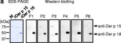
Scientific Data Images for Goat anti-Canine IgE Heavy Chain Secondary Antibody [HRP]
Goat-anti-Canine-IgE-Secondary-Antibody-[HRP]-Western-Blot-NB7346-img0001.jpg
Applications for Goat anti-Canine IgE Heavy Chain Secondary Antibody [HRP]
Application
Recommended Usage
ELISA
1:10000 - 1:100000
Immunocytochemistry/ Immunofluorescence
1:200 - 1:2000
Immunohistochemistry
1:200 - 1:2000
Western Blot
1:2000 - 1:20000
Reviewed Applications
Read 2 reviews rated 4.5 using NB7346 in the following applications:
Formulation, Preparation, and Storage
Purification
Immunogen affinity purified
Formulation
Phosphate Buffered Saline (PBS) containing 0.2%BSA
Preservative
0.05% Pro-Clean 400
Concentration
1.0 mg/ml
Shipping
The product is shipped with polar packs. Upon receipt, store it immediately at the temperature recommended below.
Stability & Storage
Store at 4C. Do not freeze.
Background: IgE Heavy Chain
Alternate Names
Constant Region of Heavy Chain of IgE, Ig Epsilon Chain C Region, Ig Epsilon Chain C Region ND, IgE, Immunoglobulin Epsilon, immunoglobulin heavy constant epsilon
Additional IgE Heavy Chain Products
Product Documents for Goat anti-Canine IgE Heavy Chain Secondary Antibody [HRP]
Certificate of Analysis
To download a Certificate of Analysis, please enter a lot or batch number in the search box below.
Product Specific Notices for Goat anti-Canine IgE Heavy Chain Secondary Antibody [HRP]
This product is for research use only and is not approved for use in humans or in clinical diagnosis. Secondary Antibodies are guaranteed for 1 year from date of receipt.
Citations for Goat anti-Canine IgE Heavy Chain Secondary Antibody [HRP]
 Powered by Bioz
Customer Reviews for Goat anti-Canine IgE Heavy Chain Secondary Antibody [HRP] (2)
4.5 out of 5
2 Customer Ratings
Have you used Goat anti-Canine IgE Heavy Chain Secondary Antibody [HRP]?
Submit a review and receive an Amazon gift card!
$25/€18/£15/$25CAN/¥2500 Yen for a review with an image
$10/€7/£6/$10CAN/¥1110 Yen for a review without an image
Submit a review
Customer Images
![Goat anti-Canine IgE Heavy Chain Secondary Antibody [HRP] NB7346](https://resources.rndsystems.com/images/reviews/review_nb7346_28356.jpg)
Showing
1
-
2 of
2 reviews
Showing All
Filter By:
-
Verified Customer | Posted 01/31/2017Western Blot detecting dog IgE standard.Western blot on nitrocellulose membrane. Sample: 0.5µg dog IgE Blocking: 5% skimmed milk powder in TBS +0.1% Tween-20 Detection: NB7346, 1:8000 diluted in 5% skimmed milk powder in TBS +0.1% Tween-20. Substrate: Clarity™ ECL Western Blotting Substrate Exposure time: 5 sec
![Goat anti-Canine IgE Heavy Chain Secondary Antibody [HRP] NB7346](data:image/png;base64,R0lGODlhAQABAAD/ACwAAAAAAQABAAACADs=)
-
Application: ELISASample Tested: Dog whole bloodSpecies: CanineVerified Customer | Posted 01/20/2017
There are no reviews that match your criteria.
Protocols
Find general support by application which include: protocols, troubleshooting, illustrated assays, videos and webinars.
- Antigen Retrieval Protocol (PIER)
- Antigen Retrieval for Frozen Sections Protocol
- Appropriate Fixation of IHC/ICC Samples
- Cellular Response to Hypoxia Protocols
- Chromogenic IHC Staining of Formalin-Fixed Paraffin-Embedded (FFPE) Tissue Protocol
- Chromogenic Immunohistochemistry Staining of Frozen Tissue
- ClariTSA™ Fluorophore Kits
- Detection & Visualization of Antibody Binding
- ELISA Sample Preparation & Collection Guide
- ELISA Troubleshooting Guide
- Fluorescent IHC Staining of Frozen Tissue Protocol
- Graphic Protocol for Heat-induced Epitope Retrieval
- Graphic Protocol for the Preparation and Fluorescent IHC Staining of Frozen Tissue Sections
- Graphic Protocol for the Preparation and Fluorescent IHC Staining of Paraffin-embedded Tissue Sections
- Graphic Protocol for the Preparation of Gelatin-coated Slides for Histological Tissue Sections
- How to Run an R&D Systems DuoSet ELISA
- How to Run an R&D Systems Quantikine ELISA
- How to Run an R&D Systems Quantikine™ QuicKit™ ELISA
- ICC Cell Smear Protocol for Suspension Cells
- ICC Immunocytochemistry Protocol Videos
- ICC for Adherent Cells
- IHC Sample Preparation (Frozen sections vs Paraffin)
- Immunocytochemistry (ICC) Protocol
- Immunocytochemistry Troubleshooting
- Immunofluorescence of Organoids Embedded in Cultrex Basement Membrane Extract
- Immunofluorescent IHC Staining of Formalin-Fixed Paraffin-Embedded (FFPE) Tissue Protocol
- Immunohistochemistry (IHC) and Immunocytochemistry (ICC) Protocols
- Immunohistochemistry Frozen Troubleshooting
- Immunohistochemistry Paraffin Troubleshooting
- Preparing Samples for IHC/ICC Experiments
- Preventing Non-Specific Staining (Non-Specific Binding)
- Primary Antibody Selection & Optimization
- Protocol for Heat-Induced Epitope Retrieval (HIER)
- Protocol for Making a 4% Formaldehyde Solution in PBS
- Protocol for VisUCyte™ HRP Polymer Detection Reagent
- Protocol for the Fluorescent ICC Staining of Cell Smears - Graphic
- Protocol for the Fluorescent ICC Staining of Cultured Cells on Coverslips - Graphic
- Protocol for the Preparation & Fixation of Cells on Coverslips
- Protocol for the Preparation and Chromogenic IHC Staining of Frozen Tissue Sections
- Protocol for the Preparation and Chromogenic IHC Staining of Frozen Tissue Sections - Graphic
- Protocol for the Preparation and Chromogenic IHC Staining of Paraffin-embedded Tissue Sections
- Protocol for the Preparation and Chromogenic IHC Staining of Paraffin-embedded Tissue Sections - Graphic
- Protocol for the Preparation and Fluorescent ICC Staining of Cells on Coverslips
- Protocol for the Preparation and Fluorescent ICC Staining of Non-adherent Cells
- Protocol for the Preparation and Fluorescent ICC Staining of Stem Cells on Coverslips
- Protocol for the Preparation and Fluorescent IHC Staining of Frozen Tissue Sections
- Protocol for the Preparation and Fluorescent IHC Staining of Paraffin-embedded Tissue Sections
- Protocol for the Preparation of Gelatin-coated Slides for Histological Tissue Sections
- Protocol for the Preparation of a Cell Smear for Non-adherent Cell ICC - Graphic
- Quantikine HS ELISA Kit Assay Principle, Alkaline Phosphatase
- Quantikine HS ELISA Kit Principle, Streptavidin-HRP Polymer
- R&D Systems Quality Control Western Blot Protocol
- Sandwich ELISA (Colorimetric) – Biotin/Streptavidin Detection Protocol
- Sandwich ELISA (Colorimetric) – Direct Detection Protocol
- TUNEL and Active Caspase-3 Detection by IHC/ICC Protocol
- The Importance of IHC/ICC Controls
- Troubleshooting Guide: ELISA
- Troubleshooting Guide: Immunohistochemistry
- Troubleshooting Guide: Western Blot Figures
- Western Blot Conditions
- Western Blot Protocol
- Western Blot Protocol for Cell Lysates
- Western Blot Troubleshooting
- Western Blot Troubleshooting Guide
- View all Protocols, Troubleshooting, Illustrated assays and Webinars
Loading...