IGFBP-4 Antibody - BSA Free
Novus Biologicals | Catalog # NBP1-80549
![Western Blot: IGFBP-4 Antibody [NBP1-80549]](https://resources.rndsystems.com/images/products/IGFBP-4-Antibody-Western-Blot-NBP1-80549-img0007.jpg "Western Blot: IGFBP-4 Antibody [NBP1-80549]")
Loading...
Key Product Details
Species Reactivity
Validated:
Human, Mouse
Cited:
Mouse
Applications
Validated:
Immunohistochemistry, Immunohistochemistry-Paraffin, Immunohistochemistry-Frozen, Western Blot, Immunocytochemistry/ Immunofluorescence
Cited:
Immunohistochemistry-Frozen
Label
Unconjugated
Antibody Source
Polyclonal Rabbit IgG
Format
BSA Free
Loading...
Product Specifications
Immunogen
Synthetic peptide directed towards the middle region of human IGFBP4. Peptide sequence RALERLAASQSRTHEDLYIIPIPNCDRNGNFHPKQCHPALDGQRGKCWCV. The peptide sequence for this immunogen was taken from within the described region.
Clonality
Polyclonal
Host
Rabbit
Isotype
IgG
Theoretical MW
28 kDa.
Disclaimer note: The observed molecular weight of the protein may vary from the listed predicted molecular weight due to post translational modifications, post translation cleavages, relative charges, and other experimental factors.
Disclaimer note: The observed molecular weight of the protein may vary from the listed predicted molecular weight due to post translational modifications, post translation cleavages, relative charges, and other experimental factors.
Description
The addition of 50% glycerol is optional for those storing this antibody at -20C and not aliquoting smaller units. However, please note that glycerol may interrupt some downstream antibody applications and should be added with caution.
Scientific Data Images for IGFBP-4 Antibody - BSA Free
Western Blot: IGFBP-4 Antibody [NBP1-80549]
Western Blot: IGFBP-4 Antibody [NBP1-80549] - Host: Mouse. Target Name: IGFBP4. Sample Tissue: Mouse Brain. Antibody Dilution: 1ug/ml![Immunocytochemistry/ Immunofluorescence: IGFBP-4 Antibody [NBP1-80549]](https://resources.rndsystems.com/images/products/IGFBP-4-Antibody-Immunocytochemistry-Immunofluorescence-NBP1-80549-img0006.jpg "Immunocytochemistry/ Immunofluorescence: IGFBP-4 Antibody [NBP1-80549]")
Immunocytochemistry/ Immunofluorescence: IGFBP-4 Antibody [NBP1-80549]
IGFBP-4-Antibody-Immunocytochemistry-Immunofluorescence-NBP1-80549-img0006.jpg![Immunohistochemistry-Paraffin: IGFBP-4 Antibody [NBP1-80549]](https://resources.rndsystems.com/images/products/IGFBP-4-Antibody-Immunohistochemistry-Paraffin-NBP1-80549-img0003.jpg "Immunohistochemistry-Paraffin: IGFBP-4 Antibody [NBP1-80549]")
Immunohistochemistry-Paraffin: IGFBP-4 Antibody [NBP1-80549]
Immunohistochemistry-Paraffin: IGFBP-4 Antibody [NBP1-80549] - Human Prostate Tissue, 5.0ug/ml.![Western Blot: IGFBP-4 Antibody [NBP1-80549]](https://resources.rndsystems.com/images/products/IGFBP-4-Antibody-Western-Blot-NBP1-80549-img0004.jpg "Western Blot: IGFBP-4 Antibody [NBP1-80549]")
Western Blot: IGFBP-4 Antibody [NBP1-80549]
Western Blot: IGFBP-4 Antibody [NBP1-80549] - Human Fetal Brain Lysate, concentration 1 ug/ml.![Western Blot: IGFBP-4 Antibody [NBP1-80549]](https://resources.rndsystems.com/images/products/IGFBP-4-Antibody-Western-Blot-NBP1-80549-img0008.jpg "Western Blot: IGFBP-4 Antibody [NBP1-80549]")
Western Blot: IGFBP-4 Antibody [NBP1-80549]
Western Blot: IGFBP-4 Antibody [NBP1-80549] - Host: Rabbit. Target Name: IGFBP4. Sample Tissue: Human MDA-MB-435s Whole Cell. Antibody Dilution: 1ug/ml![Immunocytochemistry/ Immunofluorescence: IGFBP-4 Antibody [NBP1-80549]](https://resources.rndsystems.com/images/products/IGFBP-4-Antibody-Immunocytochemistry-Immunofluorescence-NBP1-80549-img0005.jpg "Immunocytochemistry/ Immunofluorescence: IGFBP-4 Antibody [NBP1-80549]")
Immunocytochemistry/ Immunofluorescence: IGFBP-4 Antibody [NBP1-80549]
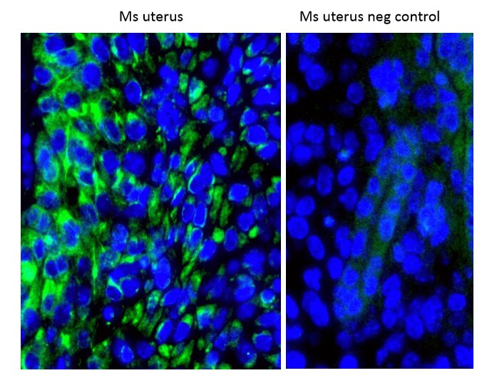
Immunocytochemistry/Immunofluorescence: IGFBP-4 Antibody [NBP1-80549] - Mouse uterus stained with at a dilution of 1:1000. HIER is applied using citrate buffer pH6. Image from verified customer review.
Immunocytochemistry/ Immunofluorescence: IGFBP-4 Antibody - BSA Free [NBP1-80549] -
Rac1 regulates vesicular exocytosis in decidual cells by controlling Rab27b.(A & B) Expression of Rab27b mRNA and protein is downregulated in Rac1-null stromal cells. (A) qPCR was performed to monitor the expression of Rab27a and Rab27b in the uteri of Rac1f/f and Rac1d/d mice on day 8 of pregnancy. Data represent mean +/- SEM from four separate samples and were analyzed by t-test. Asterisks indicate statistically significant differences (***P < 0.001). (B) IF of RAB27B in Rac1f/f and Rac1d/d uteri on day 8 of pregnancy. AMD, MD, and E denote antimesometrial decidua, mesometrial decidua, and embryo respectively. (C & D) Secretions by decidual cells are reduced in the conditioned media of Rac1-null stromal cells. Stromal cells isolated from Rac1f/f and Rac1d/d uteri on day 4 of pregnancy were cultured for 96 hours, fixed and subjected to IF using VEGFA (C, Left) and IGFBP4 (D, Left) antibodies. Conditioned media from cultured stromal cells isolated from Rac1f/f and Rac1d/d uteri were analyzed for VEGFA (C, Right) and IGFBP4 (D, Right) by ELISA. Data represent mean +/- SEM from three separate samples and were analyzed by two-way ANOVA with Bonferroni post-test. Asterisks indicate statistically significant differences (*P < 0.05, **P < 0.01, and ***P < 0.001). Image collected and cropped by CiteAb from the following open publication (https://pubmed.ncbi.nlm.nih.gov/26305333), licensed under a CC-BY license. Not internally tested by Novus Biologicals.Applications for IGFBP-4 Antibody - BSA Free
Application
Recommended Usage
Immunocytochemistry/ Immunofluorescence
1:10-1:500
Immunohistochemistry
1:10-1:500
Immunohistochemistry-Paraffin
1:10-1:500
Western Blot
1.0 ug/ml
Application Notes
Use in Immunohistochemistry-Frozen reported in scientific literature (PMID: 26305333).
Reviewed Applications
Read 1 review rated 5 using NBP1-80549 in the following applications:
Formulation, Preparation, and Storage
Purification
Affinity purified
Formulation
PBS, 2% Sucrose
Format
BSA Free
Preservative
0.09% Sodium Azide
Concentration
0.5 mg/ml
Shipping
The product is shipped with polar packs. Upon receipt, store it immediately at the temperature recommended below.
Stability & Storage
Store at 4C short term. Aliquot and store at -20C long term. Avoid freeze-thaw cycles.
Background: IGFBP-4
Long Name
Insulin-like Growth Factor Binding Protein 4
Alternate Names
IGFBP4
Gene Symbol
IGFBP4
UniProt
Additional IGFBP-4 Products
Product Documents for IGFBP-4 Antibody - BSA Free
Certificate of Analysis
To download a Certificate of Analysis, please enter a lot or batch number in the search box below.
Product Specific Notices for IGFBP-4 Antibody - BSA Free
This product is for research use only and is not approved for use in humans or in clinical diagnosis. Primary Antibodies are guaranteed for 1 year from date of receipt.
Related Research Areas
Citations for IGFBP-4 Antibody - BSA Free
 Powered by Bioz
Customer Reviews for IGFBP-4 Antibody - BSA Free (1)
5 out of 5
1 Customer Rating
Have you used IGFBP-4 Antibody - BSA Free?
Submit a review and receive an Amazon gift card!
$25/€18/£15/$25CAN/¥2500 Yen for a review with an image
$10/€7/£6/$10CAN/¥1110 Yen for a review without an image
Submit a review
Customer Images
Showing
1
-
1 of
1 review
Showing All
Filter By:
-
Application: Immunocytochemistry/ImmunofluorescenceSample Tested: mouse uterusSpecies: MouseVerified Customer | Posted 11/16/2016Used IF with Alexa fluor 488 as secondary antibody and used 1:1000 dilution of primary antibody. HIER is applied using citrate buffer pH6.

There are no reviews that match your criteria.
Protocols
Find general support by application which include: protocols, troubleshooting, illustrated assays, videos and webinars.
- Antigen Retrieval Protocol (PIER)
- Antigen Retrieval for Frozen Sections Protocol
- Appropriate Fixation of IHC/ICC Samples
- Cellular Response to Hypoxia Protocols
- Chromogenic IHC Staining of Formalin-Fixed Paraffin-Embedded (FFPE) Tissue Protocol
- Chromogenic Immunohistochemistry Staining of Frozen Tissue
- ClariTSA™ Fluorophore Kits
- Detection & Visualization of Antibody Binding
- Fluorescent IHC Staining of Frozen Tissue Protocol
- Graphic Protocol for Heat-induced Epitope Retrieval
- Graphic Protocol for the Preparation and Fluorescent IHC Staining of Frozen Tissue Sections
- Graphic Protocol for the Preparation and Fluorescent IHC Staining of Paraffin-embedded Tissue Sections
- Graphic Protocol for the Preparation of Gelatin-coated Slides for Histological Tissue Sections
- ICC Cell Smear Protocol for Suspension Cells
- ICC Immunocytochemistry Protocol Videos
- ICC for Adherent Cells
- IHC Sample Preparation (Frozen sections vs Paraffin)
- Immunocytochemistry (ICC) Protocol
- Immunocytochemistry Troubleshooting
- Immunofluorescence of Organoids Embedded in Cultrex Basement Membrane Extract
- Immunofluorescent IHC Staining of Formalin-Fixed Paraffin-Embedded (FFPE) Tissue Protocol
- Immunohistochemistry (IHC) and Immunocytochemistry (ICC) Protocols
- Immunohistochemistry Frozen Troubleshooting
- Immunohistochemistry Paraffin Troubleshooting
- Preparing Samples for IHC/ICC Experiments
- Preventing Non-Specific Staining (Non-Specific Binding)
- Primary Antibody Selection & Optimization
- Protocol for Heat-Induced Epitope Retrieval (HIER)
- Protocol for Making a 4% Formaldehyde Solution in PBS
- Protocol for VisUCyte™ HRP Polymer Detection Reagent
- Protocol for the Fluorescent ICC Staining of Cell Smears - Graphic
- Protocol for the Fluorescent ICC Staining of Cultured Cells on Coverslips - Graphic
- Protocol for the Preparation & Fixation of Cells on Coverslips
- Protocol for the Preparation and Chromogenic IHC Staining of Frozen Tissue Sections
- Protocol for the Preparation and Chromogenic IHC Staining of Frozen Tissue Sections - Graphic
- Protocol for the Preparation and Chromogenic IHC Staining of Paraffin-embedded Tissue Sections
- Protocol for the Preparation and Chromogenic IHC Staining of Paraffin-embedded Tissue Sections - Graphic
- Protocol for the Preparation and Fluorescent ICC Staining of Cells on Coverslips
- Protocol for the Preparation and Fluorescent ICC Staining of Non-adherent Cells
- Protocol for the Preparation and Fluorescent ICC Staining of Stem Cells on Coverslips
- Protocol for the Preparation and Fluorescent IHC Staining of Frozen Tissue Sections
- Protocol for the Preparation and Fluorescent IHC Staining of Paraffin-embedded Tissue Sections
- Protocol for the Preparation of Gelatin-coated Slides for Histological Tissue Sections
- Protocol for the Preparation of a Cell Smear for Non-adherent Cell ICC - Graphic
- R&D Systems Quality Control Western Blot Protocol
- TUNEL and Active Caspase-3 Detection by IHC/ICC Protocol
- The Importance of IHC/ICC Controls
- Troubleshooting Guide: Immunohistochemistry
- Troubleshooting Guide: Western Blot Figures
- Western Blot Conditions
- Western Blot Protocol
- Western Blot Protocol for Cell Lysates
- Western Blot Troubleshooting
- Western Blot Troubleshooting Guide
- View all Protocols, Troubleshooting, Illustrated assays and Webinars
Loading...