Mouse anti-Rabbit IgG (H+L) Secondary Antibody [Rhodamine] (Pre-adsorbed)
Novus Biologicals | Catalog # NBP1-73656
Key Product Details
Species Reactivity
Rabbit
Applications
Western Blot, Fluorophore-linked immunosorbent assay, Flow Cytometry, Immunocytochemistry/ Immunofluorescence, Dot Blot
Label
Rhodamine (Excitation = 570 nm, Emission = 590 nm)
Antibody Source
Polyclonal Mouse IgG
Format
Pre-adsorbed
Loading...
Product Specifications
Immunogen
Rabbit IgG whole molecule
Specificity
This antibody was pre-adsorbed against Human, Goat, and Mouse Serum Proteins. No reaction was observed against Goat, Human and Mouse Serum Proteins.
Clonality
Polyclonal
Host
Mouse
Isotype
IgG
Description
Store vial at 4C prior to restoration. For extended storage aliquot contents and freeze at -20C or below. Avoid cycles of freezing and thawing. Centrifuge product if not completely clear after standing at room temperature. This product is stable for several weeks at 4C as an undiluted liquid. Dilute only prior to immediate use.
This product was prepared from monospecific antiserum by immunoaffinity chromatography using Rabbit IgG coupled to agarose beads followed by solid phase adsorption(s) to remove any unwanted reactivities. Assay by immunoelectrophoresis resulted in a single precipitin arc against anti-Mouse Serum, Rabbit IgG and Rabbit Serum
This product was prepared from monospecific antiserum by immunoaffinity chromatography using Rabbit IgG coupled to agarose beads followed by solid phase adsorption(s) to remove any unwanted reactivities. Assay by immunoelectrophoresis resulted in a single precipitin arc against anti-Mouse Serum, Rabbit IgG and Rabbit Serum
Scientific Data Images
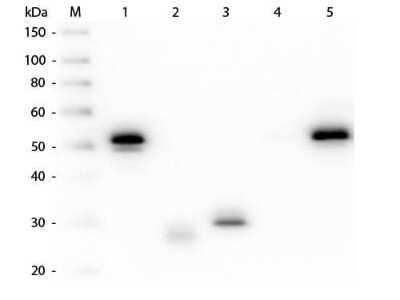
Western Blot: Mouse anti-Rabbit IgG (H+L) Secondary Antibody [Rhodamine] (Pre-adsorbed) [NBP1-73656] - Western Blot of Mouse anti-Rabbit IgG (H+L) Secondary antibody [Rhodamine] (Pre-adsorbed). Lane M: 3 ul Molecular Ladder. Lane 1: Rabbit IgG whole molecule. Lane 2: Rabbit IgG F(ab) Fragment. Lane 3: Rabbit IgG F(c) Fragment. Lane 4: Rabbit IgM Whole Molecule. Lane 5: Normal Rabbit Serum. All samples were reduced. Load: 50 ng per lane. Block for 30 min at RT. Primary Antibody: Mouse anti-Rabbit IgG (H+L) Secondary antibody [Rhodamine] (Pre-adsorbed) 1:1,000 for 60 min at RT. Secondary antibody: Anti-Mouse IgG (RABBIT) Peroxidase Conjugated Antibody 1:40,000 in blocking buffer for 30 min at RT. Predicted/Obsevered Size: 25 and 50 kDa for Rabbit IgG and Serum, 25 kDa for F(c) and F(ab), 70 and 23 kDa for IgM. Rabbit F(c) migrates slightly higher.
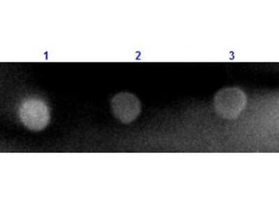
Dot Blot: Mouse anti-Rabbit IgG (H+L) Secondary Antibody [Rhodamine] (Pre-adsorbed) [NBP1-73656] - Dot Blot results of Mouse anti-Rabbit IgG (H+L) Secondary antibody [Rhodamine] (Pre-adsorbed).Dots are Rabbit IgG at (1) 100ng, (2) 33.3ng, (3) 11.1ng, (4) 3.70ng, (5) 1.23ng. Blocking buffer for 60 min at RT.Primary Antibody: none.Secondary Antibody: Mouse Anti-Rabbit IgG TRITC at 1ug/mL for 1hr at RT.Imaged with ChemiDoc, Rhodamine filter.
![Mouse anti-Rabbit IgG (H+L) Secondary Antibody [Rhodamine] (Pre-adsorbed)](https://resources.rndsystems.com/images/products/nbp1-73656_mouse-polyclonal-mouse-anti-rabbit-igg-h-l-secondary-antibody-rhodamine-pre-adsorbed-235202317492129.jpg "Mouse anti-Rabbit IgG (H+L) Secondary Antibody [Rhodamine] (Pre-adsorbed)")
Mouse anti-Rabbit IgG (H+L) Secondary Antibody [Rhodamine] (Pre-adsorbed)
Dot Blot results of Mouse Mouse anti-Rabbit IgG (H+L) Secondary Antibody [Rhodamine] (Pre-adsorbed). Dots are Rabbit IgG at (1) 100ng, (2) 33.3ng, (3) 11.1ng, (4) 3.70ng, (5) 1.23ng. blocking for 60 min at RT. Primary Antibody: none. Secondary Antibody: Mouse Anti-Rabbit IgG TRITC at 1ug/mL for 1hr at RT. Imaged with BioRad ChemiDoc, Rhodamine filter.Applications
Application
Recommended Usage
Flow Cytometry
1:500 - 1:2500
Fluorophore-linked immunosorbent assay
1:10000 - 1:50000
Immunocytochemistry/ Immunofluorescence
1:1000 - 1:5000
Western Blot
1:1000 - 1:10000
Application Notes
This product has been tested by dot blot and is designed for immunofluorescence microscopy, fluorescence based plate assays (FLISA) and fluorescent western blotting. This product is also suitable for multiplex analysis, including multicolor imaging, utilizing various commercial platforms.
Spectra Viewer
Plan Your Experiments
Use our spectra viewer to interactively plan your experiments, assessing multiplexing options. View the excitation and emission spectra for our fluorescent dye range and other commonly used dyes.
Spectra Viewer
Flow Cytometry Panel Builder
Bio-Techne Knows Flow Cytometry
Save time and reduce costly mistakes by quickly finding compatible reagents using the Panel Builder Tool.
Advanced Features
- Spectra Viewer - Custom analysis of spectra from multiple fluorochromes
- Spillover Popups - Visualize the spectra of individual fluorochromes
- Antigen Density Selector - Match fluorochrome brightness with antigen density

Formulation, Preparation, and Storage
Purification
Multi-step
Reconstitution
Reconstitute with 1.0 ml deionized water (or equivalent).
Formulation
Lyophilized from 0.02 M Potassium Phosphate, 0.15 M Sodium Chloride, pH 7.2, 10 mg/mL Bovine Serum Albumin (BSA) - Immunoglobulin and Protease free
Format
Pre-adsorbed
Preservative
0.01% Sodium Azide
Concentration
LYOPH mg/ml
Shipping
The product is shipped with polar packs. Upon receipt, store it immediately at the temperature recommended below.
Stability & Storage
Store lyophilized antibody at 4C in the dark. Aliquot reconstituted liquid and store at -20C. Avoid freeze-thaw cycles.
Calculators
Background: IgG (H+L)
The 4 IgG subclasses, sharing 95% amino acid identity, include IgG1, IgG2, IgG3, and IgG4 for humans and IgG1, IgG2a, IgG2b, and IgG3 for mice. The relative abundance of each human subclass is 60% for IgG1, 32% for IgG2, 4% for IgG3, and 4% for IgG4. In an IgG deficiency, there may be a shortage of one or more subclasses (4).
References
1. Painter RH. (1998) Encyclopedia of Immunology (Second Edition). Elsevier. 1208-1211
2. Chapter 9 - Antibodies. (2012) Immunology for Pharmacy. Mosby 70-78
3. Schroeder H, Cavacini, L. (2010) Structure and Function of Immunoglobulins. J Allergy Clin Immunol. 125(2 0 2): S41-S52. PMID: 20176268
4. Vidarsson G, Dekkers G, Rispens T. (2014) IgG subclasses and allotypes: from structure to effector functions. Front Immunol. 5:520. PMID: 25368619
Additional IgG (H+L) Products
Product Documents
Certificate of Analysis
To download a Certificate of Analysis, please enter a lot or batch number in the search box below.
Product Specific Notices
This product is for research use only and is not approved for use in humans or in clinical diagnosis. Secondary Antibodies are guaranteed for 1 year from date of receipt.
Customer Reviews
There are currently no reviews for this product. Be the first to review Mouse anti-Rabbit IgG (H+L) Secondary Antibody [Rhodamine] (Pre-adsorbed) and earn rewards!
Have you used Mouse anti-Rabbit IgG (H+L) Secondary Antibody [Rhodamine] (Pre-adsorbed)?
Submit a review and receive an Amazon gift card!
$25/€18/£15/$25CAN/¥2500 Yen for a review with an image
$10/€7/£6/$10CAN/¥1110 Yen for a review without an image
Submit a review
Protocols
Find general support by application which include: protocols, troubleshooting, illustrated assays, videos and webinars.
- 7-Amino Actinomycin D (7-AAD) Cell Viability Flow Cytometry Protocol
- Appropriate Fixation of IHC/ICC Samples
- Cellular Response to Hypoxia Protocols
- ClariTSA™ Fluorophore Kits
- Detection & Visualization of Antibody Binding
- ELISA Sample Preparation & Collection Guide
- ELISA Troubleshooting Guide
- Extracellular Membrane Flow Cytometry Protocol
- Flow Cytometry Protocol for Cell Surface Markers
- Flow Cytometry Protocol for Staining Membrane Associated Proteins
- Flow Cytometry Staining Protocols
- Flow Cytometry Troubleshooting Guide
- How to Run an R&D Systems DuoSet ELISA
- How to Run an R&D Systems Quantikine ELISA
- How to Run an R&D Systems Quantikine™ QuicKit™ ELISA
- ICC Cell Smear Protocol for Suspension Cells
- ICC Immunocytochemistry Protocol Videos
- ICC for Adherent Cells
- Immunocytochemistry (ICC) Protocol
- Immunocytochemistry Troubleshooting
- Immunofluorescence of Organoids Embedded in Cultrex Basement Membrane Extract
- Immunohistochemistry (IHC) and Immunocytochemistry (ICC) Protocols
- Intracellular Flow Cytometry Protocol Using Alcohol (Methanol)
- Intracellular Flow Cytometry Protocol Using Detergents
- Intracellular Nuclear Staining Flow Cytometry Protocol Using Detergents
- Intracellular Staining Flow Cytometry Protocol Using Alcohol Permeabilization
- Intracellular Staining Flow Cytometry Protocol Using Detergents to Permeabilize Cells
- Preparing Samples for IHC/ICC Experiments
- Preventing Non-Specific Staining (Non-Specific Binding)
- Primary Antibody Selection & Optimization
- Propidium Iodide Cell Viability Flow Cytometry Protocol
- Protocol for Liperfluo
- Protocol for VisUCyte™ HRP Polymer Detection Reagent
- Protocol for the Characterization of Human Th22 Cells
- Protocol for the Characterization of Human Th9 Cells
- Protocol for the Fluorescent ICC Staining of Cell Smears - Graphic
- Protocol for the Fluorescent ICC Staining of Cultured Cells on Coverslips - Graphic
- Protocol for the Preparation and Fluorescent ICC Staining of Cells on Coverslips
- Protocol for the Preparation and Fluorescent ICC Staining of Non-adherent Cells
- Protocol for the Preparation and Fluorescent ICC Staining of Stem Cells on Coverslips
- Protocol for the Preparation of a Cell Smear for Non-adherent Cell ICC - Graphic
- Protocol: Annexin V and PI Staining by Flow Cytometry
- Protocol: Annexin V and PI Staining for Apoptosis by Flow Cytometry
- Quantikine HS ELISA Kit Assay Principle, Alkaline Phosphatase
- Quantikine HS ELISA Kit Principle, Streptavidin-HRP Polymer
- R&D Systems Quality Control Western Blot Protocol
- Sandwich ELISA (Colorimetric) – Biotin/Streptavidin Detection Protocol
- Sandwich ELISA (Colorimetric) – Direct Detection Protocol
- TUNEL and Active Caspase-3 Detection by IHC/ICC Protocol
- The Importance of IHC/ICC Controls
- Troubleshooting Guide: ELISA
- Troubleshooting Guide: Fluorokine Flow Cytometry Kits
- Troubleshooting Guide: Western Blot Figures
- Western Blot Conditions
- Western Blot Protocol
- Western Blot Protocol for Cell Lysates
- Western Blot Troubleshooting
- Western Blot Troubleshooting Guide
- View all Protocols, Troubleshooting, Illustrated assays and Webinars
Loading...