Mouse IgG Isotype Control
Novus Biologicals | Catalog # NBP1-97019
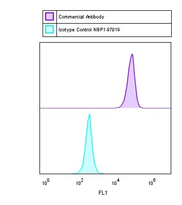
![Flow Cytometry: Mouse IgG Isotype Control [NBP1-97019]](https://resources.rndsystems.com/images/products/Mouse-IgG-Isotype-Control-Flow-Cytometry-NBP1-97019-img0004.jpg "Flow Cytometry: Mouse IgG Isotype Control [NBP1-97019]")
Key Product Details
Species Reactivity
Mouse
Applications
Immunohistochemistry, Western Blot, ELISA, Flow Cytometry, Immunoprecipitation, SDS-Page
Label
Unconjugated
Antibody Source
Mouse IgG
Loading...
Product Specifications
Host
Mouse
Isotype
IgG
Description
This product was prepared from normal mouse serum by a multi-step process which includes delipidation, salt fractionation and ion exchange chromatography followed by extensive dialysis against the buffer stated above. Assay by immunoelectrophoresis resulted in a single precipitin arc against anti-Mouse IgG and anti-Mouse Serum.
Store at 4C prior to restoration. For extended storage aliquot contents and freeze at -20C or below. Avoid cycles of freezing and thawing. Centrifuge product if not completely clear after standing at room temperature. This product is stable for several weeks at 4C as an undiluted liquid. Dilute only prior to immediate use.
Store at 4C prior to restoration. For extended storage aliquot contents and freeze at -20C or below. Avoid cycles of freezing and thawing. Centrifuge product if not completely clear after standing at room temperature. This product is stable for several weeks at 4C as an undiluted liquid. Dilute only prior to immediate use.
Scientific Data Images for Mouse IgG Isotype Control
Flow Cytometry: Mouse IgG Isotype Control [NBP1-97019]
Flow Cytometry: Mouse IgG Isotype Control [NBP1-97019]![SDS-PAGE: Mouse IgG Isotype Control [NBP1-97019]](https://resources.rndsystems.com/images/products/Mouse-IgG-Isotype-Control-SDS-Page-NBP1-97019-img0003.jpg "SDS-PAGE: Mouse IgG Isotype Control [NBP1-97019]")
SDS-PAGE: Mouse IgG Isotype Control [NBP1-97019]
SDS-Page: Mouse IgG Isotype Control [NBP1-97019] - Lane 1: 5 uL Opal Prestained Marker Lane 2: Reduced Mouse IgG Whole Molecule (BULK ORDER) Lane 3: Reduced Mouse F(c) Fragment Lane 4: Reduced Mouse F(ab) Fragment Lane 5: Mouse IgM Kappa Myeloma Protein Load: 1 ug per lane. Predicted/Observed size: IgG at 50 and 25 kDa; F(c) at 25 kDa; F(ab) at 25 kDa; IgM K at 70 and 23 kDa. Observed F(c) Fragment migrates slightly higher.![SDS-PAGE: Mouse IgG Isotype Control [NBP1-97019]](https://resources.rndsystems.com/images/products/Mouse-IgG-Isotype-Control-SDS-Page-NBP1-97019-img0005.jpg "SDS-PAGE: Mouse IgG Isotype Control [NBP1-97019]")
SDS-PAGE: Mouse IgG Isotype Control [NBP1-97019]
SDS-Page: Mouse IgG Isotype Control [NBP1-97019] - SDS-Page of Mouse IgG Isotype control. Lane 1: Mouse IgG reduced. Lane 2: Molecular Weight Marker. Lane 3: Mouse IgG non-reduced. Load: 1 ug per lane.Predicted/Observed size (non-reduced): 160 kDa, 160 kDa.Predicted/Observed size (reduced): 55 and 28 kDa, 55 and 28 kDa.Applications for Mouse IgG Isotype Control
Application
Recommended Usage
ELISA
Optimal dilutions of this antibody should be experimentally determined.
Flow Cytometry
Optimal dilutions of this antibody should be experimentally determined.
Immunohistochemistry
Optimal dilutions of this antibody should be experimentally determined.
Immunoprecipitation
Optimal dilutions of this antibody should be experimentally determined.
SDS-Page
Optimal dilutions of this antibody should be experimentally determined.
Western Blot
Optimal dilutions of this antibody should be experimentally determined.
Application Notes
This product has been tested in SDS-Page and can be utilized as a control or standard reagent in SDS, Western Blotting, and ELISA experiments.
Flow Cytometry Panel Builder
Bio-Techne Knows Flow Cytometry
Save time and reduce costly mistakes by quickly finding compatible reagents using the Panel Builder Tool.
Advanced Features
- Spectra Viewer - Custom analysis of spectra from multiple fluorochromes
- Spillover Popups - Visualize the spectra of individual fluorochromes
- Antigen Density Selector - Match fluorochrome brightness with antigen density

Formulation, Preparation, and Storage
Purification
Multi-step
Reconstitution
Reconstitute with 500 ul deionized water (or equivalent).
Formulation
Lyophilized from 0.02 M Potassium Phosphate, 0.15 M Sodium Chloride, pH 7.2
Preservative
0.01% Sodium Azide
Concentration
LYOPH mg/ml
Shipping
The product is shipped with polar packs. Upon receipt, store it immediately at the temperature recommended below.
Stability & Storage
Store lyophilized antibody at 4C. Aliquot reconstituted liquid and store at -20C. Avoid freeze-thaw cycles.
Calculators
Background: IgG
Long Name
Immunoglobulin G
Alternate Names
Immunoglobulin G, ImmunoglobulinG
Gene Symbol
IGHG1
Additional IgG Products
Product Documents for Mouse IgG Isotype Control
Certificate of Analysis
To download a Certificate of Analysis, please enter a lot or batch number in the search box below.
Product Specific Notices for Mouse IgG Isotype Control
This product is for research use only and is not approved for use in humans or in clinical diagnosis. Isotype Controls are guaranteed for 1 year from date of receipt.
Citations for Mouse IgG Isotype Control
 Powered by Bioz
Customer Reviews for Mouse IgG Isotype Control (1)
5 out of 5
1 Customer Rating
Have you used Mouse IgG Isotype Control?
Submit a review and receive an Amazon gift card!
$25/€18/£15/$25CAN/¥2500 Yen for a review with an image
$10/€7/£6/$10CAN/¥1110 Yen for a review without an image
Submit a review
Customer Images
Showing
1
-
1 of
1 review
Showing All
Filter By:
-
Verified Customer | Posted 04/13/2018

There are no reviews that match your criteria.
Protocols
Find general support by application which include: protocols, troubleshooting, illustrated assays, videos and webinars.
- 7-Amino Actinomycin D (7-AAD) Cell Viability Flow Cytometry Protocol
- Antigen Retrieval Protocol (PIER)
- Antigen Retrieval for Frozen Sections Protocol
- Appropriate Fixation of IHC/ICC Samples
- Cellular Response to Hypoxia Protocols
- Chromogenic IHC Staining of Formalin-Fixed Paraffin-Embedded (FFPE) Tissue Protocol
- Chromogenic Immunohistochemistry Staining of Frozen Tissue
- ClariTSA™ Fluorophore Kits
- Detection & Visualization of Antibody Binding
- ELISA Sample Preparation & Collection Guide
- ELISA Troubleshooting Guide
- Extracellular Membrane Flow Cytometry Protocol
- Flow Cytometry Protocol for Cell Surface Markers
- Flow Cytometry Protocol for Staining Membrane Associated Proteins
- Flow Cytometry Staining Protocols
- Flow Cytometry Troubleshooting Guide
- Fluorescent IHC Staining of Frozen Tissue Protocol
- Graphic Protocol for Heat-induced Epitope Retrieval
- Graphic Protocol for the Preparation and Fluorescent IHC Staining of Frozen Tissue Sections
- Graphic Protocol for the Preparation and Fluorescent IHC Staining of Paraffin-embedded Tissue Sections
- Graphic Protocol for the Preparation of Gelatin-coated Slides for Histological Tissue Sections
- How to Run an R&D Systems DuoSet ELISA
- How to Run an R&D Systems Quantikine ELISA
- How to Run an R&D Systems Quantikine™ QuicKit™ ELISA
- IHC Sample Preparation (Frozen sections vs Paraffin)
- Immunofluorescent IHC Staining of Formalin-Fixed Paraffin-Embedded (FFPE) Tissue Protocol
- Immunohistochemistry (IHC) and Immunocytochemistry (ICC) Protocols
- Immunohistochemistry Frozen Troubleshooting
- Immunohistochemistry Paraffin Troubleshooting
- Immunoprecipitation Protocol
- Intracellular Flow Cytometry Protocol Using Alcohol (Methanol)
- Intracellular Flow Cytometry Protocol Using Detergents
- Intracellular Nuclear Staining Flow Cytometry Protocol Using Detergents
- Intracellular Staining Flow Cytometry Protocol Using Alcohol Permeabilization
- Intracellular Staining Flow Cytometry Protocol Using Detergents to Permeabilize Cells
- Preparing Samples for IHC/ICC Experiments
- Preventing Non-Specific Staining (Non-Specific Binding)
- Primary Antibody Selection & Optimization
- Propidium Iodide Cell Viability Flow Cytometry Protocol
- Protocol for Heat-Induced Epitope Retrieval (HIER)
- Protocol for Liperfluo
- Protocol for Making a 4% Formaldehyde Solution in PBS
- Protocol for VisUCyte™ HRP Polymer Detection Reagent
- Protocol for the Characterization of Human Th22 Cells
- Protocol for the Characterization of Human Th9 Cells
- Protocol for the Preparation & Fixation of Cells on Coverslips
- Protocol for the Preparation and Chromogenic IHC Staining of Frozen Tissue Sections
- Protocol for the Preparation and Chromogenic IHC Staining of Frozen Tissue Sections - Graphic
- Protocol for the Preparation and Chromogenic IHC Staining of Paraffin-embedded Tissue Sections
- Protocol for the Preparation and Chromogenic IHC Staining of Paraffin-embedded Tissue Sections - Graphic
- Protocol for the Preparation and Fluorescent IHC Staining of Frozen Tissue Sections
- Protocol for the Preparation and Fluorescent IHC Staining of Paraffin-embedded Tissue Sections
- Protocol for the Preparation of Gelatin-coated Slides for Histological Tissue Sections
- Protocol: Annexin V and PI Staining by Flow Cytometry
- Protocol: Annexin V and PI Staining for Apoptosis by Flow Cytometry
- Quantikine HS ELISA Kit Assay Principle, Alkaline Phosphatase
- Quantikine HS ELISA Kit Principle, Streptavidin-HRP Polymer
- R&D Systems Quality Control Western Blot Protocol
- Sandwich ELISA (Colorimetric) – Biotin/Streptavidin Detection Protocol
- Sandwich ELISA (Colorimetric) – Direct Detection Protocol
- TUNEL and Active Caspase-3 Detection by IHC/ICC Protocol
- The Importance of IHC/ICC Controls
- Troubleshooting Guide: ELISA
- Troubleshooting Guide: Fluorokine Flow Cytometry Kits
- Troubleshooting Guide: Immunohistochemistry
- Troubleshooting Guide: Western Blot Figures
- Western Blot Conditions
- Western Blot Protocol
- Western Blot Protocol for Cell Lysates
- Western Blot Troubleshooting
- Western Blot Troubleshooting Guide
- View all Protocols, Troubleshooting, Illustrated assays and Webinars
Loading...