ILF1 Antibody - BSA Free
Novus Biologicals | Catalog # NBP1-87700
![Western Blot: ILF1 Antibody [NBP1-87700]](https://resources.rndsystems.com/images/products/ILF1-Antibody-Western-Blot-NBP1-87700-img0006.jpg "Western Blot: ILF1 Antibody [NBP1-87700]")
Loading...
Key Product Details
Species Reactivity
Validated:
Human
Predicted:
Mouse (90%). Backed by our 100% Guarantee.
Applications
Immunohistochemistry, Immunohistochemistry-Paraffin, Western Blot, Immunocytochemistry/ Immunofluorescence, Chromatin Immunoprecipitation-exo-Seq
Label
Unconjugated
Antibody Source
Polyclonal Rabbit IgG
Format
BSA Free
Loading...
Product Specifications
Immunogen
This antibody was developed against Recombinant Protein corresponding to amino acids: SSQPVLITVQRQLPQAIKPVTYTVATPVTTSTSQPPVVQTVHVVHQIPAVSVTSVAGLAPANTYTVSGQAVVTPAAVLAPPKAEAQENGDHREVKVKVEPIPAIGHATLGTASRIIQTAQTTPVQTVTIVQQAPLGQHQLPIK
Clonality
Polyclonal
Host
Rabbit
Isotype
IgG
Scientific Data Images for ILF1 Antibody - BSA Free
Western Blot: ILF1 Antibody [NBP1-87700]
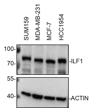
Western Blot: ILF1 Antibody [NBP1-87700] - Whole cell lysates from SUM159, MDA-MB-231, MCF-7 and HCC1954 cells were loaded with 50 ug/lane. 10% SDS-PAGE. ILF1 antibody (NBP1-87700) was used for primary antibody: 1:2000, 4C, overnight. Image from verified customer review.![Immunohistochemistry-Paraffin: ILF1 Antibody [NBP1-87700]](https://resources.rndsystems.com/images/products/ILF1-Antibody-Immunohistochemistry-Paraffin-NBP1-87700-img0002.jpg "Immunohistochemistry-Paraffin: ILF1 Antibody [NBP1-87700]")
Immunohistochemistry-Paraffin: ILF1 Antibody [NBP1-87700]
Immunohistochemistry-Paraffin: ILF1 Antibody [NBP1-87700] - Staining of human small intestine shows distinct nuclear positivity in glandular cells.![Western Blot: ILF1 Antibody [NBP1-87700]](https://resources.rndsystems.com/images/products/ILF1-Antibody-Western-Blot-NBP1-87700-img0005.jpg "Western Blot: ILF1 Antibody [NBP1-87700]")
Western Blot: ILF1 Antibody [NBP1-87700]
Western Blot: ILF1 Antibody [NBP1-87700] - Analysis in human cell line RT-4 and human cell line U-251 MG.![ILF1 Antibody - BSA Free Chromatin Immunoprecipitation-exo-Seq: ILF1 Antibody - BSA Free [NBP1-87700]](https://resources.rndsystems.com/images/products/nbp1-87700_rabbit-polyclonal-ilf1-antibody-25620259114078.jpg "Chromatin Immunoprecipitation-exo-Seq: ILF1 Antibody - BSA Free [NBP1-87700]")
Chromatin Immunoprecipitation-exo-Seq: ILF1 Antibody - BSA Free [NBP1-87700]
ChIP-Exo-Seq composite graph for Anti-FOXK2 (NBP1-87700) tested in K562 cells. Strand-specific reads (blue: forward, red: reverse) and IgG controls (black: forward, grey: reverse) are plotted against the distance from a composite set of reference binding sites. The antibody exhibits robust target enrichment compared to a non-specific IgG control and precisely reveals its structural organization around the binding site. Data generated by Prof. B. F. Pugh´s Lab at Cornell University.
Western Blot: ILF1 Antibody - BSA Free [NBP1-87700] -
Foxk1 and Foxk2 extend the cardiomyocyte proliferative window.A Experimental design: neonatal mice were administered AAV9-GFP, AAV9-Foxk1, or AAV9-Foxk2 on postnatal day 1 (P1) to overexpress Foxk1 or Foxk2 specifically in cardiomyocytes. Heart tissue was subsequently collected at P14. B Western blot analysis of FOXK1 and FOXK2 expression in P14 mouse cardiomyocytes (n = 3 mice per group). C H&E-stained heart sections (n = 5 mice per group). D, E WGA staining of heart tissue sections and quantitative analysis of the cross-sectional area of cardiomyocytes (CM size) (n = 3 per group). F Analysis of total cardiomyocyte counts per heart across different experimental groups (n = 6 per group). G Representative images of cardiomyocytes isolated from the hearts of mice treated with AAV9-Foxk1 or AAV9-Foxk2 and quantification of nucleation at P14 (n = 3 per group). H–J Representative images of co-immunostaining and quantitative analysis for alpha -actinin (red) and Aurora B (green, indicated by yellow arrows) (H), pH3 (green) (I), and Ki67 (green) (J) in heart tissue sections (n = 4 per group). Data are presented as mean +/- SEM. p values were determined by one-way ANOVA with Bonferroni multiple comparisons test (E, F, and H–J) and two-way ANOVA with Bonferroni multiple comparisons test (G). Source data are provided as a Source Data file. Image collected and cropped by CiteAb from the following open publication (https://pubmed.ncbi.nlm.nih.gov/40128196), licensed under a CC-BY license. Not internally tested by Novus Biologicals.![ILF1 Antibody - BSA Free Immunocytochemistry/ Immunofluorescence: ILF1 Antibody [NBP1-87700]](https://resources.rndsystems.com/images/products/nbp1-87700_-immunocytochemistry-immunofluorescence-639174076676011204.jpg "Immunocytochemistry/ Immunofluorescence: ILF1 Antibody [NBP1-87700]")
Immunocytochemistry/ Immunofluorescence: ILF1 Antibody [NBP1-87700]
Staining of human cell line U-2 OS shows localization to nucleoplasm & vesicles.Applications for ILF1 Antibody - BSA Free
Application
Recommended Usage
Chromatin Immunoprecipitation-exo-Seq
1-10ug per reaction
Immunocytochemistry/ Immunofluorescence
0.25-2 ug/ml
Immunohistochemistry
1:200 - 1:500
Immunohistochemistry-Paraffin
1:200 - 1:500
Western Blot
0.04-0.4 ug/ml
Application Notes
IHC-Paraffin, HIER pH 6 retrieval is recommended. ICC/IF, Fixation Permeabilization: Use PFA/Triton X-100.
Reviewed Applications
Read 1 review rated 5 using NBP1-87700 in the following applications:
Formulation, Preparation, and Storage
Purification
Affinity purified
Formulation
PBS (pH 7.2) and 40% Glycerol
Format
BSA Free
Preservative
0.02% Sodium Azide
Concentration
Concentrations vary lot to lot. See vial label for concentration. If unlisted please contact technical services.
Shipping
The product is shipped with polar packs. Upon receipt, store it immediately at the temperature recommended below.
Stability & Storage
Store at 4C short term. Aliquot and store at -20C long term. Avoid freeze-thaw cycles.
Background: ILF1
Alternate Names
Cellular transcription factor ILF-1, forkhead box K2, forkhead box protein K2, FOXK1, ILF-1, ILF1ILF, interleukin enhancer binding factor 1, Interleukin enhancer-binding factor 1
Gene Symbol
FOXK2
Additional ILF1 Products
Product Documents for ILF1 Antibody - BSA Free
Certificate of Analysis
To download a Certificate of Analysis, please enter a lot or batch number in the search box below.
Product Specific Notices for ILF1 Antibody - BSA Free
This product is for research use only and is not approved for use in humans or in clinical diagnosis. Primary Antibodies are guaranteed for 1 year from date of receipt.
Citations for ILF1 Antibody - BSA Free
 Powered by Bioz
Customer Reviews for ILF1 Antibody - BSA Free (1)
5 out of 5
1 Customer Rating
Have you used ILF1 Antibody - BSA Free?
Submit a review and receive an Amazon gift card!
$25/€18/£15/$25CAN/¥2500 Yen for a review with an image
$10/€7/£6/$10CAN/¥1110 Yen for a review without an image
Submit a review
Customer Images
Showing
1
-
1 of
1 review
Showing All
Filter By:
-
Application: Western BlotSample Tested: SUM-159PT, MDA-MB-231 and MCF-7Species: HumanVerified Customer | Posted 11/15/2022Western Blot: whole cell lysates from SUM159, MDA-MB-231, MCF-7 and HCC1954 cells were loaded with 50 ug/lane. 10% SDS-PAGE. ILF1 Antibody (NBP1-87700) was used for primary antibody: 1:2000, 4℃, overnight.

There are no reviews that match your criteria.
Protocols
Find general support by application which include: protocols, troubleshooting, illustrated assays, videos and webinars.
- Antigen Retrieval Protocol (PIER)
- Antigen Retrieval for Frozen Sections Protocol
- Appropriate Fixation of IHC/ICC Samples
- Cellular Response to Hypoxia Protocols
- Chromogenic IHC Staining of Formalin-Fixed Paraffin-Embedded (FFPE) Tissue Protocol
- Chromogenic Immunohistochemistry Staining of Frozen Tissue
- ClariTSA™ Fluorophore Kits
- Detection & Visualization of Antibody Binding
- Fluorescent IHC Staining of Frozen Tissue Protocol
- Graphic Protocol for Heat-induced Epitope Retrieval
- Graphic Protocol for the Preparation and Fluorescent IHC Staining of Frozen Tissue Sections
- Graphic Protocol for the Preparation and Fluorescent IHC Staining of Paraffin-embedded Tissue Sections
- Graphic Protocol for the Preparation of Gelatin-coated Slides for Histological Tissue Sections
- ICC Cell Smear Protocol for Suspension Cells
- ICC Immunocytochemistry Protocol Videos
- ICC for Adherent Cells
- IHC Sample Preparation (Frozen sections vs Paraffin)
- Immunocytochemistry (ICC) Protocol
- Immunocytochemistry Troubleshooting
- Immunofluorescence of Organoids Embedded in Cultrex Basement Membrane Extract
- Immunofluorescent IHC Staining of Formalin-Fixed Paraffin-Embedded (FFPE) Tissue Protocol
- Immunohistochemistry (IHC) and Immunocytochemistry (ICC) Protocols
- Immunohistochemistry Frozen Troubleshooting
- Immunohistochemistry Paraffin Troubleshooting
- Preparing Samples for IHC/ICC Experiments
- Preventing Non-Specific Staining (Non-Specific Binding)
- Primary Antibody Selection & Optimization
- Protocol for Heat-Induced Epitope Retrieval (HIER)
- Protocol for Making a 4% Formaldehyde Solution in PBS
- Protocol for VisUCyte™ HRP Polymer Detection Reagent
- Protocol for the Fluorescent ICC Staining of Cell Smears - Graphic
- Protocol for the Fluorescent ICC Staining of Cultured Cells on Coverslips - Graphic
- Protocol for the Preparation & Fixation of Cells on Coverslips
- Protocol for the Preparation and Chromogenic IHC Staining of Frozen Tissue Sections
- Protocol for the Preparation and Chromogenic IHC Staining of Frozen Tissue Sections - Graphic
- Protocol for the Preparation and Chromogenic IHC Staining of Paraffin-embedded Tissue Sections
- Protocol for the Preparation and Chromogenic IHC Staining of Paraffin-embedded Tissue Sections - Graphic
- Protocol for the Preparation and Fluorescent ICC Staining of Cells on Coverslips
- Protocol for the Preparation and Fluorescent ICC Staining of Non-adherent Cells
- Protocol for the Preparation and Fluorescent ICC Staining of Stem Cells on Coverslips
- Protocol for the Preparation and Fluorescent IHC Staining of Frozen Tissue Sections
- Protocol for the Preparation and Fluorescent IHC Staining of Paraffin-embedded Tissue Sections
- Protocol for the Preparation of Gelatin-coated Slides for Histological Tissue Sections
- Protocol for the Preparation of a Cell Smear for Non-adherent Cell ICC - Graphic
- R&D Systems Quality Control Western Blot Protocol
- TUNEL and Active Caspase-3 Detection by IHC/ICC Protocol
- The Importance of IHC/ICC Controls
- Troubleshooting Guide: Immunohistochemistry
- Troubleshooting Guide: Western Blot Figures
- Western Blot Conditions
- Western Blot Protocol
- Western Blot Protocol for Cell Lysates
- Western Blot Troubleshooting
- Western Blot Troubleshooting Guide
- View all Protocols, Troubleshooting, Illustrated assays and Webinars
Loading...