![Immunohistochemistry-Paraffin: iNOS Antibody (K13-A) [NBP1-33780]](https://resources.rndsystems.com/images/products/iNOS-Antibody-K13-A-Immunohistochemistry-Paraffin-NBP1-33780-img0011.jpg "Immunohistochemistry-Paraffin: iNOS Antibody (K13-A) [NBP1-33780]")
Loading...
Key Product Details
Species Reactivity
Validated:
Human, Mouse, Rat, Porcine
Cited:
Human, Mouse, Rat
Applications
Validated:
Immunohistochemistry, Immunohistochemistry-Paraffin, Immunohistochemistry-Frozen, Western Blot
Cited:
Immunohistochemistry, Immunohistochemistry-Paraffin, Immunohistochemistry-Frozen, Western Blot, Immunocytochemistry/ Immunofluorescence, IF/IHC, Knockdown Validated
Label
Unconjugated
Antibody Source
Recombinant Monoclonal Rabbit IgG Clone # K13-A
Loading...
Product Specifications
Immunogen
Peptide derived from human iNOS sequence. Antibody recognizes the epitope between Ser1118 - Gly1129.
Epitope
Ser1118 - Gly1129
Reactivity Notes
This antibody also recognizes nNOS (neuronal nitric oxide synthase). Porcine reactivity reported per verified customer review.
Clonality
Monoclonal
Host
Rabbit
Isotype
IgG
Description
This antibody is immunoaffinity purified with immunogenic peptide as a ligand.
Scientific Data Images for iNOS Antibody (K13-A)
Immunohistochemistry-Paraffin: iNOS Antibody (K13-A) [NBP1-33780]
Immunohistochemistry-Paraffin: iNOS Antibody (K13-A) [NBP1-33780] - Liver tissue stained with anti-iNOS antibody shows strong positive immunostaining of hepatocytes. Formalin fixed, paraffin embedded human tissues (4 um sections) stained according to IHC-P Protocol for NBP1-33780.![Immunohistochemistry-Paraffin: iNOS Antibody (K13-A) [NBP1-33780]](https://resources.rndsystems.com/images/products/iNOS-Antibody-K13-A-Immunohistochemistry-Paraffin-NBP1-33780-img0012.jpg "Immunohistochemistry-Paraffin: iNOS Antibody (K13-A) [NBP1-33780]")
Immunohistochemistry-Paraffin: iNOS Antibody (K13-A) [NBP1-33780]
Immunohistochemistry-Paraffin: iNOS Antibody (K13-A) [NBP1-33780] - Lung adenocarcinoma tissue stained with anti-iNOS antibody shows strong positive immunostaining of hepatocytes. Formalin fixed, paraffin embedded human tissues (4 um sections) stained according to IHC-P Protocol for NBP1-33780.![Immunohistochemistry-Paraffin: iNOS Antibody (K13-A) [NBP1-33780]](https://resources.rndsystems.com/images/products/iNOS-Antibody-K13-A-Immunohistochemistry-Paraffin-NBP1-33780-img0010.jpg "Immunohistochemistry-Paraffin: iNOS Antibody (K13-A) [NBP1-33780]")
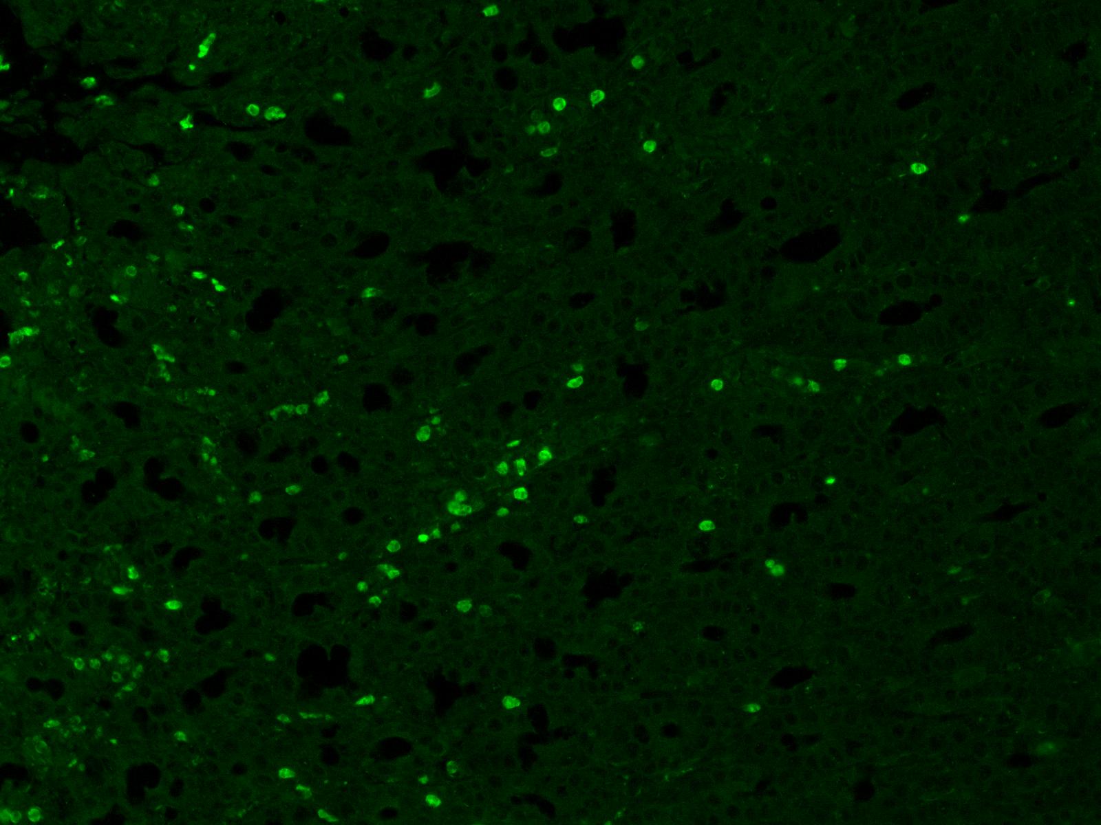
Immunohistochemistry-Paraffin: iNOS Antibody (K13-A) [NBP1-33780]
Immunohistochemistry-Paraffin: iNOS Antibody (K13-A) [NBP1-33780] - Inflamed porcine intestine. Antibody dilution 1:200, overnight 4C. Secondary anti-rabbit Alexa 488 (1:1000) 2 hr incubation. Image from verified customer review. [NBP1-33780] -")
Western Blot: iNOS Antibody (K13-A) [NBP1-33780] -
Effects of omega-3 supplementation on the immunocontent of macrophages M1 markers: nitric oxide synthase 2 (NOS-2) (A) and CD86 (B), and M2 marker: Arginase-1 (C) on the mouse paw muscle 48 h after the model induction in Saline/Sham, Saline/CPIP, Omega-3/CPIP, and Corn oil/CPIP groups. Data are expressed as mean ± SD of 6 animals per group, statistically assessed by the one-way ANOVA followed by Tukey’s test. ##p < 0.01 vs. the Saline/Sham group. [NBP1-33780] -")
Western Blot: iNOS Antibody (K13-A) [NBP1-33780] -
Western Blot: iNOS Antibody (K13-A) [NBP1-33780] - Effects of omega-3 supplementation on the immunocontent of macrophages M1 markers: nitric oxide synthase 2 (NOS-2) (A) & CD86 (B), & M2 marker: Arginase-1 (C) on the mouse paw muscle 48 h after the model induction in Saline/Sham, Saline/CPIP, Omega-3/CPIP, & Corn oil/CPIP groups. Data are expressed as mean ± SD of 6 animals per group, statistically assessed by the one-way ANOVA followed by Tukey’s test. ##p < 0.01 vs. the Saline/Sham group. Image collected & cropped by CiteAb from the following publication (https://pubmed.ncbi.nlm.nih.gov/35391753), licensed under a CC-BY license. Not internally tested by Novus Biologicals.Applications for iNOS Antibody (K13-A)
Application
Recommended Usage
Immunohistochemistry
1:10-1:500
Immunohistochemistry-Paraffin
1:100-1:200
Western Blot
1:2000
Application Notes
Use in IHC-P reported in scientific literature (PMID:34496231).
Reviewed Applications
Read 1 review rated 5 using NBP1-33780 in the following applications:
Formulation, Preparation, and Storage
Purification
Immunogen affinity purified
Formulation
20mM Tris-HCl (pH 8.0) and 20mg/ml BSA
Preservative
0.05% Sodium Azide
Concentration
Please see the vial label for concentration. If unlisted please contact technical services.
Shipping
The product is shipped with polar packs. Upon receipt, store it immediately at the temperature recommended below.
Stability & Storage
Store at 4C. Do not freeze.
Background: iNOS
The 131 kDa enzyme, iNOS, is found in a variety of cell types including macrophages, hepatocytes, synoviocytes, and smooth muscle cells. While constitutively expressed in kidneys, in other tissues iNOS is induced by bacterial lipopolysaccharides (LPS), growth factors, and cytokines such as IFN-gamma, TNF, IL-1 and IL-2. iNOS is not regulated by the level of intracellular Ca2+ and is constantly active as a dimer when expressed. iNOS activity is elevated in a variety of diseases including atherosclerosis, heart failure, sepsis, solid tumors, and type 2 diabetes. Acting as a critical mediator of inflammation and apoptosis, iNOS inhibitors have been shown to alleviate obesity and stress inducted insulin resistance in mouse models (2,3).
References
1. Forstermann U, and Sessa WC. (2012) Nitric oxide synthases: regulation and function. Eur Heart J. 33(7): 829-837. PMID: 21890489
2. Aktan F. (2004) iNOS-mediated nitric oxide production and its regulation. Life Sci. 75(6):639-53. PMID: 15172174
3. Cinelli MA, Do HT, Miley GP, Silverman RB. (2020) Inducible nitric oxide synthase: Regulation, structure, and inhibition. Med Res Rev. 40(1):158-189. PMID: 31192483
Long Name
Inducible Nitic Oxide Synthase
Alternate Names
NOS2, NOS2A
Entrez Gene IDs
4843 (Human)
Gene Symbol
NOS2
UniProt
Additional iNOS Products
Product Documents for iNOS Antibody (K13-A)
Certificate of Analysis
To download a Certificate of Analysis, please enter a lot or batch number in the search box below.
Product Specific Notices for iNOS Antibody (K13-A)
This antibody is immunoaffinity purified with immunogenic peptide as a ligand.
This product is for research use only and is not approved for use in humans or in clinical diagnosis. Primary Antibodies are guaranteed for 1 year from date of receipt.
Citations for iNOS Antibody (K13-A)
 Powered by Bioz
Customer Reviews for iNOS Antibody (K13-A) (1)
5 out of 5
1 Customer Rating
Have you used iNOS Antibody (K13-A)?
Submit a review and receive an Amazon gift card!
$25/€18/£15/$25CAN/¥2500 Yen for a review with an image
$10/€7/£6/$10CAN/¥1110 Yen for a review without an image
Submit a review
Customer Images
Showing
1
-
1 of
1 review
Showing All
Filter By:
-
Application: Immunohistochemistry-ParaffinSample Tested: paraffin sections of inflamed intestineSpecies: PorcineVerified Customer | Posted 04/18/2018Antibody dilution 1:200, overnight 4C. Secondary anti-rabbit Alexa 488 (1:1000) 2 hr incubation.

There are no reviews that match your criteria.
Protocols
View specific protocols for iNOS Antibody (K13-A) (NBP1-33780):
Immunohistochemistry-Paraffin protocol for iNOS Antibody (NBP1-33780):
Immunohistochemistry-Paraffin
1. Deparaffinize the section in 3 changes of xylene, 5 minutes each.
2. Wash the section in 96%, 80% and 70% ethyl alcohol for 10 minutes each.
3. Rinse in distilled water.
4. Block the endogenous peroxidase by incubating the tissue in 3% hydrogen peroxide (H2O2) for 10 minutes.
5. Wash in distilled water.
6. For antigen retrieval: Immerse the slide in Tris-EDTA buffer*, pH 9.0 and incubate at 95-97C in water bath for 25 minutes. (Alternatively adjust to your own protocol, keeping the required pH)
7. Remove the staining to room temperature and let the slide to cool (in Tris-EDTA buffer, pH 9.0) for 15 minutes.
8. Rinse in distilled water, 2 x 5 minutes.
9. Wash in PBS (phosphate buffer saline, pH 7.0-7.5) supplemented with 0.05% of Tween-20 (buffer A), 2 x 5 minutes.
10. Incubate the section with primary antibody at the dilution 1:100 - 1:200 for 1 hour in the closed wet chamber.
11. Wash 3 x 5 minutes with buffer A.
12. Apply the secondary antibody (the protocol depends on the supplier), and proceed with immunohistochemistry protocol (HRP - Peroxide - DAB).
13. Wash 3 x 5 minutes with buffer A.
14. Apply the chromogen (DAB), 1-3 minutes.
15. Wash in water, 2 x 5 minutes.
16. Stain in hematoxylin for 5 minutes.
17. Wash in water, 2 x 5 minutes.
18. Stain in hematoxylin for 5 minutes.
19. Wash in distilled water, 3 x 2 minutes.
20. Mount the slide for observation.
* Tris-EDTA Buffer (10mM Tris Base, 1mM EDTA solution, pH 9.0)
Tris -- 1.21 g; EDTA -- 0.37 g; Distilled water -- 1000 ml
Mix to dissolve in 700 ml of distilled water. Adjust pH to 9.0 with 1M HCl and mix well.
Adjust the final volume to 1 liter with distilled water.
Store this solution at room temperature for 3 months or at 4C for longer storage.
Find general support by application which include: protocols, troubleshooting, illustrated assays, videos and webinars.
- Antigen Retrieval Protocol (PIER)
- Antigen Retrieval for Frozen Sections Protocol
- Appropriate Fixation of IHC/ICC Samples
- Cellular Response to Hypoxia Protocols
- Chromogenic IHC Staining of Formalin-Fixed Paraffin-Embedded (FFPE) Tissue Protocol
- Chromogenic Immunohistochemistry Staining of Frozen Tissue
- ClariTSA™ Fluorophore Kits
- Detection & Visualization of Antibody Binding
- Fluorescent IHC Staining of Frozen Tissue Protocol
- Graphic Protocol for Heat-induced Epitope Retrieval
- Graphic Protocol for the Preparation and Fluorescent IHC Staining of Frozen Tissue Sections
- Graphic Protocol for the Preparation and Fluorescent IHC Staining of Paraffin-embedded Tissue Sections
- Graphic Protocol for the Preparation of Gelatin-coated Slides for Histological Tissue Sections
- IHC Sample Preparation (Frozen sections vs Paraffin)
- Immunofluorescent IHC Staining of Formalin-Fixed Paraffin-Embedded (FFPE) Tissue Protocol
- Immunohistochemistry (IHC) and Immunocytochemistry (ICC) Protocols
- Immunohistochemistry Frozen Troubleshooting
- Immunohistochemistry Paraffin Troubleshooting
- Preparing Samples for IHC/ICC Experiments
- Preventing Non-Specific Staining (Non-Specific Binding)
- Primary Antibody Selection & Optimization
- Protocol for Heat-Induced Epitope Retrieval (HIER)
- Protocol for Making a 4% Formaldehyde Solution in PBS
- Protocol for VisUCyte™ HRP Polymer Detection Reagent
- Protocol for the Preparation & Fixation of Cells on Coverslips
- Protocol for the Preparation and Chromogenic IHC Staining of Frozen Tissue Sections
- Protocol for the Preparation and Chromogenic IHC Staining of Frozen Tissue Sections - Graphic
- Protocol for the Preparation and Chromogenic IHC Staining of Paraffin-embedded Tissue Sections
- Protocol for the Preparation and Chromogenic IHC Staining of Paraffin-embedded Tissue Sections - Graphic
- Protocol for the Preparation and Fluorescent IHC Staining of Frozen Tissue Sections
- Protocol for the Preparation and Fluorescent IHC Staining of Paraffin-embedded Tissue Sections
- Protocol for the Preparation of Gelatin-coated Slides for Histological Tissue Sections
- R&D Systems Quality Control Western Blot Protocol
- TUNEL and Active Caspase-3 Detection by IHC/ICC Protocol
- The Importance of IHC/ICC Controls
- Troubleshooting Guide: Immunohistochemistry
- Troubleshooting Guide: Western Blot Figures
- Western Blot Conditions
- Western Blot Protocol
- Western Blot Protocol for Cell Lysates
- Western Blot Troubleshooting
- Western Blot Troubleshooting Guide
- View all Protocols, Troubleshooting, Illustrated assays and Webinars
FAQs for iNOS Antibody (K13-A)
Showing
1
-
3 of
3 FAQs
Showing All
-
Q: Hi, I would like to know if this antibody (NBP1-33780) works on mouse tissue in Immunohistochemistry. Did you test the mouse cross-reactivity on IHC ?
A: At this time we have only tested NBP1-33780 on human tissues in IHC, but we do expect it to work on mouse tissues in IHC based on sequence homology.
-
Q: We need to evaluate the level of iNOS in Rat sciatic nerve by Western Blot. I see that you have few antibodies suitable for that, could you please recommend the best what you have for our purposes?
A:
We have 4 iNOS antibodies that are suitable for your experiment. The best choice for you will depend on your specific preferences, in this case the main difference will be the immunogen location. If the immunogen is not important to you, then I would recommend choosing NB300-605 as it gives you the best value and we are able to provide the exact immunogen.
-
Q: What is the concentration of NBP1-33780?
A: The concentration of the lot number DB003-02-5B is 4.9 mg/ml
-
Q: Hi, I would like to know if this antibody (NBP1-33780) works on mouse tissue in Immunohistochemistry. Did you test the mouse cross-reactivity on IHC ?
A: At this time we have only tested NBP1-33780 on human tissues in IHC, but we do expect it to work on mouse tissues in IHC based on sequence homology.
-
Q: We need to evaluate the level of iNOS in Rat sciatic nerve by Western Blot. I see that you have few antibodies suitable for that, could you please recommend the best what you have for our purposes?
A:
We have 4 iNOS antibodies that are suitable for your experiment. The best choice for you will depend on your specific preferences, in this case the main difference will be the immunogen location. If the immunogen is not important to you, then I would recommend choosing NB300-605 as it gives you the best value and we are able to provide the exact immunogen.
-
Q: What is the concentration of NBP1-33780?
A: The concentration of the lot number DB003-02-5B is 4.9 mg/ml
Loading...