Involucrin Antibody (SY5) - BSA Free
Novus Biologicals | Catalog # NB100-2727
![Western Blot: Involucrin Antibody (SY5)BSA Free [NB100-2727]](https://resources.rndsystems.com/images/products/Involucrin-Antibody-SY5-Western-Blot-NB100-2727-img0003.jpg "Western Blot: Involucrin Antibody (SY5)BSA Free [NB100-2727]")
Key Product Details
Species Reactivity
Human, Porcine, Canine, Primate
Applications
Validated:
Immunohistochemistry, Immunohistochemistry-Paraffin, Western Blot, Flow Cytometry, Immunocytochemistry/ Immunofluorescence, Immunoprecipitation, CyTOF-ready
Cited:
Immunohistochemistry-Paraffin
Label
Unconjugated
Antibody Source
Monoclonal Mouse IgG1 kappa Clone # SY5
Format
BSA Free
Loading...
Product Specifications
Immunogen
Purified involucrin from human keratinocytes.
Epitope
The epitope maps between codon 421-568 of human involucrin.
Reactivity Notes
There is weak reactivity with Canine, but it does not react with Mouse.
Localization
Cytoplasmic
Clonality
Monoclonal
Host
Mouse
Isotype
IgG1 kappa
Theoretical MW
118 kDa.
Disclaimer note: The observed molecular weight of the protein may vary from the listed predicted molecular weight due to post translational modifications, post translation cleavages, relative charges, and other experimental factors.
Disclaimer note: The observed molecular weight of the protein may vary from the listed predicted molecular weight due to post translational modifications, post translation cleavages, relative charges, and other experimental factors.
Scientific Data Images for Involucrin Antibody (SY5) - BSA Free
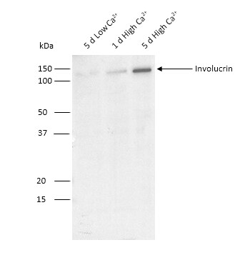
Western Blot: Involucrin Antibody (SY5)BSA Free [NB100-2727]
Western Blot: Involucrin Antibody (SY5) [NB100-2727] - Involucrin from primary human epidermal keratinocyte lysates grown in low calcium (0.07 mM) for 5 days or differentiated with high calcium (1.5 mM) for 1-5 days. Block in 5% nonfat dry milk in TBST incubated at RT for 1 hr, antibody 1:500 in TBST, overnight at 4C. Image from verified customer review.![Immunohistochemistry-Paraffin: Involucrin Antibody (SY5) - BSA Free [NB100-2727]](https://resources.rndsystems.com/images/products/Involucrin-Antibody-SY5-Immunohistochemistry-Paraffin-NB100-2727-img0002.jpg "Immunohistochemistry-Paraffin: Involucrin Antibody (SY5) - BSA Free [NB100-2727]")
Immunohistochemistry-Paraffin: Involucrin Antibody (SY5) - BSA Free [NB100-2727]
Immunohistochemistry-Paraffin: Involucrin Antibody (SY5) [NB100-2727] - Staining of human Tonsil with Involucrin Monoclonal Antibody (SY5).Applications for Involucrin Antibody (SY5) - BSA Free
Application
Recommended Usage
Flow Cytometry
0.5 - 1.0 ug/10^6 cells
Immunocytochemistry/ Immunofluorescence
1 - 2 ug/ml
Immunohistochemistry
0.1-0.2 ug/ml
Immunohistochemistry-Paraffin
1:10-1:500
Immunoprecipitation
1:10 - 1:500
Western Blot
1:100 - 1:2000
Application Notes
Localized to upper spinous and granular layers in normal skin. IHC: Staining of formalin-fixed tissues is enhanced by digestion with trypsin or Protease XXV at 1mg/ml PBS for 5 min at 37 degrees Celsius. Note than enzyme digestion is better than citrate buffer based epitope unmasking. This antibody is CyTOF ready. Positive control(s): MCF-7 cells.
Reviewed Applications
Read 1 review rated 4 using NB100-2727 in the following applications:
Flow Cytometry Panel Builder
Bio-Techne Knows Flow Cytometry
Save time and reduce costly mistakes by quickly finding compatible reagents using the Panel Builder Tool.
Advanced Features
- Spectra Viewer - Custom analysis of spectra from multiple fluorochromes
- Spillover Popups - Visualize the spectra of individual fluorochromes
- Antigen Density Selector - Match fluorochrome brightness with antigen density

Formulation, Preparation, and Storage
Purification
Protein A purified
Formulation
PBS
Format
BSA Free
Preservative
0.02% Sodium Azide
Concentration
1 mg/ml
Shipping
The product is shipped with polar packs. Upon receipt, store it immediately at the temperature recommended below.
Stability & Storage
Store at 4C short term. Aliquot and store at -20C long term. Avoid freeze-thaw cycles.
Background: Involucrin
Additional Involucrin Products
Product Documents for Involucrin Antibody (SY5) - BSA Free
Certificate of Analysis
To download a Certificate of Analysis, please enter a lot or batch number in the search box below.
Product Specific Notices for Involucrin Antibody (SY5) - BSA Free
This product is for research use only and is not approved for use in humans or in clinical diagnosis. Primary Antibodies are guaranteed for 1 year from date of receipt.
Citations for Involucrin Antibody (SY5) - BSA Free
 Powered by Bioz
Customer Reviews for Involucrin Antibody (SY5) - BSA Free (1)
4 out of 5
1 Customer Rating
Have you used Involucrin Antibody (SY5) - BSA Free?
Submit a review and receive an Amazon gift card!
$25/€18/£15/$25CAN/¥2500 Yen for a review with an image
$10/€7/£6/$10CAN/¥1110 Yen for a review without an image
Submit a review
Customer Images
Showing
1
-
1 of
1 review
Showing All
Filter By:
-
Application: Western BlotSample Tested: Human keratinocytes and Cultured Human KeratinocytesSpecies: HumanVerified Customer | Posted 04/25/2018Western blot of Involucrin from primary human epidermal keratinocyte lysates grown in low calcium (0.07 mM) for 5 days or differentiated with high calcium (1.5 mM) for 1-5 days.Block in 5% nonfat dry milk in TBST incubated at RT for 1 hr 1:500 in TBST, overnight at 4C

There are no reviews that match your criteria.
Protocols
Find general support by application which include: protocols, troubleshooting, illustrated assays, videos and webinars.
- 7-Amino Actinomycin D (7-AAD) Cell Viability Flow Cytometry Protocol
- Antigen Retrieval Protocol (PIER)
- Antigen Retrieval for Frozen Sections Protocol
- Appropriate Fixation of IHC/ICC Samples
- Cellular Response to Hypoxia Protocols
- Chromogenic IHC Staining of Formalin-Fixed Paraffin-Embedded (FFPE) Tissue Protocol
- Chromogenic Immunohistochemistry Staining of Frozen Tissue
- ClariTSA™ Fluorophore Kits
- Detection & Visualization of Antibody Binding
- Extracellular Membrane Flow Cytometry Protocol
- Flow Cytometry Protocol for Cell Surface Markers
- Flow Cytometry Protocol for Staining Membrane Associated Proteins
- Flow Cytometry Staining Protocols
- Flow Cytometry Troubleshooting Guide
- Fluorescent IHC Staining of Frozen Tissue Protocol
- Graphic Protocol for Heat-induced Epitope Retrieval
- Graphic Protocol for the Preparation and Fluorescent IHC Staining of Frozen Tissue Sections
- Graphic Protocol for the Preparation and Fluorescent IHC Staining of Paraffin-embedded Tissue Sections
- Graphic Protocol for the Preparation of Gelatin-coated Slides for Histological Tissue Sections
- ICC Cell Smear Protocol for Suspension Cells
- ICC Immunocytochemistry Protocol Videos
- ICC for Adherent Cells
- IHC Sample Preparation (Frozen sections vs Paraffin)
- Immunocytochemistry (ICC) Protocol
- Immunocytochemistry Troubleshooting
- Immunofluorescence of Organoids Embedded in Cultrex Basement Membrane Extract
- Immunofluorescent IHC Staining of Formalin-Fixed Paraffin-Embedded (FFPE) Tissue Protocol
- Immunohistochemistry (IHC) and Immunocytochemistry (ICC) Protocols
- Immunohistochemistry Frozen Troubleshooting
- Immunohistochemistry Paraffin Troubleshooting
- Immunoprecipitation Protocol
- Intracellular Flow Cytometry Protocol Using Alcohol (Methanol)
- Intracellular Flow Cytometry Protocol Using Detergents
- Intracellular Nuclear Staining Flow Cytometry Protocol Using Detergents
- Intracellular Staining Flow Cytometry Protocol Using Alcohol Permeabilization
- Intracellular Staining Flow Cytometry Protocol Using Detergents to Permeabilize Cells
- Preparing Samples for IHC/ICC Experiments
- Preventing Non-Specific Staining (Non-Specific Binding)
- Primary Antibody Selection & Optimization
- Propidium Iodide Cell Viability Flow Cytometry Protocol
- Protocol for Heat-Induced Epitope Retrieval (HIER)
- Protocol for Liperfluo
- Protocol for Making a 4% Formaldehyde Solution in PBS
- Protocol for VisUCyte™ HRP Polymer Detection Reagent
- Protocol for the Characterization of Human Th22 Cells
- Protocol for the Characterization of Human Th9 Cells
- Protocol for the Fluorescent ICC Staining of Cell Smears - Graphic
- Protocol for the Fluorescent ICC Staining of Cultured Cells on Coverslips - Graphic
- Protocol for the Preparation & Fixation of Cells on Coverslips
- Protocol for the Preparation and Chromogenic IHC Staining of Frozen Tissue Sections
- Protocol for the Preparation and Chromogenic IHC Staining of Frozen Tissue Sections - Graphic
- Protocol for the Preparation and Chromogenic IHC Staining of Paraffin-embedded Tissue Sections
- Protocol for the Preparation and Chromogenic IHC Staining of Paraffin-embedded Tissue Sections - Graphic
- Protocol for the Preparation and Fluorescent ICC Staining of Cells on Coverslips
- Protocol for the Preparation and Fluorescent ICC Staining of Non-adherent Cells
- Protocol for the Preparation and Fluorescent ICC Staining of Stem Cells on Coverslips
- Protocol for the Preparation and Fluorescent IHC Staining of Frozen Tissue Sections
- Protocol for the Preparation and Fluorescent IHC Staining of Paraffin-embedded Tissue Sections
- Protocol for the Preparation of Gelatin-coated Slides for Histological Tissue Sections
- Protocol for the Preparation of a Cell Smear for Non-adherent Cell ICC - Graphic
- Protocol: Annexin V and PI Staining by Flow Cytometry
- Protocol: Annexin V and PI Staining for Apoptosis by Flow Cytometry
- R&D Systems Quality Control Western Blot Protocol
- TUNEL and Active Caspase-3 Detection by IHC/ICC Protocol
- The Importance of IHC/ICC Controls
- Troubleshooting Guide: Fluorokine Flow Cytometry Kits
- Troubleshooting Guide: Immunohistochemistry
- Troubleshooting Guide: Western Blot Figures
- Western Blot Conditions
- Western Blot Protocol
- Western Blot Protocol for Cell Lysates
- Western Blot Troubleshooting
- Western Blot Troubleshooting Guide
- View all Protocols, Troubleshooting, Illustrated assays and Webinars
Loading...