IRE1 alpha Antibody - BSA Free
Novus Biologicals | Catalog # NB100-2324
![Immunohistochemistry: IRE1 alpha Antibody [NB100-2324]](https://resources.rndsystems.com/images/products/IRE1-alpha-Antibody-Immunohistochemistry-NB100-2324-img0010.jpg "Immunohistochemistry: IRE1 alpha Antibody [NB100-2324]")
Key Product Details
Validated by
Knockout/Knockdown
Species Reactivity
Validated:
Human, Mouse, Rat
Cited:
Human, Mouse, Rat
Applications
Validated:
Knockout Validated, Immunohistochemistry, Immunohistochemistry-Paraffin, Western Blot, Immunocytochemistry/ Immunofluorescence, Knockdown Validated
Cited:
Immunohistochemistry-Paraffin, Western Blot, Immunocytochemistry/ Immunofluorescence, Knockdown Validated
Label
Unconjugated
Antibody Source
Polyclonal Rabbit IgG
Format
BSA Free
Loading...
Product Specifications
Immunogen
This IRE1 alpha antibody was raised against a synthetic peptide within the human IRE1 alpha protein (within residues 700-800). [Swiss-Prot #O75460]
Localization
Endoplasmic reticulum
Clonality
Polyclonal
Host
Rabbit
Isotype
IgG
Theoretical MW
110 kDa.
Disclaimer note: The observed molecular weight of the protein may vary from the listed predicted molecular weight due to post translational modifications, post translation cleavages, relative charges, and other experimental factors.
Disclaimer note: The observed molecular weight of the protein may vary from the listed predicted molecular weight due to post translational modifications, post translation cleavages, relative charges, and other experimental factors.
Description
NB100-2324 is useful for the detection of endogenous total IRE1 Alpha protein. For detecting phospho-IRE1 Alpha (Ser-724), we suggest NB100-2323 and it is recommended to normalize phospho-IRE1 alpha band intensity/immunoreactivity with total-IRE1 alpha using NB100-2324 or NB110-59971.
Scientific Data Images for IRE1 alpha Antibody - BSA Free
Immunohistochemistry: IRE1 alpha Antibody [NB100-2324]
IRE1-alpha-Antibody-Immunohistochemistry-NB100-2324-img0010.jpg![Western Blot: IRE1 alpha Antibody [NB100-2324]](https://resources.rndsystems.com/images/products/IRE1-alpha-Antibody-Western-Blot-NB100-2324-img0006.jpg "Western Blot: IRE1 alpha Antibody [NB100-2324]")
![Western Blot: IRE1 alpha Antibody [NB100-2324]](https://resources.rndsystems.com/images/products/IRE1-alpha-Antibody-Western-Blot-NB100-2324-img0009.jpg "Western Blot: IRE1 alpha Antibody [NB100-2324]")
![Immunocytochemistry/ Immunofluorescence: IRE1 alpha Antibody [NB100-2324]](https://resources.rndsystems.com/images/products/IRE1-alpha-Antibody-Immunocytochemistry-Immunofluorescence-NB100-2324-img0005.jpg "Immunocytochemistry/ Immunofluorescence: IRE1 alpha Antibody [NB100-2324]")
Immunocytochemistry/ Immunofluorescence: IRE1 alpha Antibody [NB100-2324]
Immunocytochemistry/Immunofluorescence: IRE1 alpha Antibody [NB100-2324] - IRE-1 alpha antibody was tested in HeLa cells at 1:200 against Dylight 488 (Green). Alpha tubulin and nuclei were counterstained against Dylight 568 (Red) and DAPI (Blue).![Immunohistochemistry-Paraffin: IRE1 alpha Antibody [NB100-2324]](https://resources.rndsystems.com/images/products/IRE1-alpha-Antibody-Immunohistochemistry-Paraffin-NB100-2324-img0007.jpg "Immunohistochemistry-Paraffin: IRE1 alpha Antibody [NB100-2324]")
Immunohistochemistry-Paraffin: IRE1 alpha Antibody [NB100-2324]
Immunohistochemistry-Paraffin: IRE1 alpha Antibody [NB100-2324] - Analysis of a formalin fixed mouse colon tissue section using total IRE1 alpha antibody (NB100-2324) at 1:300. Note a strong cytoplasmic signal in various cells of colonic villi (columnar epithelial and goblet cells) and the muscular layer. A few cells, potentially with active UPR, showed nuclear positivity also while the signal was negligible in cells at base of crypts and lamina propria.![Immunohistochemistry-Paraffin: IRE1 alpha Antibody [NB100-2324]](https://resources.rndsystems.com/images/products/IRE1-alpha-Antibody-Immunohistochemistry-Paraffin-NB100-2324-img0008.jpg "Immunohistochemistry-Paraffin: IRE1 alpha Antibody [NB100-2324]")
Immunohistochemistry-Paraffin: IRE1 alpha Antibody [NB100-2324]
Immunohistochemistry-Paraffin: IRE1 alpha Antibody [NB100-2324] - Analysis of a formalin fixed mouse colon tissue section using total IRE1 alpha antibody (NB100-2324) at 1:300. Note a strong cytoplasmic signal in various cells of colonic villi (columnar epithelial and goblet cells) and the muscular layer. Some cells, most likely with active UPR, depicted nuclear positivity also while the signal was negligible in crypts cells and lamina propria.
Western Blot: IRE1 alpha Antibody [NB100-2324] -
The inositol-requiring enzyme 1 alpha (IRE1 alpha )/ X-box-binding-protein 1 spliced isoform (XBP1s) arm of unfolded protein response (UPR) is induced by angiotensin II in VSMCs. (A–C) The rat aortic VSMCs infected with adenovirus encoding GRP78 or control GFP (100 moi) for 48 h were stimulated with 100 nM AngII (AII) for 1–6 h & immunoblotting was performed as indicated. (A) Representative blots from 4 independent experiments. (B) Signal intensity was used to calculate the expression ratio of XBP1s to GAPDH. (C) Signal intensity was used to calculate the IRE1 alpha Ser724 phosphorylation ratio to the total IRE1 alpha. The bars in the graphs show the mean ± SD from 4 independent experiments. * indicates p < 0.05. Image collected & cropped by CiteAb from the following publication (https://pubmed.ncbi.nlm.nih.gov/32679678), licensed under a CC-BY license. Not internally tested by Novus Biologicals.
Western Blot: IRE1 alpha Antibody [NB100-2324] -
Abnormal Paneth cells show ER stress.(A, B) Representative transmission electron microscopy images of Paneth cells at the base of ileal crypts in (A) ICR & (B) SAMP1/YitFc mice. Scale bars indicate 2 μm. (C, D) Quantitative analysis of (C) granule number & (D) ER lumen diameter in Paneth cells (n = 3/each week for SAMP1/YitFc mice). For the measurements, three Paneth cells were randomly selected from each mouse. (E) SDS–PAGE Western blot analysis of ER stress markers, pIRE1 alpha, ATF4, cleaved-ATF6, & GRP78 in ileal crypts (n = 4/each group). Total-IRE1 alpha & HPRT1 was used as loading control. (F) Relative expression level of ER stress markers calculated from the band intensity. Error bars represent mean ± SEM. (C, D, F) Statistical significance was evaluated by t test in (C, D), & one-way ANOVA followed by Tukey’s post hoc test in (F). P < 0.05 was considered statistically significant. *P < 0.05, †P < 0.01, §P < 0.001. E, ER; G, granules; N, nucleus; n.s., not significant. Image collected & cropped by CiteAb from the following publication (https://pubmed.ncbi.nlm.nih.gov/32345659), licensed under a CC-BY license. Not internally tested by Novus Biologicals.
Western Blot: IRE1 alpha Antibody [NB100-2324] -
Western Blot: IRE1 alpha Antibody [NB100-2324] - Zn (II)-curc induces endoplasmic reticulum (ER) stress in mutant p53H273-carrying cells. (a) Representative photomicrographs of ER-Red Fluorescence staining in U373 cells untreated (Mock) or treated with Zn (II)-curc (100 µg/mL) for 16 h (Original magnification: 40×). (b) Quantization of ER content in U373 cells from ER-Red Fluorescence-stained cells. Mean fluorescence intensity (MFI) of each individual cell was normalized to cell size & expressed as fold-change compared with untreated cells at the same time point. Histograms represent the mean ± SD of three independent experiments. * p ≤ 0.05. (c) Western blot analysis of p53, BiP, total (tot) IRE1 alpha, phosphorylated (p) IRE1 alpha, & XBP1 spliced (s) protein levels evaluated in U373 & HT29 cells untreated or treated with Zn (II)-curc (100 µg/mL) for 24 h. (d) Densitometric analysis was performed using Image J software to calculate the ratio of the protein levels, as detected in (c), vs. beta -actin. Histograms represent the mean ± SD of three independent experiments. * p ≤ 0.05. (e) Total mRNA was extracted from U373 & HT29 cells untreated or treated with Zn (II)-curc (100 µg/mL) for 24 h. Spliced (s) Xbp1 gene expression was assayed by the polymerase chain reaction (PCR) of reverse-transcribed cDNA. Densitometric analysis was performed using Image J software to calculate the Xbp1s/28S ratio. Histograms represent the mean ± SD of three independent experiments. * p ≤ 0.05. (f) p53 gene expression was assayed by PCR as in (e). The p53/28S ratio is indicated. Image collected & cropped by CiteAb from the following publication (https://pubmed.ncbi.nlm.nih.gov/32138264), licensed under a CC-BY license. Not internally tested by Novus Biologicals.Applications for IRE1 alpha Antibody - BSA Free
Application
Recommended Usage
Immunocytochemistry/ Immunofluorescence
1:100 - 1:250
Immunohistochemistry
1:250 - 1:500
Immunohistochemistry-Paraffin
1:250 - 1:500
Knockout Validated
reported in scientific literature (PMID 31159306)
Western Blot
1:1000 - 1:2000
Application Notes
NB100-2324 is useful for the detection of endogenous total IRE1 Alpha protein. For detecting phospho-IRE1 Alpha (Ser-724), we suggest NB100-2323 and it is recommended to normalize phospho-IRE1 alpha band intensity/immunoreactivity with total-IRE1 alpha using NB100-2324 or NB110-59971. In Western blot a band at ~110 kDa is observed. In ICC/IF endoplasmic reticulum staining was observed in HeLa cells. The observed molecular weight of the protein may vary from the listed predicted molecular weight due to post translational modifications, post translation cleavages, relative charges, and other experimental factors.
Reviewed Applications
Read 2 reviews rated 4.5 using NB100-2324 in the following applications:
Formulation, Preparation, and Storage
Purification
Immunogen affinity purified
Formulation
PBS
Format
BSA Free
Preservative
0.05% Sodium Azide
Concentration
1 mg/ml
Shipping
The product is shipped with polar packs. Upon receipt, store it immediately at the temperature recommended below.
Stability & Storage
Store at -20C. Avoid freeze-thaw cycles.
Background: IRE1 alpha
Transcriptional activation of UPR-responsive genes are regulated by the ATF6 and IRE1-XBP1 pathways, which are often regulated by HIFs and contribute to cell survival under ER hypoxic stress (3). UPR signaling can a) inhibit protein translation to restore cell function, b) activate signaling to increase production of molecular chaperones for protein folding, and c) initiate ubiquitination signaling that leads to degradation of misfolded proteins in ER.
IRE1 alpha acts as the sensor of unfolded proteins in the ER. IRE1 alpha not only promotes cell survival but can initiate apoptosis when accumulation of unfolded proteins in the ER causes stress. IRE1 alpha is essential for viability under stress conditions that cause unfolded proteins to accumulate in the ER. IRE1 alpha is a transmembrane protein that has both serine-threonine kinase and endoribonuclease activities and has a theoretical molecular weight of 110 kDa. When detecting phospho-IRE1 alpha, it is recommended to normalize its band intensity with total IRE1 alpha.
References
1. Zheng, W., Xie, W., Yin, D., Luo, R., Liu, M., & Guo, F. (2019). ATG5 and ATG7 induced autophagy interplays with UPR via PERK signaling. Cell Commun Signal, 17(1), 42. doi:10.1186/s12964-019-0353-3
2. Cho, Y. M., Kim, D. H., Lee, K. H., Jeong, S. W., & Kwon, O. J. (2018). The IRE1alpha-XBP1s pathway promotes insulin-stimulated glucose uptake in adipocytes by increasing PPARgamma activity. Exp Mol Med, 50(8), 102. doi:10.1038/s12276-018-0131-0
3. Xia, Z., Wu, S., Wei, X., Liao, Y., Yi, P., Liu, Y.,... Liu, J. (2019). Hypoxic ER stress suppresses beta-catenin expression and promotes cooperation between the transcription factors XBP1 and HIF1alpha for cell survival. J Biol Chem, 294(37), 13811-13821. doi:10.1074/jbc.RA119.008353
Long Name
Serine/threonine-protein kinase/endoribonuclease IRE1
Alternate Names
ERN1, hIRE1p, IRE1, Ire1-alpha
Gene Symbol
ERN1
Additional IRE1 alpha Products
Product Documents for IRE1 alpha Antibody - BSA Free
Certificate of Analysis
To download a Certificate of Analysis, please enter a lot or batch number in the search box below.
Product Specific Notices for IRE1 alpha Antibody - BSA Free
This product is for research use only and is not approved for use in humans or in clinical diagnosis. Primary Antibodies are guaranteed for 1 year from date of receipt.
Citations for IRE1 alpha Antibody - BSA Free
 Powered by Bioz
Customer Reviews for IRE1 alpha Antibody - BSA Free (2)
4.5 out of 5
2 Customer Ratings
Have you used IRE1 alpha Antibody - BSA Free?
Submit a review and receive an Amazon gift card!
$25/€18/£15/$25CAN/¥2500 Yen for a review with an image
$10/€7/£6/$10CAN/¥1110 Yen for a review without an image
Submit a review
Customer Images
Showing
1
-
2 of
2 reviews
Showing All
Filter By:
-
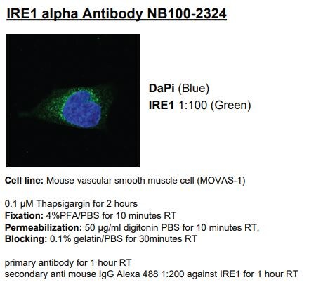
Application: ImmunocytochemistrySample Tested: mouse vascular smooth muscle cellSpecies: MouseVerified Customer | Posted 11/19/2018

-
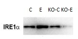
Application: Western BlotSample Tested: LiverSpecies: MouseVerified Customer | Posted 03/07/2018ALOX15 knockout blocks alcohol-induced up-regulation of IRE1a in mouse liver.
There are no reviews that match your criteria.
Protocols
View specific protocols for IRE1 alpha Antibody - BSA Free (NB100-2324):
Immunocytochemistry Protocol
Culture cells to appropriate density on suitable glass coverslips in 35 mm culture dishes or 6-well plates.
1. Remove culture medium and add 10% formalin to the dish. Fix at room temperature for 5-10 minutes.
2. Remove the formalin and add 0.5% Triton-X 100 in TBS to permeabilize the cells. Incubate for 5-10 minutes.
3. Remove the permeabilization buffer and add wash buffer (i.e. PBS or PBS with 0.1% Tween-20). Be sure to not let the specimen dry out. Gently wash three times for 10 minutes.
4. Alternatively, cells can be fixed with -20C methanol for 10 min at room temperature. Remove the methanol and rehydrate in PBS for 10 min before proceeding.
5. To block nonspecific antibody binding incubate in 10% normal goat serum for 1 hour at room temperature.
6. Add primary antibody at appropriate dilution and incubate at room temperature for 1 hour or at 4 degrees C overnight.
7. Remove primary antibody and replace with wash buffer. Gently wash three times for 10 minutes.
8. Add secondary antibody at the appropriate dilution. Incubate for 1 hour at room temperature.
9. Remove antibody and replace with wash buffer. Gently wash three times for 10 minutes.
10. Nuclei can be staining with 4',6' diamino phenylindole (DAPI) at 0.1 ug/ml, or coverslips can be directly mounted in media containing DAPI.
11. Cells can now be viewed with a fluorescence microscope.
*The above information is only intended as a guide. The researcher should determine what protocol best meets their needs. Please follow proper laboratory procedures for the disposal of formalin.
Find general support by application which include: protocols, troubleshooting, illustrated assays, videos and webinars.
- Antigen Retrieval Protocol (PIER)
- Antigen Retrieval for Frozen Sections Protocol
- Appropriate Fixation of IHC/ICC Samples
- Cellular Response to Hypoxia Protocols
- Chromogenic IHC Staining of Formalin-Fixed Paraffin-Embedded (FFPE) Tissue Protocol
- Chromogenic Immunohistochemistry Staining of Frozen Tissue
- ClariTSA™ Fluorophore Kits
- Detection & Visualization of Antibody Binding
- Fluorescent IHC Staining of Frozen Tissue Protocol
- Graphic Protocol for Heat-induced Epitope Retrieval
- Graphic Protocol for the Preparation and Fluorescent IHC Staining of Frozen Tissue Sections
- Graphic Protocol for the Preparation and Fluorescent IHC Staining of Paraffin-embedded Tissue Sections
- Graphic Protocol for the Preparation of Gelatin-coated Slides for Histological Tissue Sections
- ICC Cell Smear Protocol for Suspension Cells
- ICC Immunocytochemistry Protocol Videos
- ICC for Adherent Cells
- IHC Sample Preparation (Frozen sections vs Paraffin)
- Immunocytochemistry (ICC) Protocol
- Immunocytochemistry Troubleshooting
- Immunofluorescence of Organoids Embedded in Cultrex Basement Membrane Extract
- Immunofluorescent IHC Staining of Formalin-Fixed Paraffin-Embedded (FFPE) Tissue Protocol
- Immunohistochemistry (IHC) and Immunocytochemistry (ICC) Protocols
- Immunohistochemistry Frozen Troubleshooting
- Immunohistochemistry Paraffin Troubleshooting
- Preparing Samples for IHC/ICC Experiments
- Preventing Non-Specific Staining (Non-Specific Binding)
- Primary Antibody Selection & Optimization
- Protocol for Heat-Induced Epitope Retrieval (HIER)
- Protocol for Making a 4% Formaldehyde Solution in PBS
- Protocol for VisUCyte™ HRP Polymer Detection Reagent
- Protocol for the Fluorescent ICC Staining of Cell Smears - Graphic
- Protocol for the Fluorescent ICC Staining of Cultured Cells on Coverslips - Graphic
- Protocol for the Preparation & Fixation of Cells on Coverslips
- Protocol for the Preparation and Chromogenic IHC Staining of Frozen Tissue Sections
- Protocol for the Preparation and Chromogenic IHC Staining of Frozen Tissue Sections - Graphic
- Protocol for the Preparation and Chromogenic IHC Staining of Paraffin-embedded Tissue Sections
- Protocol for the Preparation and Chromogenic IHC Staining of Paraffin-embedded Tissue Sections - Graphic
- Protocol for the Preparation and Fluorescent ICC Staining of Cells on Coverslips
- Protocol for the Preparation and Fluorescent ICC Staining of Non-adherent Cells
- Protocol for the Preparation and Fluorescent ICC Staining of Stem Cells on Coverslips
- Protocol for the Preparation and Fluorescent IHC Staining of Frozen Tissue Sections
- Protocol for the Preparation and Fluorescent IHC Staining of Paraffin-embedded Tissue Sections
- Protocol for the Preparation of Gelatin-coated Slides for Histological Tissue Sections
- Protocol for the Preparation of a Cell Smear for Non-adherent Cell ICC - Graphic
- R&D Systems Quality Control Western Blot Protocol
- TUNEL and Active Caspase-3 Detection by IHC/ICC Protocol
- The Importance of IHC/ICC Controls
- Troubleshooting Guide: Immunohistochemistry
- Troubleshooting Guide: Western Blot Figures
- Western Blot Conditions
- Western Blot Protocol
- Western Blot Protocol for Cell Lysates
- Western Blot Troubleshooting
- Western Blot Troubleshooting Guide
- View all Protocols, Troubleshooting, Illustrated assays and Webinars
FAQs for IRE1 alpha Antibody - BSA Free
Showing
1
-
2 of
2 FAQs
Showing All
-
Q: I have a customer who is interested in antibody NB100-2324 for use with rat samples. It is listed as working with rat and for Western blot applications. Can you tell me which rat tissues were tested in Western blot?
A: Previous productions were reported to work in rat. We have not tested the current lot directly on any rat samples so I do not have data on what tissues, or lysates were effective. We will still guarantee rat as a reactive species, as it is stated on our datasheet.
-
Q: We hope to know which position the antibody NB 100-2324 recognize within the endogenous IRE1 alpha. Thanks. Is the region 700 to 800 within the luminal domain, or transmembrane or cytosolic domain? Is this region highly conserved between Human and mouse?
A:
It looks like the 700-800 region is cytoplasmic. The homology in this region is 93%, and this product has already been validated to work in both human and mouse species.
https://www.uniprot.org/uniprot/O75460 -
Q: I have a customer who is interested in antibody NB100-2324 for use with rat samples. It is listed as working with rat and for Western blot applications. Can you tell me which rat tissues were tested in Western blot?
A: Previous productions were reported to work in rat. We have not tested the current lot directly on any rat samples so I do not have data on what tissues, or lysates were effective. We will still guarantee rat as a reactive species, as it is stated on our datasheet.
-
Q: We hope to know which position the antibody NB 100-2324 recognize within the endogenous IRE1 alpha. Thanks. Is the region 700 to 800 within the luminal domain, or transmembrane or cytosolic domain? Is this region highly conserved between Human and mouse?
A:
It looks like the 700-800 region is cytoplasmic. The homology in this region is 93%, and this product has already been validated to work in both human and mouse species.
https://www.uniprot.org/uniprot/O75460
Loading...