Kappa Light Chain Antibody (HP6053 + L1C1)
Novus Biologicals | Catalog # NBP2-34258
Key Product Details
Species Reactivity
Human
Applications
Immunohistochemistry, Immunohistochemistry-Paraffin, Western Blot, Immunocytochemistry/ Immunofluorescence
Label
Unconjugated
Antibody Source
Monoclonal Mouse IgG1 Kappa/IgG1 Kappa Clone # HP6053 + L1C1
Loading...
Product Specifications
Immunogen
Purified human Ig kappa chain (HP6053) & Human B-Lymphoma Cells (L1C1)
Localization
Cell Surface, cytoplasmic and secreted
Marker
B-Cell Marker
Specificity
This monoclonal antibody is specific to kappa light chain of immunoglobulin and shows no cross-reaction with lambda light chain or any of the five heavy chains. In mammals, the two light chains in an antibody are always identical, with only one type of light chain, kappa or lambda. The ratio of Kappa to Lambda is 70:30. However, with the occurrence of multiple myeloma or other B-cell malignancies this ratio is disturbed. Antibody to the kappa light chain is reportedly useful in the identification of leukemias, plasmacytomas, and certain non-Hodgkins lymphomas. Demonstration of clonality in lymphoid infiltrates indicates that the infiltrate is malignant.
Clonality
Monoclonal
Host
Mouse
Isotype
IgG1 Kappa/IgG1 Kappa
Theoretical MW
22.5 kDa.
Disclaimer note: The observed molecular weight of the protein may vary from the listed predicted molecular weight due to post translational modifications, post translation cleavages, relative charges, and other experimental factors.
Disclaimer note: The observed molecular weight of the protein may vary from the listed predicted molecular weight due to post translational modifications, post translation cleavages, relative charges, and other experimental factors.
Description
200ug/ml of antibody purified from Bioreactor Concentrate by Protein A or G. Prepared in 10 mM PBS with 0.05% BSA & 0.05% azide. Also available WITHOUT BSA & azide at 1.0 mg/ml. (NBP2-34640)
Antibody with azide - store at 2 to 8C. Antibody without azide - store at -20 to -80C.
Antibody with azide - store at 2 to 8C. Antibody without azide - store at -20 to -80C.
Scientific Data Images for Kappa Light Chain Antibody (HP6053 + L1C1)
![Western Blot: Kappa Light Chain Antibody (HP6053 + L1C1) [NBP2-34258]](https://resources.rndsystems.com/images/products/Kappa-Light-Chain-Antibody-HP6053-+-L1C1-Western-Blot-NBP2-34258-img0002.jpg "Western Blot: Kappa Light Chain Antibody (HP6053 + L1C1) [NBP2-34258]")
Western Blot: Kappa Light Chain Antibody (HP6053 + L1C1) [NBP2-34258]
Western Blot: Kappa Light Chain Antibody (HP6053 + L1C1) [NBP2-34258] - Analysis of Raji Cell Lysate using kappa Light Chain Monclonal Antibody (HP6053+ L1C1)![Immunocytochemistry/ Immunofluorescence: Kappa Light Chain Antibody (HP6053 + L1C1) [NBP2-34258]](https://resources.rndsystems.com/images/products/Kappa-Light-Chain-Antibody-HP6053-+-L1C1-Immunocytochemistry-Immunofluorescence-NBP2-34258-img0004.jpg "Immunocytochemistry/ Immunofluorescence: Kappa Light Chain Antibody (HP6053 + L1C1) [NBP2-34258]")
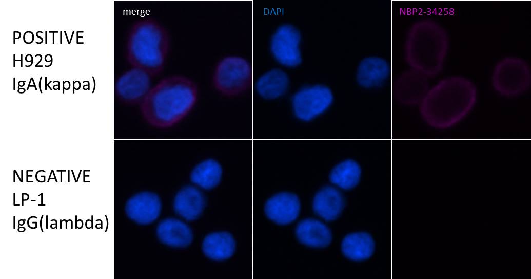
Immunocytochemistry/ Immunofluorescence: Kappa Light Chain Antibody (HP6053 + L1C1) [NBP2-34258]
Immunocytochemistry/Immunofluorescence: Kappa Light Chain Antibody (HP6053 + L1C1) [NBP2-34258] - LP-1 (multiple myeloma) and H929 (multiple myeloma). Image from verified customer review. [NBP2-34258] -")
Immunohistochemistry-Paraffin: Kappa Light Chain Antibody (HP6053 + L1C1) [NBP2-34258] -
Immunohistochemistry-Paraffin: Kappa Light Chain Antibody (HP6053 + L1C1) [NBP2-34258] - Formalin-fixed, paraffin-embedded human Tonsil stained with Kappa Light Chain Antibody (HP6053 + L1C1).Applications for Kappa Light Chain Antibody (HP6053 + L1C1)
Application
Recommended Usage
Immunohistochemistry-Paraffin
1-2 ug/ml
Western Blot
1-2 ug/ml
Application Notes
Immunohistochemistry (Formalin-fixed): 1-2ug/ml for 30 min at RT. Staining of formalin-fixed tissues requires heating tissue sections in 10mM Tris with 1mM EDTA, pH 9.0, for 45 min at 95C followed by cooling at RT for 20 minutes.
Optimal dilution for a specific application should be determined.
Use in ICC/IF reported by a customer review.
Optimal dilution for a specific application should be determined.
Use in ICC/IF reported by a customer review.
Reviewed Applications
Read 1 review rated 5 using NBP2-34258 in the following applications:
Formulation, Preparation, and Storage
Purification
Protein A or G purified
Formulation
10 mM PBS with 0.05% BSA
Preservative
0.05% Sodium Azide
Concentration
0.2 mg/ml
Shipping
The product is shipped with polar packs. Upon receipt, store it immediately at the temperature recommended below.
Stability & Storage
Store at 4C.
Background: Kappa Light Chain
Alternate Names
HCAK1, IGKCD, immunoglobulin kappa constant, Km, MGC111575, MGC62011, MGC72072, MGC88770, MGC88771, MGC88809
Gene Symbol
IGKC
UniProt
Additional Kappa Light Chain Products
Product Documents for Kappa Light Chain Antibody (HP6053 + L1C1)
Certificate of Analysis
To download a Certificate of Analysis, please enter a lot or batch number in the search box below.
Product Specific Notices for Kappa Light Chain Antibody (HP6053 + L1C1)
This product is for research use only and is not approved for use in humans or in clinical diagnosis. Primary Antibodies are guaranteed for 1 year from date of receipt.
Customer Reviews for Kappa Light Chain Antibody (HP6053 + L1C1) (1)
5 out of 5
1 Customer Rating
Have you used Kappa Light Chain Antibody (HP6053 + L1C1)?
Submit a review and receive an Amazon gift card!
$25/€18/£15/$25CAN/¥2500 Yen for a review with an image
$10/€7/£6/$10CAN/¥1110 Yen for a review without an image
Submit a review
Customer Images
Showing
1
-
1 of
1 review
Showing All
Filter By:
-
Application: ImmunocytochemistrySample Tested: LP-1 (multiple myeloma) and H929 (multiple myeloma)Species: HumanVerified Customer | Posted 07/17/2019Immunocytochemistry staining LP-1 and H929 with NBP2-342581. Cells immersion-fixed in 2%PFA 10 minutes, wash PBS X 3 2. Permeabilized 0.5% Triton X-100 5 minutes; wash PBS X 3 3. Block 10% donkey serum 1 hour 4. 1/200 NBP2-34258 O/N @4oC; wash PBS X 3 5. 1/1000 donkey F(ab’)2 Alexa Fluor 647 anti-mouse IgG (H+L) 1 hour @room temp (Jackson #715-606-150); wash PBS X 3 6. Olympus/BIOVIEW Cy5 filter 600ms exposure

There are no reviews that match your criteria.
Protocols
Find general support by application which include: protocols, troubleshooting, illustrated assays, videos and webinars.
- Antigen Retrieval Protocol (PIER)
- Antigen Retrieval for Frozen Sections Protocol
- Appropriate Fixation of IHC/ICC Samples
- Cellular Response to Hypoxia Protocols
- Chromogenic IHC Staining of Formalin-Fixed Paraffin-Embedded (FFPE) Tissue Protocol
- Chromogenic Immunohistochemistry Staining of Frozen Tissue
- ClariTSA™ Fluorophore Kits
- Detection & Visualization of Antibody Binding
- Fluorescent IHC Staining of Frozen Tissue Protocol
- Graphic Protocol for Heat-induced Epitope Retrieval
- Graphic Protocol for the Preparation and Fluorescent IHC Staining of Frozen Tissue Sections
- Graphic Protocol for the Preparation and Fluorescent IHC Staining of Paraffin-embedded Tissue Sections
- Graphic Protocol for the Preparation of Gelatin-coated Slides for Histological Tissue Sections
- ICC Cell Smear Protocol for Suspension Cells
- ICC Immunocytochemistry Protocol Videos
- ICC for Adherent Cells
- IHC Sample Preparation (Frozen sections vs Paraffin)
- Immunocytochemistry (ICC) Protocol
- Immunocytochemistry Troubleshooting
- Immunofluorescence of Organoids Embedded in Cultrex Basement Membrane Extract
- Immunofluorescent IHC Staining of Formalin-Fixed Paraffin-Embedded (FFPE) Tissue Protocol
- Immunohistochemistry (IHC) and Immunocytochemistry (ICC) Protocols
- Immunohistochemistry Frozen Troubleshooting
- Immunohistochemistry Paraffin Troubleshooting
- Preparing Samples for IHC/ICC Experiments
- Preventing Non-Specific Staining (Non-Specific Binding)
- Primary Antibody Selection & Optimization
- Protocol for Heat-Induced Epitope Retrieval (HIER)
- Protocol for Making a 4% Formaldehyde Solution in PBS
- Protocol for VisUCyte™ HRP Polymer Detection Reagent
- Protocol for the Fluorescent ICC Staining of Cell Smears - Graphic
- Protocol for the Fluorescent ICC Staining of Cultured Cells on Coverslips - Graphic
- Protocol for the Preparation & Fixation of Cells on Coverslips
- Protocol for the Preparation and Chromogenic IHC Staining of Frozen Tissue Sections
- Protocol for the Preparation and Chromogenic IHC Staining of Frozen Tissue Sections - Graphic
- Protocol for the Preparation and Chromogenic IHC Staining of Paraffin-embedded Tissue Sections
- Protocol for the Preparation and Chromogenic IHC Staining of Paraffin-embedded Tissue Sections - Graphic
- Protocol for the Preparation and Fluorescent ICC Staining of Cells on Coverslips
- Protocol for the Preparation and Fluorescent ICC Staining of Non-adherent Cells
- Protocol for the Preparation and Fluorescent ICC Staining of Stem Cells on Coverslips
- Protocol for the Preparation and Fluorescent IHC Staining of Frozen Tissue Sections
- Protocol for the Preparation and Fluorescent IHC Staining of Paraffin-embedded Tissue Sections
- Protocol for the Preparation of Gelatin-coated Slides for Histological Tissue Sections
- Protocol for the Preparation of a Cell Smear for Non-adherent Cell ICC - Graphic
- R&D Systems Quality Control Western Blot Protocol
- TUNEL and Active Caspase-3 Detection by IHC/ICC Protocol
- The Importance of IHC/ICC Controls
- Troubleshooting Guide: Immunohistochemistry
- Troubleshooting Guide: Western Blot Figures
- Western Blot Conditions
- Western Blot Protocol
- Western Blot Protocol for Cell Lysates
- Western Blot Troubleshooting
- Western Blot Troubleshooting Guide
- View all Protocols, Troubleshooting, Illustrated assays and Webinars
Loading...