KAT3B/p300 Antibody (RW105) - BSA Free
Novus Biologicals | Catalog # NB100-616
Key Product Details
Validated by
Species Reactivity
Validated:
Cited:
Applications
Validated:
Cited:
Label
Antibody Source
Format
Product Specifications
Immunogen
Reactivity Notes
Localization
Specificity
Clonality
Host
Isotype
Scientific Data Images for KAT3B/p300 Antibody (RW105) - BSA Free
![Immunocytochemistry/ Immunofluorescence: KAT3B/p300 Antibody (RW105) - BSA Free [NB100-616]](https://resources.rndsystems.com/images/products/KAT3B-p300-Antibody-RW105-Immunocytochemistry-Immunofluorescence-NB100-616-img0005.jpg "Immunocytochemistry/ Immunofluorescence: KAT3B/p300 Antibody (RW105) - BSA Free [NB100-616]")
Immunocytochemistry/ Immunofluorescence: KAT3B/p300 Antibody (RW105) - BSA Free [NB100-616]
Immunocytochemistry/Immunofluorescence: KAT3B/p300 Antibody (RW105) [NB100-616] - HeLa cells were fixed in 4% paraformaldehyde for 10 min and permeabilized in 0.05% Triton X-100 in PBS for 5 minutes. The cells were incubated with anti-KAT3B Antibody (RW105)) at 5ug/ml for 60 minutes at room temperature and detected with an anti-mouse Dylight 488 (Green) at a 1:1000 dilution for 60 minutes. Nuclei were counterstained with DAPI (Blue). Cells were imaged using a 100X objective.![Western Blot: KAT3B/p300 Antibody (RW105)BSA Free [NB100-616]](https://resources.rndsystems.com/images/products/KAT3B-p300-Antibody-RW105-Western-Blot-NB100-616-img0002.jpg "Western Blot: KAT3B/p300 Antibody (RW105)BSA Free [NB100-616]")
Western Blot: KAT3B/p300 Antibody (RW105)BSA Free [NB100-616]
Western Blot: KAT3B/p300 Antibody (RW105) [NB100-616] - p300 detected in a HeLa nuclear extract using NB 100-616 (1:250). ECL: 20 minute exposure.![Immunocytochemistry/ Immunofluorescence: KAT3B/p300 Antibody (RW105) - BSA Free [NB100-616]](https://resources.rndsystems.com/images/products/KAT3B-p300-Antibody-RW105-Immunocytochemistry-Immunofluorescence-NB100-616-img0008.jpg "Immunocytochemistry/ Immunofluorescence: KAT3B/p300 Antibody (RW105) - BSA Free [NB100-616]")
Immunocytochemistry/ Immunofluorescence: KAT3B/p300 Antibody (RW105) - BSA Free [NB100-616]
Immunocytochemistry/Immunofluorescence: KAT3B/p300 Antibody (RW105) [NB100-616] - HeLa cells were fixed in 4% paraformaldehyde for 10 minutes and permeabilized in 0.5% Triton X-100 in PBS for 5 minutes. The cells were incubated with anti-KAT3B/p300 Antibody (RW105) NB100-616 at 2 ug/ml overnight at 4C and detected with an anti-mouse Dylight 488 (Green) at a 1:1000 dilution for 60 minutes. Nuclei were counterstained with DAPI (Blue). Cells were imaged using a 100X objective and digitally deconvolved.![Flow Cytometry: KAT3B/p300 Antibody (RW105) - BSA Free [NB100-616]](https://resources.rndsystems.com/images/products/KAT3B-p300-Antibody-RW105-Flow-Cytometry-NB100-616-img0006.jpg "Flow Cytometry: KAT3B/p300 Antibody (RW105) - BSA Free [NB100-616]")
Flow Cytometry: KAT3B/p300 Antibody (RW105) - BSA Free [NB100-616]
Flow Cytometry: KAT3B/p300 Antibody (RW105) [NB100-616] - An intracellular stain was performed on RAW 246.7 cells with KAT3B/p300 antibody (RW105) NB100-616PE (blue) and a matched isotype control NBP2-27287PE (orange). Cells were fixed with 4% PFA and then permeablized with 0.1% saponin. Cells were incubated in an antibody dilution of 2 ug/mL for 30 minutes at room temperature. Both antibodies were conjugated to Phycoerythrin. Image using the PE format of this antibody.![Flow (Intracellular): KAT3B/p300 Antibody (RW105) - BSA Free [NB100-616]](https://resources.rndsystems.com/images/products/KAT3B-p300-Antibody-RW105-Flow-Intracellular-NB100-616-img0003.jpg "Flow (Intracellular): KAT3B/p300 Antibody (RW105) - BSA Free [NB100-616]")
Flow (Intracellular): KAT3B/p300 Antibody (RW105) - BSA Free [NB100-616]
Flow (Intracellular): KAT3B/p300 Antibody (RW105) [NB100-616] - An intracellular stain was performed on THP-1 cells with KAT3B/p300 (RW105) antibody NB100-616 (blue) and a matched isotype control NBP2-27287 (orange). Cells were fixed with 4% PFA and permeabilized with 0.1% Saponin. Cells were incubated in an antibody dilution of 1 ug/mL for 30 minutes at room temperature, followed by mouse F(ab)2 IgG (H+L) APC-conjugated secondary antibody (F0101B, R&D Systems).![Flow Cytometry: KAT3B/p300 Antibody (RW105) - BSA Free [NB100-616]](https://resources.rndsystems.com/images/products/KAT3B-p300-Antibody-RW105-Flow-Cytometry-NB100-616-img0007.jpg "Flow Cytometry: KAT3B/p300 Antibody (RW105) - BSA Free [NB100-616]")
Flow Cytometry: KAT3B/p300 Antibody (RW105) - BSA Free [NB100-616]
Flow Cytometry: KAT3B/p300 Antibody (RW105) [NB100-616] - An intracellular stain was performed on Raw264.7 cells with KAT3B/p300 Antibody (RW105) NB100-616 (blue) and a matched isotype control (orange). Cells were fixed with 4% PFA and then permeabilized with 0.1% saponin. Cells were incubated in an antibody dilution of 1.0 ug/mL for 30 minutes at room temperature, followed by Mouse IgG (H+L) Cross-Adsorbed Secondary Antibody, Dylight 550 (35503, Thermo Fisher). [NB100-616] -")
Western Blot: KAT3B/p300 Antibody (RW105) [NB100-616] -
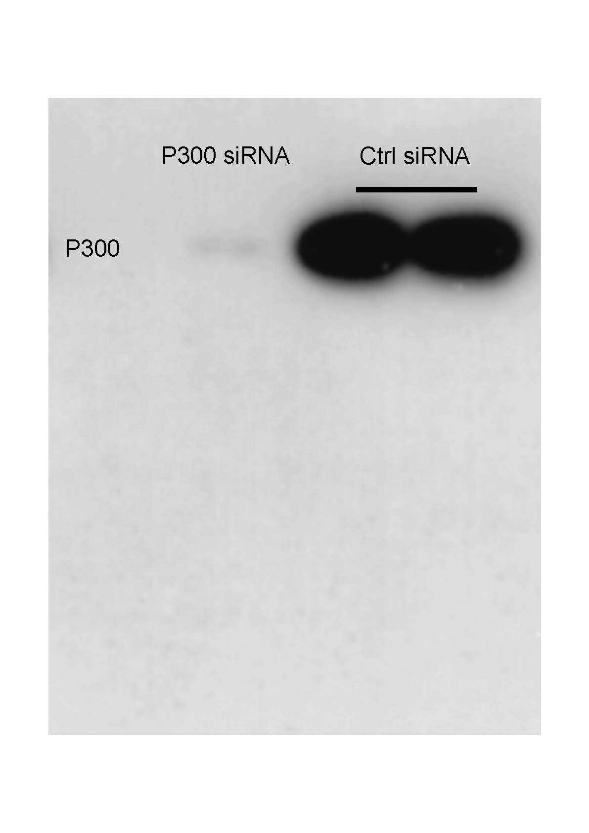
Western Blot: KAT3B/p300 Antibody (RW105) [NB100-616] - 293T cells were treated with different siRNAs against p300 alone for 2 days and core histones were prepared and subjected to WB analysis as indicated. The knockdown efficiency of p300 was confirmed by WB analysis of cell lysates. Western Blot protocol is 6% gel for running, 40V overnight transfer membrane. primary Ab 1:500, secondary Ab 1:5000. Image from verified customer review.Applications for KAT3B/p300 Antibody (RW105) - BSA Free
Electron Microscopy
Flow (Intracellular)
Immunocytochemistry/ Immunofluorescence
Immunoprecipitation
In vitro assay
Knockdown Validated
Western Blot
Reviewed Applications
Read 1 review rated 5 using NB100-616 in the following applications:
Flow Cytometry Panel Builder
Bio-Techne Knows Flow Cytometry
Save time and reduce costly mistakes by quickly finding compatible reagents using the Panel Builder Tool.
Advanced Features
- Spectra Viewer - Custom analysis of spectra from multiple fluorochromes
- Spillover Popups - Visualize the spectra of individual fluorochromes
- Antigen Density Selector - Match fluorochrome brightness with antigen density

Formulation, Preparation, and Storage
Purification
Formulation
Format
Preservative
Concentration
Shipping
Stability & Storage
Background: p300
Long Name
Alternate Names
Gene Symbol
Additional p300 Products
Product Documents for KAT3B/p300 Antibody (RW105) - BSA Free
Certificate of Analysis
To download a Certificate of Analysis, please enter a lot or batch number in the search box below.
Product Specific Notices for KAT3B/p300 Antibody (RW105) - BSA Free
This product is for research use only and is not approved for use in humans or in clinical diagnosis. Primary Antibodies are guaranteed for 1 year from date of receipt.
Related Research Areas
Citations for KAT3B/p300 Antibody (RW105) - BSA Free
 Powered by Bioz
Customer Reviews for KAT3B/p300 Antibody (RW105) - BSA Free (1)
Have you used KAT3B/p300 Antibody (RW105) - BSA Free?
Submit a review and receive an Amazon gift card!
$25/€18/£15/$25CAN/¥2500 Yen for a review with an image
$10/€7/£6/$10CAN/¥1110 Yen for a review without an image
Submit a review
Customer Images
-
Application: Knockdown ValidatedSample Tested: Mouse brainSpecies: MouseVerified Customer | Posted 06/23/2023293T cells were treated with different siRNAs against p300 alone for 2 days and core histones were prepared and subjected to WB analysis as indicated. The knockdown efficiency of p300 was confirmed by WB analysis of cell lysates.WB protocol is 6% gel for running, 40V overnight transfer membrane. primary Ab 1:500, secondary Ab 1:5000.
 Bio-Techne ResponseThis review was submitted through the legacy Novus Innovators Program, reflecting a new species or application tested on a primary antibody.
There are no reviews that match your criteria.
Protocols
View specific protocols for KAT3B/p300 Antibody (RW105) - BSA Free (NB100-616):
Sample Preparation.
1. Grow cells to 60-85% confluency. Flow cytometry requires between 2 x 10⁵ and 1 x 10⁶ cells for optimal performance.
2. If cells are adherent, harvest gently by washing once with staining buffer and then scraping. Avoid using trypsin as this can disrupt certain epitopes of interest. If enzymatic harvest is required, use Accutase, Collagenase, or TrypLE Express for a less damaging option.
3. Reserve 100 uL for counting, then transfer cell volume into a 50 mL conical tube and centrifuge for 8 minutes at 400 RCF.
a. Count cells using a hemocytometer and a 1:1 trypan blue exclusion stain to determine cell viability before starting the flow protocol. If cells appear blue, do not proceed.
4. Re-suspend cells to a concentration of 1 x 10⁶ cells/mL in staining buffer (NBP2-26247).
5. Aliquot out 1 mL samples in accordance with your experimental samples.
Tip: When cell surface and intracellular staining are required in the same sample, it is advisable that the cell surface staining be performed first since the fixation and permeablization steps might reduce the availability of surface antigens.
Intracellular Staining.
Tip: When performing intracellular staining, it is important to use appropriate fixation and permeabilization reagents based upon the target and its subcellular location. Generally, our Intracellular Flow Assay Kit (NBP2-29450) is a good place to start as it contains an optimized combination of reagents for intracellular staining as well as an inhibitor of intracellular protein transport (necessary if staining secreted proteins). Certain targets may require more gentle or transient permeabilization protocols such as the commonly employed methanol or saponin-based methods.
Protocol for Cytoplasmic Targets:
Optional: Perform cell surface staining as described in the previous section.
1. Fix the cells by adding 100 uL fixation solution (such as 4% PFA) to each sample for 10-15 minutes.
2. Permeabilize cells by adding 100 uL of a permeabization buffer to every 1 x 10⁶ cells present in the sample. Mix well and incubate at room temperature for 15 minutes.
a. For cytoplasmic targets, use a gentle permeabilization solution such as 1X PBS + 0.5% Saponin or 1X PBS + 0.5% Tween-20.
b. To maintain the permeabilized state throughout your experiment, use staining buffer + 0.1% of the permeabilization reagent (i.e. 0.1% Tween-20 or 0.1% Saponin).
3. Following the 15 minute incubation, add 2 mL of the staining buffer + 0.1% permeabilizer to each sample.
4. Centrifuge for 5 minutes at 400 RCF.
5. Discard supernatant and re-suspend in 1 mL of staining buffer + 0.1% permeabilizer.
6. Stain each sample at 1 uL/ 1 x 10⁶ cells of primary antibody or 1-3 uL/ 1 x 10⁶ cells for directly conjugated antibodies. Mix well and incubate at room temperature for 30 minutes- 1 hour. Gently mix samples every 10-15 minutes.
7. Following the primary/conjugate incubation, add 2 mL/sample of staining buffer +0.1% permeabilizer and centrifuge for 5 minutes at 400 RCF.
8. Remove supernatant and re-suspend each sample in 2 mL staining buffer + 0.1% permeabilizer, repeat wash for 5 minutes at 400 RCF.
9. If using a directly conjugated antibody, after the second wash, re-suspend cell pellet to a final volume of 500 uL per sample and proceed with flow analysis.
Culture cells to appropriate density in 35 mm culture dishes or 6-well plates.
1. Remove culture medium and wash the cells briefly in PBS. Add 10% formalin to the dish and fix at room temperature for 10 minutes.
2. Remove the formalin and wash the cells in PBS.
3. Permeablize the cells with 0.1% Triton X100 or other suitable detergent for 10 min.
4. Remove the permeablization buffer and wash three times for 10 minutes each in PBS. Be sure to not let the specimen dry out.
5. To block nonspecific antibody binding, incubate in 10% normal goat serum from 1 hour to overnight at room temperature.
6. Add primary antibody at appropriate dilution and incubate overnight at 4C.
7. Remove primary antibody and replace with PBS. Wash three times for 10 minutes each.
8. Add secondary antibody at appropriate dilution. Incubate for 1 hour at room temperature.
9. Remove secondary antibody and replace with PBS. Wash three times for 10 minutes each.
10. Counter stain DNA with DAPi if required.
1. Perform SDS-PAGE on samples to be analyzed, loading 10-25 ug of total protein per lane.
2. Transfer proteins to PVDF membrane according to the instructions provided by the manufacturer of the membrane and transfer apparatus.
3. Stain the membrane with Ponceau S (or similar product) to assess transfer success, and mark molecular weight standards where appropriate.
4. Rinse the blot TBS -0.05% Tween 20 (TBST).
5. Block the membrane in 5% Non-fat milk in TBST (blocking buffer) for at least 1 hour.
6. Wash the membrane in TBST three times for 10 minutes each.
7. Dilute primary antibody in blocking buffer and incubate overnight at 4C with gentle rocking.
8. Wash the membrane in TBST three times for 10 minutes each.
9. Incubate the membrane in diluted HRP conjugated secondary antibody in blocking buffer (as per manufacturer's instructions) for 1 hour at room temperature.
10. Wash the blot in TBST three times for 10 minutes each (this step can be repeated as required to reduce background).
11. Apply the detection reagent of choice in accordance with the manufacturer's instructions.
Find general support by application which include: protocols, troubleshooting, illustrated assays, videos and webinars.
- 7-Amino Actinomycin D (7-AAD) Cell Viability Flow Cytometry Protocol
- Appropriate Fixation of IHC/ICC Samples
- Cellular Response to Hypoxia Protocols
- ChIP Protocol Video
- Chromatin Immunoprecipitation (ChIP) Protocol
- Chromatin Immunoprecipitation Protocol
- ClariTSA™ Fluorophore Kits
- Detection & Visualization of Antibody Binding
- Extracellular Membrane Flow Cytometry Protocol
- Flow Cytometry Protocol for Cell Surface Markers
- Flow Cytometry Protocol for Staining Membrane Associated Proteins
- Flow Cytometry Staining Protocols
- Flow Cytometry Troubleshooting Guide
- ICC Cell Smear Protocol for Suspension Cells
- ICC Immunocytochemistry Protocol Videos
- ICC for Adherent Cells
- Immunocytochemistry (ICC) Protocol
- Immunocytochemistry Troubleshooting
- Immunofluorescence of Organoids Embedded in Cultrex Basement Membrane Extract
- Immunohistochemistry (IHC) and Immunocytochemistry (ICC) Protocols
- Immunoprecipitation Protocol
- Intracellular Flow Cytometry Protocol Using Alcohol (Methanol)
- Intracellular Flow Cytometry Protocol Using Detergents
- Intracellular Nuclear Staining Flow Cytometry Protocol Using Detergents
- Intracellular Staining Flow Cytometry Protocol Using Alcohol Permeabilization
- Intracellular Staining Flow Cytometry Protocol Using Detergents to Permeabilize Cells
- Preparing Samples for IHC/ICC Experiments
- Preventing Non-Specific Staining (Non-Specific Binding)
- Primary Antibody Selection & Optimization
- Propidium Iodide Cell Viability Flow Cytometry Protocol
- Protocol for Liperfluo
- Protocol for VisUCyte™ HRP Polymer Detection Reagent
- Protocol for the Characterization of Human Th22 Cells
- Protocol for the Characterization of Human Th9 Cells
- Protocol for the Fluorescent ICC Staining of Cell Smears - Graphic
- Protocol for the Fluorescent ICC Staining of Cultured Cells on Coverslips - Graphic
- Protocol for the Preparation and Fluorescent ICC Staining of Cells on Coverslips
- Protocol for the Preparation and Fluorescent ICC Staining of Non-adherent Cells
- Protocol for the Preparation and Fluorescent ICC Staining of Stem Cells on Coverslips
- Protocol for the Preparation of a Cell Smear for Non-adherent Cell ICC - Graphic
- Protocol: Annexin V and PI Staining by Flow Cytometry
- Protocol: Annexin V and PI Staining for Apoptosis by Flow Cytometry
- R&D Systems Quality Control Western Blot Protocol
- TUNEL and Active Caspase-3 Detection by IHC/ICC Protocol
- The Importance of IHC/ICC Controls
- Troubleshooting Guide: Fluorokine Flow Cytometry Kits
- Troubleshooting Guide: Western Blot Figures
- Western Blot Conditions
- Western Blot Protocol
- Western Blot Protocol for Cell Lysates
- Western Blot Troubleshooting
- Western Blot Troubleshooting Guide
- View all Protocols, Troubleshooting, Illustrated assays and Webinars
FAQs for KAT3B/p300 Antibody (RW105) - BSA Free
-
Q: I'm in the market for a monoclonal antibody for p300/KAT3B for WB, that would react with mouse p300. In your inventory you have some listed that I saw meet these criteria. I'm interested in knowing more about the RW antibodies: RW105, RW109 and RW128. Do you know if in addition to WB, any of these can also be used in IP or ChIP?
A: Thank you for contacting Novus Biologicals regarding our p300 antibodies. The three clones you are interested in are all guaranteed to work in mouse, and in WB and IP. We haven't yet tested these in ChIP, but if you wanted to try them and let us know how those results turn out you actually qualify for our Innovators Reward Program.


