![Western Blot: KDM2A/FBXL11 Antibody [NB100-74602]](https://resources.rndsystems.com/images/products/KDM2A-FBXL11-Antibody-Western-Blot-NB100-74602-img0013.jpg "Western Blot: KDM2A/FBXL11 Antibody [NB100-74602]")
Loading...
Key Product Details
Validated by
Biological Validation
Species Reactivity
Validated:
Human
Cited:
Human
Applications
Validated:
Immunohistochemistry, Immunohistochemistry-Paraffin, Western Blot, Immunoprecipitation, Chromatin Immunoprecipitation (ChIP)
Cited:
Western Blot, Block/Neutralize, Chemotaxis, Knockdown Validated
Label
Unconjugated
Antibody Source
Polyclonal Rabbit IgG
Loading...
Product Specifications
Immunogen
The immunogen recognized by this antibody maps to a region between residue 825 and 875 of human JmjC domain-containing histone demethylation protein 1A using the numbering given in entry NP_036440.1 (GeneID 22992).
Localization
Nuclear
Clonality
Polyclonal
Host
Rabbit
Isotype
IgG
Theoretical MW
133 kDa.
Disclaimer note: The observed molecular weight of the protein may vary from the listed predicted molecular weight due to post translational modifications, post translation cleavages, relative charges, and other experimental factors.
Disclaimer note: The observed molecular weight of the protein may vary from the listed predicted molecular weight due to post translational modifications, post translation cleavages, relative charges, and other experimental factors.
Scientific Data Images for KDM2A/FBXL11 Antibody
Western Blot: KDM2A/FBXL11 Antibody [NB100-74602]
Western Blot: KDM2A/FBXL11 Antibody [NB100-74602] - Detection of Human JHDM1A by Western Blot. Samples: Whole cell lysate from HeLa (50, 15, 5 ug) and 293T (50 ug) cells prepared using NETN lysis buffer. Antibodies: Affinity purified rabbit anti-JHDM1A antibody NB100-74602 used for WB at 0.1 ug/ml. Detection: Chemiluminescence with an exposure time of 3 minutes.![Immunohistochemistry-Paraffin: KDM2A/FBXL11 Antibody [NB100-74602]](https://resources.rndsystems.com/images/products/KDM2A-FBXL11-Antibody-Immunohistochemistry-NB100-74602-img0011.jpg "Immunohistochemistry-Paraffin: KDM2A/FBXL11 Antibody [NB100-74602]")
Immunohistochemistry-Paraffin: KDM2A/FBXL11 Antibody [NB100-74602]
Immunohistochemistry-Paraffin: KDM2A/FBXL11 Antibody [NB100-74602] - Section of human lung cancer. Antibody: Affinity purified rabbit anti- JHDM1A used at a dilution of 1:1,000 (1ug/ml). Detection: DAB![Immunohistochemistry-Paraffin: KDM2A/FBXL11 Antibody [NB100-74602]](https://resources.rndsystems.com/images/products/KDM2A-FBXL11-Antibody-Immunohistochemistry-Paraffin-NB100-74602-img0007.jpg "Immunohistochemistry-Paraffin: KDM2A/FBXL11 Antibody [NB100-74602]")
Immunohistochemistry-Paraffin: KDM2A/FBXL11 Antibody [NB100-74602]
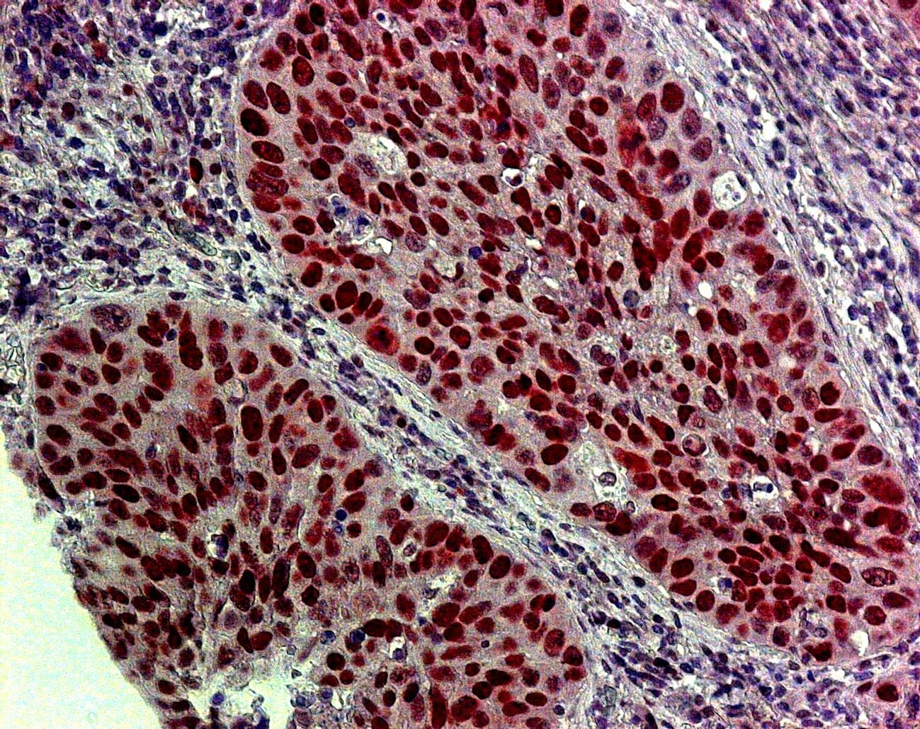
Immunohistochemistry-Paraffin: KDM2A/FBXL11 Antibody [NB100-74602] - Analysis of human lung tumor tissue using anti-KDM2A antibody. Image submitted by a verified customer review.![Immunoprecipitation: KDM2A/FBXL11 Antibody [NB100-74602]](https://resources.rndsystems.com/images/products/KDM2A-FBXL11-Antibody-Immunoprecipitation-NB100-74602-img0014.jpg "Immunoprecipitation: KDM2A/FBXL11 Antibody [NB100-74602]")
Immunoprecipitation: KDM2A/FBXL11 Antibody [NB100-74602]
Immunoprecipitation: KDM2A/FBXL11 Antibody [NB100-74602] - Detection of human JHDM1A by western blot of immunoprecipitates. Samples: Whole cell lysate (0.5 or 1.0 mg per IP reaction; 20% of IP loaded) from HeLa cells prepared using NETN lysis buffer. Antibodies: Affinity purified rabbit anti-JHDM1A antibody NB100-74602 (lot NB100-74602-2) used for IP at 6 ug per reaction. JHDM1A was also immunoprecipitated by a previous lot of this antibody (lot NB100-74602-1). For blotting immunoprecipitated JHDM1A, NB100-74602 was used at 0.4 ug/ml. Detection: Chemiluminescence with an exposure time of 3 minutes.
Knockdown Validated: KDM2A/FBXL11 Antibody [NB100-74602] -
Transcriptional and stability analysis of F508del-CFTR after demethylases downregulation. (A) CFTR mRNA levels determined by quantitative real-time PCR in F508del-CFTR expressing CFBE41o- cells were transfected with two different siRNAs for each target (JMJD6, KDM2A, KDM3B) for 48 h. CFTR mRNA expression was normalized to 18S RNA and reported relative to its expression in control (SCR) cells that was arbitrarily set to 1 (means +/- SD values, n = 5). (B) F508del-CFTR overexpressing CFBE41o- cells were transfected with siRNA targeting JMJD6, KDM2A, KDM3B or with scrambled siRNAs for 48 h. Subsequently, protein synthesis was inhibited by adding 100 ug/mL cycloheximide (CHX) and cells were harvested after 30, 60 and 90 min. Protein lysates were analyzed by western blot with an anti-CFTR antibody. Calnexin was used as a loading control. The lower-right graph represents the densitometric quantification of the immunostained bands of F508del-CFTR band B normalized by the value at time = 0 (mean +/- SD, n = 4; * p < 0.05 vs. the value of the same time-point of SCR). Image collected and cropped by CiteAb from the following open publication (https://pubmed.ncbi.nlm.nih.gov/36077010), licensed under a CC-BY license. Not internally tested by Novus Biologicals.
Western Blot: KDM2A/FBXL11 Antibody [NB100-74602] -
Mutational analysis of CFTR methylated lysine and effect of demethylases downregulation on chaperone machinery. (A) CFBE41o- cells were transiently transfected with F508del-CFTR, or other derivative mutants as indicated. After 24 h, cells were treated with 3 μM VX-809 and the amounts of F508del-CFTR band B and band C were assessed by western blotting. alpha -tubulin was used as loading control (upper panel). In the lower panel, the graphs show the ratio of F508del-CFTR band C/band B for the cells treated with VX-809, obtained by the densitometric quantification of the immunostained band C normalized by band B expression, and expressed as a percentage of the control cells (Ctrl) (means +/- SD values, n = 4; * p < 0.05 vs. Ctrl). (B) F508del-CFTR expressing CFBE41o- cells were transfected with siRNA (#1 and #2 guides) targeting KDM2A or KDM3B or with a scrambled (SCR) siRNA for 48 h. In the left and central panels, the expression of the indicated chaperones was analysed by western blotting. alpha -tubulin was used as loading control. In the right panel, the graphs report the densitometric quantification of the immunostained bands of the experiment detailed in the left and central panels normalized by alpha -tubulin expression, and expressed as a percentage of the control cells (SCR). (Means +/- SD values, n = 3; * p < 0.05 vs. SCR). Image collected and cropped by CiteAb from the following open publication (https://pubmed.ncbi.nlm.nih.gov/36077010), licensed under a CC-BY license. Not internally tested by Novus Biologicals.Applications for KDM2A/FBXL11 Antibody
Application
Recommended Usage
Immunohistochemistry-Paraffin
1:100-1:500
Immunoprecipitation
2-10 ug/mg lysate
Western Blot
1:2000-1:10000
Application Notes
Epitope retrieval with citrate buffer pH 6.0 is recommended for FFPE tissue sections. Use in chromatin immunoprecipitation reported in scientific literature (PMID: 30132864). KDM2A/FBXL11 antibody validated for IHC-P from a verified customer review.
Reviewed Applications
Read 2 reviews rated 5 using NB100-74602 in the following applications:
Formulation, Preparation, and Storage
Purification
Immunogen affinity purified
Formulation
TBS and 0.1% BSA
Preservative
0.09% Sodium Azide
Concentration
0.2 mg/ml
Shipping
The product is shipped with polar packs. Upon receipt, store it immediately at the temperature recommended below.
Stability & Storage
Store at 4C. Do not freeze.
Background: KDM2A/FBXL11
Long Name
Lysine-specific demethylase 2A
Alternate Names
CXXC8, EC 1.14.11.27, F-box protein FBL7, FBL11, FBL7, FBXL11, JHDM1A, KDM2A, KIAA1004
Entrez Gene IDs
22992 (Human)
Gene Symbol
KDM2A
UniProt
Additional KDM2A/FBXL11 Products
Product Documents for KDM2A/FBXL11 Antibody
Certificate of Analysis
To download a Certificate of Analysis, please enter a lot or batch number in the search box below.
Product Specific Notices for KDM2A/FBXL11 Antibody
This product is for research use only and is not approved for use in humans or in clinical diagnosis. Primary Antibodies are guaranteed for 1 year from date of receipt.
Citations for KDM2A/FBXL11 Antibody
 Powered by Bioz
Customer Reviews for KDM2A/FBXL11 Antibody (2)
5 out of 5
2 Customer Ratings
Have you used KDM2A/FBXL11 Antibody?
Submit a review and receive an Amazon gift card!
$25/€18/£15/$25CAN/¥2500 Yen for a review with an image
$10/€7/£6/$10CAN/¥1110 Yen for a review without an image
Submit a review
Customer Images
Showing
1
-
2 of
2 reviews
Showing All
Filter By:
-
Application: ImmunocytochemistrySample Tested: Human lung tumorSpecies: HumanVerified Customer | Posted 10/08/2015IHC staining on humna lung cancer tissue section

-
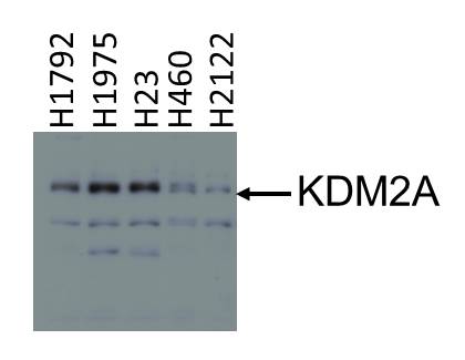
Application: Western BlotSample Tested: Lung cell lines whole cell lysateSpecies: HumanVerified Customer | Posted 10/08/2015Lung cell lines whole cell lysate
There are no reviews that match your criteria.
Protocols
Find general support by application which include: protocols, troubleshooting, illustrated assays, videos and webinars.
- Antigen Retrieval Protocol (PIER)
- Antigen Retrieval for Frozen Sections Protocol
- Appropriate Fixation of IHC/ICC Samples
- Cellular Response to Hypoxia Protocols
- ChIP Protocol Video
- Chromatin Immunoprecipitation (ChIP) Protocol
- Chromatin Immunoprecipitation Protocol
- Chromogenic IHC Staining of Formalin-Fixed Paraffin-Embedded (FFPE) Tissue Protocol
- Chromogenic Immunohistochemistry Staining of Frozen Tissue
- ClariTSA™ Fluorophore Kits
- Detection & Visualization of Antibody Binding
- Fluorescent IHC Staining of Frozen Tissue Protocol
- Graphic Protocol for Heat-induced Epitope Retrieval
- Graphic Protocol for the Preparation and Fluorescent IHC Staining of Frozen Tissue Sections
- Graphic Protocol for the Preparation and Fluorescent IHC Staining of Paraffin-embedded Tissue Sections
- Graphic Protocol for the Preparation of Gelatin-coated Slides for Histological Tissue Sections
- IHC Sample Preparation (Frozen sections vs Paraffin)
- Immunofluorescent IHC Staining of Formalin-Fixed Paraffin-Embedded (FFPE) Tissue Protocol
- Immunohistochemistry (IHC) and Immunocytochemistry (ICC) Protocols
- Immunohistochemistry Frozen Troubleshooting
- Immunohistochemistry Paraffin Troubleshooting
- Immunoprecipitation Protocol
- Preparing Samples for IHC/ICC Experiments
- Preventing Non-Specific Staining (Non-Specific Binding)
- Primary Antibody Selection & Optimization
- Protocol for Heat-Induced Epitope Retrieval (HIER)
- Protocol for Making a 4% Formaldehyde Solution in PBS
- Protocol for VisUCyte™ HRP Polymer Detection Reagent
- Protocol for the Preparation & Fixation of Cells on Coverslips
- Protocol for the Preparation and Chromogenic IHC Staining of Frozen Tissue Sections
- Protocol for the Preparation and Chromogenic IHC Staining of Frozen Tissue Sections - Graphic
- Protocol for the Preparation and Chromogenic IHC Staining of Paraffin-embedded Tissue Sections
- Protocol for the Preparation and Chromogenic IHC Staining of Paraffin-embedded Tissue Sections - Graphic
- Protocol for the Preparation and Fluorescent IHC Staining of Frozen Tissue Sections
- Protocol for the Preparation and Fluorescent IHC Staining of Paraffin-embedded Tissue Sections
- Protocol for the Preparation of Gelatin-coated Slides for Histological Tissue Sections
- R&D Systems Quality Control Western Blot Protocol
- TUNEL and Active Caspase-3 Detection by IHC/ICC Protocol
- The Importance of IHC/ICC Controls
- Troubleshooting Guide: Immunohistochemistry
- Troubleshooting Guide: Western Blot Figures
- Western Blot Conditions
- Western Blot Protocol
- Western Blot Protocol for Cell Lysates
- Western Blot Troubleshooting
- Western Blot Troubleshooting Guide
- View all Protocols, Troubleshooting, Illustrated assays and Webinars
Loading...