![Western Blot: KDM2A/FBXL11 AntibodyBSA Free [NBP1-78305]](https://resources.rndsystems.com/images/products/KDM2A-FBXL11-Antibody-Western-Blot-NBP1-78305-img0006.jpg "Western Blot: KDM2A/FBXL11 AntibodyBSA Free [NBP1-78305]")
Key Product Details
Species Reactivity
Human, Mouse
Applications
Immunohistochemistry, Immunohistochemistry-Paraffin, Western Blot, Immunocytochemistry/ Immunofluorescence
Label
Unconjugated
Antibody Source
Polyclonal Rabbit IgG
Format
BSA Free
Loading...
Product Specifications
Immunogen
A synthetic peptide made to an N-terminal portion of the human KDM2A/FBXL11 protein (between residues 350-500) [UniProt Q9Y2K7]
Localization
Nucleus, nucleoplasm. Note: Punctate expression throughout the nucleoplasm and enriched in the perinucleolar region. Specifically nucleates at CpG islands where it's presence results in chromatin depleted in H3K36me2.
Clonality
Polyclonal
Host
Rabbit
Isotype
IgG
Scientific Data Images for KDM2A/FBXL11 Antibody - BSA Free
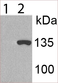
Western Blot: KDM2A/FBXL11 AntibodyBSA Free [NBP1-78305]
Western Blot: KDM2A/FBXL11 Antibody [NBP1-78305] - 1) 293T. 2) 293T transiently transfected to express human KDM2A. WB image submitted by a verified customer review.![Immunocytochemistry/ Immunofluorescence: KDM2A/FBXL11 Antibody - BSA Free [NBP1-78305]](https://resources.rndsystems.com/images/products/KDM2A-FBXL11-Antibody---BSA-Free-Immunocytochemistry-Immunofluorescence-NBP1-78305-img0008.jpg "Immunocytochemistry/ Immunofluorescence: KDM2A/FBXL11 Antibody - BSA Free [NBP1-78305]")
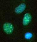
Immunocytochemistry/ Immunofluorescence: KDM2A/FBXL11 Antibody - BSA Free [NBP1-78305]
Immunocytochemistry/Immunofluorescence: KDM2A/FBXL11 Antibody - BSA Free [NBP1-78305] - Mouse MS1 cells were fixed in 4% paraformaldehyde for 10 minutes and permeabilized in 0.05% Triton X-100 in PBS for 5 minutes. The cells were incubated with KCM2A/FBXL11 Antibody (NBP1-78305) at 2ug/ml overnight at 4C and detected with an anti-rabbit DyLight 488 (Green) at a 1:1000 dilution for 60 minutes. Nuclei were counterstained with DAPI (Blue). Cells were imaged using a 100X objective and digitally deconvolved.![Immunohistochemistry: KDM2A/FBXL11 Antibody - BSA Free [NBP1-78305]](https://resources.rndsystems.com/images/products/KDM2A-FBXL11-Antibody-Immunohistochemistry-NBP1-78305-img0004.jpg "Immunohistochemistry: KDM2A/FBXL11 Antibody - BSA Free [NBP1-78305]")
Immunohistochemistry: KDM2A/FBXL11 Antibody - BSA Free [NBP1-78305]
Immunohistochemistry: KDM2A/FBXL11 Antibody [NBP1-78305] - IHC analysis of KDM2A / FBXL11 in human kidney cancer xenograft using DAB with hematoxylin counterstain.![Immunocytochemistry/ Immunofluorescence: KDM2A/FBXL11 Antibody - BSA Free [NBP1-78305]](https://resources.rndsystems.com/images/products/KDM2A-FBXL11-Antibody-Immunocytochemistry-Immunofluorescence-NBP1-78305-img0005.jpg "Immunocytochemistry/ Immunofluorescence: KDM2A/FBXL11 Antibody - BSA Free [NBP1-78305]")
Immunocytochemistry/ Immunofluorescence: KDM2A/FBXL11 Antibody - BSA Free [NBP1-78305]
Immunocytochemistry/Immunofluorescence: KDM2A/FBXL11 Antibody [NBP1-78305] - Human astrocytoma cell line lentivirally transduced and expressing KDM2A. Cells were fixed with 4% paraformaldehyde and stained with anti-KDM2A, followed by Alexa Fluor 488-anti-rabbit secondary antibody. DAPI staining shows nuclei. ICC/IF image submitted by a verified customer review.![Immunocytochemistry/ Immunofluorescence: KDM2A/FBXL11 Antibody - BSA Free [NBP1-78305]](https://resources.rndsystems.com/images/products/KDM2A-FBXL11-Antibody---BSA-Free-Immunocytochemistry-Immunofluorescence-NBP1-78305-img0007.jpg "Immunocytochemistry/ Immunofluorescence: KDM2A/FBXL11 Antibody - BSA Free [NBP1-78305]")
Immunocytochemistry/ Immunofluorescence: KDM2A/FBXL11 Antibody - BSA Free [NBP1-78305]
Immunocytochemistry/Immunofluorescence: KDM2A/FBXL11 Antibody - BSA Free [NBP1-78305] - Caco-2 cells were fixed in 4% paraformaldehyde for 10 minutes and permeabilized in 0.05% Triton X-100 in PBS for 5 minutes. The cells were incubated with KDM2A/FBXL11 Antibody (NBP1-78305) at 1ug/ml overnight at 4C and detected with an anti-rabbit DyLight 488 (Green) at a 1:1000 dilution for 60 minutes. Nuclei were counterstained with DAPI (Blue). Cells were imaged using a 40X objective.Applications for KDM2A/FBXL11 Antibody - BSA Free
Application
Recommended Usage
Immunocytochemistry/ Immunofluorescence
1:100-1:400
Immunohistochemistry
1:300
Immunohistochemistry-Paraffin
1:300
Western Blot
reported by customer review
Application Notes
This KDM2A/FBXL11 antibody is useful for Immunocytochemistry/Immunofluorescence where nuclear staining is observed in HeLa cells and IHC-paraffin embedded sections where nuclear staining is seen in human kidney cancer xenograft. Prior to immunostaining paraffin tissues, antigen retrieval with sodium citrate buffer (pH 6.0) is recommended.
Reviewed Applications
Read 3 reviews rated 4.7 using NBP1-78305 in the following applications:
Formulation, Preparation, and Storage
Purification
Immunogen affinity purified
Formulation
PBS and 30% Glycerol
Format
BSA Free
Preservative
0.05% Sodium Azide
Concentration
1.2 mg/ml
Shipping
The product is shipped with polar packs. Upon receipt, store it immediately at the temperature recommended below.
Stability & Storage
Store at 4C short term. Aliquot and store at -20C long term. Avoid freeze-thaw cycles.
Background: KDM2A/FBXL11
Long Name
Lysine-specific demethylase 2A
Alternate Names
CXXC8, EC 1.14.11.27, F-box protein FBL7, FBL11, FBL7, FBXL11, JHDM1A, KDM2A, KIAA1004
Entrez Gene IDs
22992 (Human)
Gene Symbol
KDM2A
UniProt
Additional KDM2A/FBXL11 Products
Product Documents for KDM2A/FBXL11 Antibody - BSA Free
Certificate of Analysis
To download a Certificate of Analysis, please enter a lot or batch number in the search box below.
Product Specific Notices for KDM2A/FBXL11 Antibody - BSA Free
This product is for research use only and is not approved for use in humans or in clinical diagnosis. Primary Antibodies are guaranteed for 1 year from date of receipt.
Customer Reviews for KDM2A/FBXL11 Antibody - BSA Free (3)
4.7 out of 5
3 Customer Ratings
Have you used KDM2A/FBXL11 Antibody - BSA Free?
Submit a review and receive an Amazon gift card!
$25/€18/£15/$25CAN/¥2500 Yen for a review with an image
$10/€7/£6/$10CAN/¥1110 Yen for a review without an image
Submit a review
Customer Images
Showing
1
-
3 of
3 reviews
Showing All
Filter By:
-
Application: Western BlotSample Tested: 293TSpecies: HumanVerified Customer | Posted 12/31/20191. 293T 2. 293T transiently transfected to express human KDM2A

-
Application: ImmunocytochemistrySample Tested: AGS human gastric adenocarcinoma cell line and brain tumor cell lineSpecies: HumanVerified Customer | Posted 12/31/2019Human astrocytoma cell line lentivirally transduced and expressing KDM2A. Cells were fixed with 4% paraformaldehyde and stained with anti-KDM2A, followed by Alexa488-anti-rabbit secondary antibody. DAPI staining shows nuclei.
-
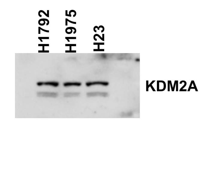
Application: Western BlotSample Tested: Human lung cancer cell lines whole cell lysateSpecies: HumanVerified Customer | Posted 05/18/2016Western blot shows detection of Rabbit Anti-Human KDM2A polyclonal Antibody in lung cancer cell lines (dilution 1:1000)
There are no reviews that match your criteria.
Protocols
View specific protocols for KDM2A/FBXL11 Antibody - BSA Free (NBP1-78305):
KDM2A/FBXL11 Antibody:
Immunocytochemistry Protocol
Culture cells to appropriate density in 35 mm culture dishes or 6-well plates.
1. Remove culture medium and add 10% formalin to the dish. Fix at room temperature for 30 minutes.
2. Remove the formalin and add ice cold methanol. Incubate for 5-10 minutes.
3. Remove methanol and add washing solution (i.e. PBS). Be sure to not let the specimen dry out. Wash three times for 10 minutes.
4. To block nonspecific antibody binding incubate in 10% normal goat serum from 1 hour to overnight at room temperature.
5. Add primary antibody at appropriate dilution and incubate at room temperature from 2 hours to overnight at room temperature.
6. Remove primary antibody and replace with washing solution. Wash three times for 10 minutes.
7. Add secondary antibody at appropriate dilution. Incubate for 1 hour at room temperature.
8. Remove antibody and replace with wash solution, then wash for 10 minutes. Add Hoechst 33258 to wash solution at 1:25,0000 and incubate for 10 minutes. Wash a third time for 10 minutes.
9. Cells can be viewed directly after washing. The plates can also be stored in PBS containing Azide covered in Parafilm (TM). Cells can also be cover-slipped using Fluoromount, with appropriate sealing.
*The above information is only intended as a guide. The researcher should determine what protocol best meets their needs. Please follow safe laboratory procedures.
KDM2A/FBXL11 Antibody:
Immunohistochemistry-Paraffin Embedded Sections
Antigen Unmasking:
Bring slides to a boil in 10 mM sodium citrate buffer (pH 6.0) then maintain at a sub-boiling temperature for 10 minutes. Cool slides on bench-top for 30 minutes.
Staining:
1. Wash sections in deionized water three times for 5 minutes each.
2. Wash sections in wash buffer for 5 minutes.
3. Block each section with 100-400 ul blocking solution for 1 hour at room temperature.
4. Remove blocking solution and add 100-400 ul diluted primary antibody. Incubate overnight at 4C.
5. Remove antibody solution and wash sections in wash buffer three times for 5 minutes each.
6. Add 100-400 ul biotinylated diluted secondary antibody. Incubate 30 minutes at room temperature.
7. Remove secondary antibody solution and wash sections three times with wash buffer for 5 minutes each.
8. Add 100-400 ul Streptavidin-HRP reagent to each section and incubate for 30 minutes at room temperature.
9. Wash sections three times in wash buffer for 5 minutes each.
10. Add 100-400 ul DAB substrate to each section and monitor staining closely.
11. As soon as the sections develop, immerse slides in deionized water.
12. Counterstain sections in hematoxylin.
13. Wash sections in deionized water two times for 5 minutes each.
14. Dehydrate sections.
15. Mount coverslips.
*The above information is only intended as a guide. The researcher should determine what protocol best meets their needs. Please follow safe laboratory procedures.
Find general support by application which include: protocols, troubleshooting, illustrated assays, videos and webinars.
- Antigen Retrieval Protocol (PIER)
- Antigen Retrieval for Frozen Sections Protocol
- Appropriate Fixation of IHC/ICC Samples
- Cellular Response to Hypoxia Protocols
- Chromogenic IHC Staining of Formalin-Fixed Paraffin-Embedded (FFPE) Tissue Protocol
- Chromogenic Immunohistochemistry Staining of Frozen Tissue
- ClariTSA™ Fluorophore Kits
- Detection & Visualization of Antibody Binding
- Fluorescent IHC Staining of Frozen Tissue Protocol
- Graphic Protocol for Heat-induced Epitope Retrieval
- Graphic Protocol for the Preparation and Fluorescent IHC Staining of Frozen Tissue Sections
- Graphic Protocol for the Preparation and Fluorescent IHC Staining of Paraffin-embedded Tissue Sections
- Graphic Protocol for the Preparation of Gelatin-coated Slides for Histological Tissue Sections
- ICC Cell Smear Protocol for Suspension Cells
- ICC Immunocytochemistry Protocol Videos
- ICC for Adherent Cells
- IHC Sample Preparation (Frozen sections vs Paraffin)
- Immunocytochemistry (ICC) Protocol
- Immunocytochemistry Troubleshooting
- Immunofluorescence of Organoids Embedded in Cultrex Basement Membrane Extract
- Immunofluorescent IHC Staining of Formalin-Fixed Paraffin-Embedded (FFPE) Tissue Protocol
- Immunohistochemistry (IHC) and Immunocytochemistry (ICC) Protocols
- Immunohistochemistry Frozen Troubleshooting
- Immunohistochemistry Paraffin Troubleshooting
- Preparing Samples for IHC/ICC Experiments
- Preventing Non-Specific Staining (Non-Specific Binding)
- Primary Antibody Selection & Optimization
- Protocol for Heat-Induced Epitope Retrieval (HIER)
- Protocol for Making a 4% Formaldehyde Solution in PBS
- Protocol for VisUCyte™ HRP Polymer Detection Reagent
- Protocol for the Fluorescent ICC Staining of Cell Smears - Graphic
- Protocol for the Fluorescent ICC Staining of Cultured Cells on Coverslips - Graphic
- Protocol for the Preparation & Fixation of Cells on Coverslips
- Protocol for the Preparation and Chromogenic IHC Staining of Frozen Tissue Sections
- Protocol for the Preparation and Chromogenic IHC Staining of Frozen Tissue Sections - Graphic
- Protocol for the Preparation and Chromogenic IHC Staining of Paraffin-embedded Tissue Sections
- Protocol for the Preparation and Chromogenic IHC Staining of Paraffin-embedded Tissue Sections - Graphic
- Protocol for the Preparation and Fluorescent ICC Staining of Cells on Coverslips
- Protocol for the Preparation and Fluorescent ICC Staining of Non-adherent Cells
- Protocol for the Preparation and Fluorescent ICC Staining of Stem Cells on Coverslips
- Protocol for the Preparation and Fluorescent IHC Staining of Frozen Tissue Sections
- Protocol for the Preparation and Fluorescent IHC Staining of Paraffin-embedded Tissue Sections
- Protocol for the Preparation of Gelatin-coated Slides for Histological Tissue Sections
- Protocol for the Preparation of a Cell Smear for Non-adherent Cell ICC - Graphic
- R&D Systems Quality Control Western Blot Protocol
- TUNEL and Active Caspase-3 Detection by IHC/ICC Protocol
- The Importance of IHC/ICC Controls
- Troubleshooting Guide: Immunohistochemistry
- Troubleshooting Guide: Western Blot Figures
- Western Blot Conditions
- Western Blot Protocol
- Western Blot Protocol for Cell Lysates
- Western Blot Troubleshooting
- Western Blot Troubleshooting Guide
- View all Protocols, Troubleshooting, Illustrated assays and Webinars
Loading...