Key Product Details
Species Reactivity
All Species
Applications
Western Blot, Immunocytochemistry/ Immunofluorescence
Label
FITC (Excitation = 495 nm, Emission = 519 nm)
Antibody Source
Polyclonal Rabbit IgG
Loading...
Product Specifications
Immunogen
KLH (Keyhole Limpet Hemocyanin)
Localization
Secreted protein; extracellular space
Clonality
Polyclonal
Host
Rabbit
Isotype
IgG
Description
Store vial at 4C prior to restoration. For extended storage aliquot contents and freeze at -20C or below. Avoid cycles of freezing and thawing. Centrifuge product if not completely clear after standing at room temperature. This product is stable for several weeks at 4C as an undiluted liquid. Dilute only prior to immediate use.
Stabilizer: 10 mg/ml Bovine Serum Albumin (BSA)-Immunoglobulin and Protease free
Color: Green Excitation Wavelength: 495 Emission Wavelength: 528
Conjugation Chemistry: Isothiocyanate
This product was prepared from monospecific antiserum by immunoaffinity chromatography using KLH coupled to agarose beads followed by solid phase adsorption(s) to remove any unwanted reactivities. Assay by immunoelectrophoresis resulted in a single precipitin arc against anti-rabbit serum, anti-Fluorescein and KLH.
Scientific Data Images for KLH Antibody [FITC]
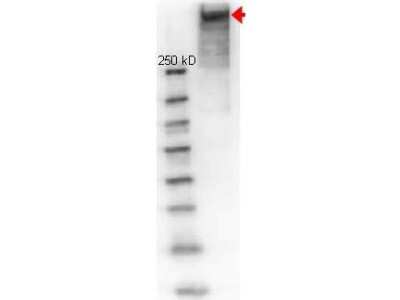
Western Blot: KLH Antibody [Fluorescein] [NBP1-97356] - Lane 1: molecular weight. Lane 2: reduced KLH. Load: 1 ug. Primary antibody: KLH antibody at 1:1000 for overnight at 4C. Secondary antibody: HRP Goat-anti Rabbit secondary antibody at 1:40,000 for 45 min at RT. Block: 1% BSA-TBS overnight at 4C. Predicted/Observed size: 350-390 kDa, 350-390 kDa for KLH. Other band(s): none.
Applications for KLH Antibody [FITC]
Application
Recommended Usage
Immunocytochemistry/ Immunofluorescence
1:500-1:2500
Western Blot
1:100-1:2000
Application Notes
This product is designed for immunofluorescence microscopy, fluorescence based plate assays (FLISA) and fluorescent western blotting. This product is also suitable for multiplex analysis, including multicolor imaging, utilizing various commercial platforms.
Formulation, Preparation, and Storage
Purification
Immunogen affinity purified
Reconstitution
Reconstitute with 100 ul deionized water (or equivalent)
Formulation
Lyophilized from 0.02 M Potassium Phosphate, 0.15 M Sodium Chloride, pH 7.2, 10 mg/mL Bovine Serum Albumin (BSA) - Immunoglobulin and Protease free
Preservative
0.01% Sodium Azide
Concentration
LYOPH mg/ml
Shipping
The product is shipped with polar packs. Upon receipt, store it immediately at the temperature recommended below.
Stability & Storage
Store lyophilized antibody at 4C in the dark. Aliquot reconstituted liquid and store at -20C. Avoid freeze-thaw cycles.
Calculators
Background: KLH
Alternate Names
keyhole limpet hemocyanin (KLH)
Additional KLH Products
Product Documents for KLH Antibody [FITC]
Certificate of Analysis
To download a Certificate of Analysis, please enter a lot or batch number in the search box below.
Product Specific Notices for KLH Antibody [FITC]
This product is for research use only and is not approved for use in humans or in clinical diagnosis. Primary Antibodies are guaranteed for 1 year from date of receipt.
Customer Reviews for KLH Antibody [FITC]
There are currently no reviews for this product. Be the first to review KLH Antibody [FITC] and earn rewards!
Have you used KLH Antibody [FITC]?
Submit a review and receive an Amazon gift card!
$25/€18/£15/$25CAN/¥2500 Yen for a review with an image
$10/€7/£6/$10CAN/¥1110 Yen for a review without an image
Submit a review
Protocols
Find general support by application which include: protocols, troubleshooting, illustrated assays, videos and webinars.
- Appropriate Fixation of IHC/ICC Samples
- Cellular Response to Hypoxia Protocols
- ClariTSA™ Fluorophore Kits
- Detection & Visualization of Antibody Binding
- ICC Cell Smear Protocol for Suspension Cells
- ICC Immunocytochemistry Protocol Videos
- ICC for Adherent Cells
- Immunocytochemistry (ICC) Protocol
- Immunocytochemistry Troubleshooting
- Immunofluorescence of Organoids Embedded in Cultrex Basement Membrane Extract
- Immunohistochemistry (IHC) and Immunocytochemistry (ICC) Protocols
- Preparing Samples for IHC/ICC Experiments
- Preventing Non-Specific Staining (Non-Specific Binding)
- Primary Antibody Selection & Optimization
- Protocol for VisUCyte™ HRP Polymer Detection Reagent
- Protocol for the Fluorescent ICC Staining of Cell Smears - Graphic
- Protocol for the Fluorescent ICC Staining of Cultured Cells on Coverslips - Graphic
- Protocol for the Preparation and Fluorescent ICC Staining of Cells on Coverslips
- Protocol for the Preparation and Fluorescent ICC Staining of Non-adherent Cells
- Protocol for the Preparation and Fluorescent ICC Staining of Stem Cells on Coverslips
- Protocol for the Preparation of a Cell Smear for Non-adherent Cell ICC - Graphic
- R&D Systems Quality Control Western Blot Protocol
- TUNEL and Active Caspase-3 Detection by IHC/ICC Protocol
- The Importance of IHC/ICC Controls
- Troubleshooting Guide: Western Blot Figures
- Western Blot Conditions
- Western Blot Protocol
- Western Blot Protocol for Cell Lysates
- Western Blot Troubleshooting
- Western Blot Troubleshooting Guide
- View all Protocols, Troubleshooting, Illustrated assays and Webinars
Loading...