![Western Blot: KMT1A/SUV39H1 Antibody [NBP1-21367]](https://resources.rndsystems.com/images/products/KMT1A-SUV39H1-Antibody-NBP1-21367-img0006.jpg "Western Blot: KMT1A/SUV39H1 Antibody [NBP1-21367]")
Loading...
Key Product Details
Species Reactivity
Validated:
Human, Mouse
Cited:
Human, Mouse
Predicted:
Bovine (100%), Orangutan (100%). Backed by our 100% Guarantee.
Applications
Validated:
Immunohistochemistry, Immunohistochemistry-Paraffin, Western Blot, Immunocytochemistry/ Immunofluorescence, Immunoprecipitation
Cited:
Immunohistochemistry-Paraffin, Western Blot, Immunocytochemistry/ Immunofluorescence, IF/IHC
Label
Unconjugated
Antibody Source
Polyclonal Rabbit IgG
Loading...
Product Specifications
Immunogen
A synthetic peptide made to an N-terminal portion of the human KMT1A/SUV39H1 protein (between residues 1-100) [UniProt O43463]
Reactivity Notes
Mouse reactivity reported in scientific literature (PMID: 28425504).
Localization
Nucleus.
Clonality
Polyclonal
Host
Rabbit
Isotype
IgG
Scientific Data Images for KMT1A/SUV39H1 Antibody
Western Blot: KMT1A/SUV39H1 Antibody [NBP1-21367]
Western Blot: KMT1A/SUV39H1 Antibody [NBP1-21367] - Detection of human SUV39H1 by western blot and immunoprecipitation. Samples: Whole cell lysate (5, 15 and 50 ug for WB; 1 mg for IP, 20% of IP loaded) from HeLa cells. Antibodies: Affinity purified rabbit anti-SUV39H1 antibody NBP1-21367 used for WB at 0.04 ug/ml (A) and 1 ug/ml (B) and used for IP at 10 ug/mg lysate. SUV39H1 was also immunoprecipitated by a rabbit anti-SUV39H1 antibody recognizing a downstream epitope (B). Detection: Chemiluminescence with exposure times of 3 minutes (A) and 30 seconds (B).![Immunohistochemistry-Paraffin: KMT1A/SUV39H1 Antibody [NBP1-21367]](https://resources.rndsystems.com/images/products/KMT1A-SUV39H1-Antibody-Immunohistochemistry-Paraffin-NBP1-21367-img0003.jpg "Immunohistochemistry-Paraffin: KMT1A/SUV39H1 Antibody [NBP1-21367]")
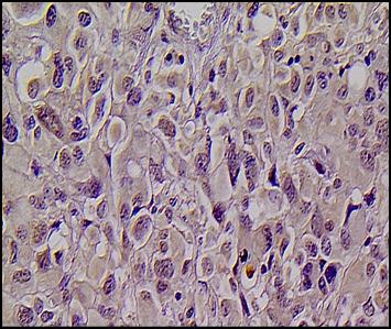
Immunohistochemistry-Paraffin: KMT1A/SUV39H1 Antibody [NBP1-21367]
Immunohistochemistry-Paraffin: KMT1A/SUV39H1 Antibody [NBP1-21367] - SUV39H1 expression in human glioma tissue grade IV. Image submitted by a verified customer review.![Western Blot: KMT1A/SUV39H1 Antibody [NBP1-21367]](https://resources.rndsystems.com/images/products/KMT1A-SUV39H1-Antibody-Western-Blot-NBP1-21367-img0004.jpg "Western Blot: KMT1A/SUV39H1 Antibody [NBP1-21367]")
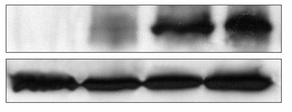
Western Blot: KMT1A/SUV39H1 Antibody [NBP1-21367]
Western Blot: KMT1A/SUV39H1 Antibody [NBP1-21367] - Up:SUV39H1 expression in normal brain, human glioma tissue grades II, III and IV respectively. Down: Actin expression levels. Image submitted by a verified customer review.![Western Blot: KMT1A/SUV39H1 Antibody [NBP1-21367]](https://resources.rndsystems.com/images/products/KMT1A-SUV39H1-Antibody-Western-Blot-NBP1-21367-img0005.jpg "Western Blot: KMT1A/SUV39H1 Antibody [NBP1-21367]")
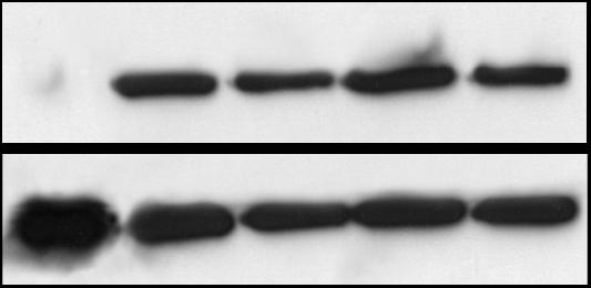
Western Blot: KMT1A/SUV39H1 Antibody [NBP1-21367]
Western Blot: KMT1A/SUV39H1 Antibody [NBP1-21367] - Up: SUV39H1 expression in C6 astrocytic cell line and human glioma cell lines GOS-3, T98G, U87MG and 1321N1. Down: Actin levels. Image submitted by a verified customer review.Applications for KMT1A/SUV39H1 Antibody
Application
Recommended Usage
Immunohistochemistry
1:10-1:500
Immunohistochemistry-Paraffin
1:10-1:1500
Immunoprecipitation
5- 10 ug/mg lysate
Western Blot
1:2000-1:10000
Application Notes
IHC-P, WB reactivitiy reported in scientific literature (PMID: 23943221), and verified customer reviews. Use in ICC/IF reported in scientific literature (PMID:28425504).
Reviewed Applications
Read 4 reviews rated 3.8 using NBP1-21367 in the following applications:
Formulation, Preparation, and Storage
Purification
Immunogen affinity purified
Formulation
TBS and 0.1% BSA
Preservative
0.09% Sodium Azide
Concentration
0.2 mg/ml
Shipping
The product is shipped with polar packs. Upon receipt, store it immediately at the temperature recommended below.
Stability & Storage
Store at 4C short term. Aliquot and store at -20C long term. Avoid freeze-thaw cycles.
Background: KMT1A/SUV39H1
Long Name
Histone-lysine N-methyltransferase SUV39H1
Alternate Names
EC 2.1.1.355, H3-K9-HMTase 1, KMT1A, SUV39H, SUV39H1
Entrez Gene IDs
6839 (Human)
Gene Symbol
SUV39H1
UniProt
Additional KMT1A/SUV39H1 Products
Product Documents for KMT1A/SUV39H1 Antibody
Certificate of Analysis
To download a Certificate of Analysis, please enter a lot or batch number in the search box below.
Product Specific Notices for KMT1A/SUV39H1 Antibody
This product is for research use only and is not approved for use in humans or in clinical diagnosis. Primary Antibodies are guaranteed for 1 year from date of receipt.
Citations for KMT1A/SUV39H1 Antibody
 Powered by Bioz
Customer Reviews for KMT1A/SUV39H1 Antibody (4)
3.8 out of 5
4 Customer Ratings
Have you used KMT1A/SUV39H1 Antibody?
Submit a review and receive an Amazon gift card!
$25/€18/£15/$25CAN/¥2500 Yen for a review with an image
$10/€7/£6/$10CAN/¥1110 Yen for a review without an image
Submit a review
Customer Images
Showing
1
-
4 of
4 reviews
Showing All
Filter By:
-
Application: Western BlotSample Tested:Species: HumanVerified Customer | Posted 01/29/2014Up: SUV39H1 expression in C6 astrocytic cell line and human glioma cell lines GOS-3, T98G, U87MG and 1321N1. Down: Actin levels

-
Application: Western BlotSample Tested:Species: HumanVerified Customer | Posted 01/29/2014Up:SUV39H1 expression in normal brain, human glioma tissue grades II, III and IV respectively. Down: Actin expression levels
-
Application: Immunohistochemistry-ParaffinSample Tested:Species: HumanVerified Customer | Posted 01/29/2014SUV39H1 expression in human glioma tissue grade IV
-
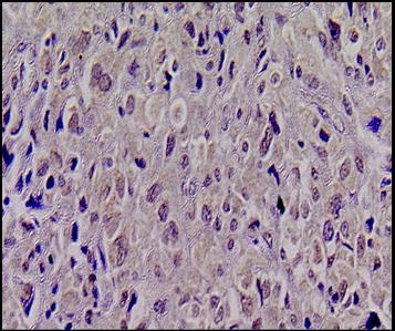
Application: Immunohistochemistry-ParaffinSample Tested: SUV39H1 expression in human glioma tissue grade IISpecies: HumanVerified Customer | Posted 01/29/2014SUV39H1 expression in human glioma tissue grade II
There are no reviews that match your criteria.
Protocols
View specific protocols for KMT1A/SUV39H1 Antibody (NBP1-21367):
Western Blot Protocol for KMT1A/SUV39H1 Antibody (NBP1-21367):
Western Blot Protocol
1. Perform SDS-PAGE on samples to be analyzed, loading 40 ug of total protein per lane.
2. Transfer proteins to membrane according to the instructions provided by the manufacturer of the membrane and transfer apparatus.
3. Stain according to standard Ponceau S procedure (or similar product) to assess transfer success, and mark molecular weight standards where appropriate.
4. Rinse the blot.
5. Block the membrane using standard blocking buffer for at least 1 hour.
6. Wash the membrane in wash buffer three times for 10 minutes each.
7. Dilute primary antibody in blocking buffer and incubate 1 hour at room temperature.
8. Wash the membrane in wash buffer three times for 10 minutes each.
9. Apply the diluted HRP conjugated secondary antibody in blocking buffer (as per manufacturers instructions) and incubate 1 hour at room temperature.
10. Wash the blot in wash buffer three times for 10 minutes each (this step can be repeated as required to reduce background).
11. Apply the detection reagent of choice in accordance with the manufacturers instructions.
Note: Tween-20 can be added to the blocking or antibody dilution buffer at a final concentration of 0.05-0.2%.
Immunohistochemistry-Paraffin Embedded Sections
Antigen Unmasking:
Bring slides to a boil in 10 mM sodium citrate buffer (pH 6.0) then maintain at a sub-boiling temperature for 10 minutes. Cool slides on bench-top for 30 minutes.
Staining:
1. Wash sections in deionized water three times for 5 minutes each.
2. Wash sections in wash buffer for 5 minutes.
3. Block each section with 100-400 ul blocking solution for 1 hour at room temperature.
4. Remove blocking solution and add 100-400 ul diluted primary antibody. Incubate overnight at 4 C.
5. Remove antibody solution and wash sections in wash buffer three times for 5 minutes each.
6. Add 100-400 ul biotinylated diluted secondary antibody. Incubate 30 minutes at room temperature.
7. Remove secondary antibody solution and wash sections three times with wash buffer for 5 minutes each.
8. Add 100-400 ul Streptavidin-HRP reagent to each section and incubate for 30 minutes at room temperature.
9. Wash sections three times in wash buffer for 5 minutes each.
10. Add 100-400 ul DAB substrate to each section and monitor staining closely.
11. As soon as the sections develop, immerse slides in deionized water.
12. Counterstain sections in hematoxylin.
13. Wash sections in deionized water two times for 5 minutes each.
14. Dehydrate sections.
15. Mount coverslips.
*The above information is only intended as a guide. The researcher should determine what protocol best meets their needs. Please follow safe laboratory procedures.
Find general support by application which include: protocols, troubleshooting, illustrated assays, videos and webinars.
- Antigen Retrieval Protocol (PIER)
- Antigen Retrieval for Frozen Sections Protocol
- Appropriate Fixation of IHC/ICC Samples
- Cellular Response to Hypoxia Protocols
- Chromogenic IHC Staining of Formalin-Fixed Paraffin-Embedded (FFPE) Tissue Protocol
- Chromogenic Immunohistochemistry Staining of Frozen Tissue
- ClariTSA™ Fluorophore Kits
- Detection & Visualization of Antibody Binding
- Fluorescent IHC Staining of Frozen Tissue Protocol
- Graphic Protocol for Heat-induced Epitope Retrieval
- Graphic Protocol for the Preparation and Fluorescent IHC Staining of Frozen Tissue Sections
- Graphic Protocol for the Preparation and Fluorescent IHC Staining of Paraffin-embedded Tissue Sections
- Graphic Protocol for the Preparation of Gelatin-coated Slides for Histological Tissue Sections
- ICC Cell Smear Protocol for Suspension Cells
- ICC Immunocytochemistry Protocol Videos
- ICC for Adherent Cells
- IHC Sample Preparation (Frozen sections vs Paraffin)
- Immunocytochemistry (ICC) Protocol
- Immunocytochemistry Troubleshooting
- Immunofluorescence of Organoids Embedded in Cultrex Basement Membrane Extract
- Immunofluorescent IHC Staining of Formalin-Fixed Paraffin-Embedded (FFPE) Tissue Protocol
- Immunohistochemistry (IHC) and Immunocytochemistry (ICC) Protocols
- Immunohistochemistry Frozen Troubleshooting
- Immunohistochemistry Paraffin Troubleshooting
- Immunoprecipitation Protocol
- Preparing Samples for IHC/ICC Experiments
- Preventing Non-Specific Staining (Non-Specific Binding)
- Primary Antibody Selection & Optimization
- Protocol for Heat-Induced Epitope Retrieval (HIER)
- Protocol for Making a 4% Formaldehyde Solution in PBS
- Protocol for VisUCyte™ HRP Polymer Detection Reagent
- Protocol for the Fluorescent ICC Staining of Cell Smears - Graphic
- Protocol for the Fluorescent ICC Staining of Cultured Cells on Coverslips - Graphic
- Protocol for the Preparation & Fixation of Cells on Coverslips
- Protocol for the Preparation and Chromogenic IHC Staining of Frozen Tissue Sections
- Protocol for the Preparation and Chromogenic IHC Staining of Frozen Tissue Sections - Graphic
- Protocol for the Preparation and Chromogenic IHC Staining of Paraffin-embedded Tissue Sections
- Protocol for the Preparation and Chromogenic IHC Staining of Paraffin-embedded Tissue Sections - Graphic
- Protocol for the Preparation and Fluorescent ICC Staining of Cells on Coverslips
- Protocol for the Preparation and Fluorescent ICC Staining of Non-adherent Cells
- Protocol for the Preparation and Fluorescent ICC Staining of Stem Cells on Coverslips
- Protocol for the Preparation and Fluorescent IHC Staining of Frozen Tissue Sections
- Protocol for the Preparation and Fluorescent IHC Staining of Paraffin-embedded Tissue Sections
- Protocol for the Preparation of Gelatin-coated Slides for Histological Tissue Sections
- Protocol for the Preparation of a Cell Smear for Non-adherent Cell ICC - Graphic
- R&D Systems Quality Control Western Blot Protocol
- TUNEL and Active Caspase-3 Detection by IHC/ICC Protocol
- The Importance of IHC/ICC Controls
- Troubleshooting Guide: Immunohistochemistry
- Troubleshooting Guide: Western Blot Figures
- Western Blot Conditions
- Western Blot Protocol
- Western Blot Protocol for Cell Lysates
- Western Blot Troubleshooting
- Western Blot Troubleshooting Guide
- View all Protocols, Troubleshooting, Illustrated assays and Webinars
Loading...