L1CAM Antibody (UJ127.11) - BSA Free
Novus Biologicals | Catalog # NB100-2682
![Western Blot: L1CAM Antibody (UJ127.11)BSA Free [NB100-2682]](https://resources.rndsystems.com/images/products/L1CAM-Antibody-UJ127-11-Western-Blot-NB100-2682-img0006.jpg "Western Blot: L1CAM Antibody (UJ127.11)BSA Free [NB100-2682]")
Key Product Details
Validated by
Biological Validation
Species Reactivity
Validated:
Human, Mouse
Cited:
Human
Applications
Validated:
Immunohistochemistry, Immunohistochemistry-Paraffin, Immunohistochemistry-Frozen, Western Blot, ELISA, Flow Cytometry, Flow (Cell Surface), Immunofluorescence, Immunocytochemistry/ Immunofluorescence, Immunoprecipitation, CyTOF-ready
Cited:
Immunohistochemistry-Paraffin, Western Blot, IF/IHC
Label
Unconjugated
Antibody Source
Monoclonal Mouse IgG1 kappa Clone # UJ127.11
Format
BSA Free
Loading...
Product Specifications
Immunogen
Homogenous suspension of 16 week human foetal brain.
Localization
Cell Membrane
Marker
Axon Marker
Clonality
Monoclonal
Host
Mouse
Isotype
IgG1 kappa
Scientific Data Images for L1CAM Antibody (UJ127.11) - BSA Free
Western Blot: L1CAM Antibody (UJ127.11)BSA Free [NB100-2682]
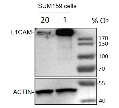
Western Blot: L1CAM Antibody (UJ127.11) [NB100-2682] - SUM159 cells were exposed to 20% or 1% O2 for 48 hours, whole cell lysates were loaded with 50 ug/lane. 10% SDS-PAGE. L1CAM Antibody (NB100-2682) primary antibody: 1:1000, 4C, overnight. Western blot image submitted by a verified customer review.![Immunocytochemistry/ Immunofluorescence: L1CAM Antibody (UJ127.11) - BSA Free [NB100-2682]](https://resources.rndsystems.com/images/products/L1CAM-Antibody-UJ127-11-Immunocytochemistry-Immunofluorescence-NB100-2682-img0005.jpg "Immunocytochemistry/ Immunofluorescence: L1CAM Antibody (UJ127.11) - BSA Free [NB100-2682]")
Immunocytochemistry/ Immunofluorescence: L1CAM Antibody (UJ127.11) - BSA Free [NB100-2682]
Immunocytochemistry/Immunofluorescence: L1CAM Antibody (UJ127.11) [NB100-2682] - The left panel (A) shows untreated Neuro2a cells and the right panel (B) shows Neuro2a cells that were serum starved then treated with 1mM cAMP overnight to induce axon growth. Cells were fixed in 4% paraformaldehyde for 10 minutes and then permeabilized for 5 minutes using 1X PBS + 0.05% Triton-X100. The cells were incubated with anti- NB100-2682 at 5 ug/ml overnight at 4C and detected with an anti-mouse Dylight 488 (Green) at a 1:1000 dilution for 60 minutes. Nuclei were counterstained with DAPI (Blue). Cells were imaged using a 100X objective and digitally deconvolved.![Immunohistochemistry-Frozen: L1CAM Antibody (UJ127.11) - BSA Free [NB100-2682]](https://resources.rndsystems.com/images/products/L1CAM-Antibody-UJ127-11-Immunohistochemistry-Frozen-NB100-2682-img0007.jpg "Immunohistochemistry-Frozen: L1CAM Antibody (UJ127.11) - BSA Free [NB100-2682]")
Immunohistochemistry-Frozen: L1CAM Antibody (UJ127.11) - BSA Free [NB100-2682]
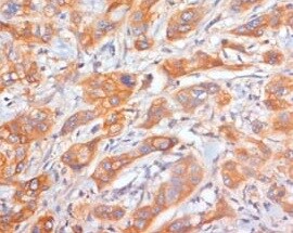
Immunohistochemistry-Frozen: L1CAM Antibody (UJ127.11) [NB100-2682] - Human pancreatic ductal adenocarcinoma (PDAC) tissue section stained with L1CAM antibody. IHC-Fr image submitted by a verified customer review.![Flow Cytometry: L1CAM Antibody (UJ127.11) - BSA Free [NB100-2682]](https://resources.rndsystems.com/images/products/L1CAM-Antibody-UJ127-11-Flow-Cytometry-NB100-2682-img0001.jpg "Flow Cytometry: L1CAM Antibody (UJ127.11) - BSA Free [NB100-2682]")
Flow Cytometry: L1CAM Antibody (UJ127.11) - BSA Free [NB100-2682]
Flow Cytometry: L1CAM Antibody (UJ127.11) [NB100-2682] - A surface stain was performed on HeLa cells with L1CAM Antibody (UJ127.11) NB100-2682 (blue) and a matched isotype control (orange). Cells were incubated in an antibody dilution of 2.5 ug/mL for 20 minutes at room temperature, followed by Mouse IgG (H+L) Cross-Adsorbed Secondary Antibody, Dylight 550 (35503, Thermo Fisher).![Western Blot: L1CAM Antibody (UJ127.11)BSA Free [NB100-2682]](https://resources.rndsystems.com/images/products/L1CAM-Antibody-UJ127-11-Western-Blot-NB100-2682-img0002.jpg "Western Blot: L1CAM Antibody (UJ127.11)BSA Free [NB100-2682]")
Western Blot: L1CAM Antibody (UJ127.11)BSA Free [NB100-2682]
Western Blot: L1CAM Antibody (UJ127.11) [NB100-2682] - Total protein from human HeLa, A431 and SHSY-5Y cell lines and human Brain was separated on a 7.5% gel by SDS-PAGE, transferred to PVDF membrane and blocked in 5% non-fat milk in TBST. The membrane was probed with 2.0 ug/ml anti-L1CAM (NB100-2682) in blocking buffer and detected with an anti-mouse HRP secondary antibody using NovaLume chemiluminescence detection reagent (NPB2-61915).![Immunocytochemistry/ Immunofluorescence: L1CAM Antibody (UJ127.11) - BSA Free [NB100-2682]](https://resources.rndsystems.com/images/products/L1CAM-Antibody-UJ127-11-Immunocytochemistry-Immunofluorescence-NB100-2682-img0004.jpg "Immunocytochemistry/ Immunofluorescence: L1CAM Antibody (UJ127.11) - BSA Free [NB100-2682]")
Immunocytochemistry/ Immunofluorescence: L1CAM Antibody (UJ127.11) - BSA Free [NB100-2682]
Immunocytochemistry/Immunofluorescence: L1CAM Antibody (UJ127.11) [NB100-2682] - The left panel (A) shows untreated Neuro2a cells and the right panel (B) shows Neuro2a cells that were serum starved then treated with 1mM cAMP overnight to induce axon growth. Cells were fixed in 4% paraformaldehyde for 10 minutes and then permeabilized for 5 minutes using 1X PBS + 0.05% Triton-X100. The cells were incubated with anti- NB100-2682 at 5 ug/ml overnight at 4C and detected with an anti-mouse Dylight 488 (Green) at a 1:1000 dilution for 60 minutes. Nuclei were counterstained with DAPI (Blue). Cells were imaged using a 40X objective.![Immunohistochemistry-Paraffin: L1CAM Antibody (UJ127.11) - BSA Free [NB100-2682]](https://resources.rndsystems.com/images/products/L1CAM-Antibody-UJ127-11-Immunohistochemistry-Paraffin-NB100-2682-img0003.jpg "Immunohistochemistry-Paraffin: L1CAM Antibody (UJ127.11) - BSA Free [NB100-2682]")
Immunohistochemistry-Paraffin: L1CAM Antibody (UJ127.11) - BSA Free [NB100-2682]
Immunohistochemistry-Paraffin: L1CAM Antibody (UJ127.11) [NB100-2682] - Analysis of a FFPE tissue section of human brain using 1:200 dilution of L1CAM [UJ127.11] antibody. The staining was developed using HRP labeled anti-mouse secondary antibody and DAB reagent, and nuclei of cells were counter-stained with hematoxylin. Cytoplasmic and membrane staining as observed in the cerebral cortex - BSA Free [NB100-2682] -")
Western Blot: L1CAM Antibody (UJ127.11) - BSA Free [NB100-2682] -
Loss of alpha -syn decreases EMT-like markers and motility in SK-MEL-28 cells. (A–D) Representative western blots of E-Cad, L1CAM, N-Cad, vimentin, alpha -syn, and alpha -tubulin in lysates of the control, KO, and KI cells cultured in vitro. (E) Quantitative analysis of relative protein levels. E-Cad, L1CAM, N-Cad, and vimentin band intensities are normalized to alpha -tubulin. All the experiments were repeated with at least three biological replicates (n = 3). (F) Representative light microscope images of phagokinetic tracks created by control, KO8, and KI8 cells on colloidal gold-coated wells. Black arrows mark individual phagokinetic tracks in respective cell lines. Images were acquired by using a ×10 objective, and the cleared phagokinetic area per cell was measured using ImageJ software. (G) Bar graphs show quantification of the cleared areas from three independent experiments. At least 33 tracks per experimental condition were randomly chosen for quantification. 100 tracks for n = 3 per experimental group were used for statistical analysis. (E,G) Values are mean +/- s.d. **, p = 0.0015–0.0027; *, p = 0.0073–0.0134 determined using a one-way ANOVA, Dunnett post hoc test. Image collected and cropped by CiteAb from the following open publication (https://pubmed.ncbi.nlm.nih.gov/37286800), licensed under a CC-BY license. Not internally tested by Novus Biologicals. - BSA Free [NB100-2682] -")
Western Blot: L1CAM Antibody (UJ127.11) - BSA Free [NB100-2682] -
Loss of alpha -syn decreases L1CAM and N-cadherin and motility in SK-MEL-29 cells. (A) Representative Western blots of L1CAM, N-Cad, alpha -syn, and alpha -tubulin in lysates of the control and alpha -syn KO cells cultured in vitro. (B) Representative Western blots of vimentin, alpha -syn, and alpha -tubulin. Quantitative data showing the relative protein levels of L1CAM (C), N-Cad (D), and vimentin (E) normalized to alpha -tubulin. All the experiments were repeated with at least with three biological replicates (n = 3). (F) Representative brightfield microscope images of phagokinetic tracks created by control and KO alpha -syn cells on colloidal gold-coated wells were acquired using a light microscope with a ×10 objective. The individual phagokinetic tracks represented by black arrow marks were measured using ImageJ software and represented in (G) At least 33 tracks per experimental condition were randomly chosen for quantification. A total of 100 tracks for n = 3 per experimental group were used for statistical analysis. Values in plots (C–E, G) are mean +/- s.d. P-values were determined by a one-way ANOVA, Dunnett post hoc test. Image collected and cropped by CiteAb from the following open publication (https://pubmed.ncbi.nlm.nih.gov/37286800), licensed under a CC-BY license. Not internally tested by Novus Biologicals. - BSA Free [NB100-2682] -")
Western Blot: L1CAM Antibody (UJ127.11) - BSA Free [NB100-2682] -
Expressing alpha -syn in SH-SY5Y cells increases L1CAM and motility. (A) Representative western blots of L1CAM, vimentin, N-Cad, alpha -syn, and alpha -tubulin in lysates of control and SH/ alpha S cells cultured in vitro. Quantitative analyses representing the relative protein levels were measured by normalizing the band intensities of L1CAM and N-Cad (B) and vimentin (C) to alpha -tubulin. All the experiments were repeated with at least with three biological replicates (n = 3–6). (D) Representative brightfield microscope images of phagokinetic tracks created by control and alpha -syn overexpressing SH-SY5Y cells on colloidal gold-coated wells. The images were acquired using a light microscope with a ×10 objective. The individual phagokinetic tracks represented by black arrow marks were measured using ImageJ software and represented in (E). At least 33 tracks per experimental condition were randomly chosen for quantification. A total of 100 tracks for n = 3 per experimental group were used for statistical analysis. Values in plots (B,C,E) are mean +/- s.d. P-values were determined by a two-sided Student’s t test. Image collected and cropped by CiteAb from the following open publication (https://pubmed.ncbi.nlm.nih.gov/37286800), licensed under a CC-BY license. Not internally tested by Novus Biologicals.Applications for L1CAM Antibody (UJ127.11) - BSA Free
Application
Recommended Usage
ELISA
1:100 - 1:2000
Flow (Cell Surface)
2.5 ug/mL
Flow Cytometry
2.5 ug/mL
Immunocytochemistry/ Immunofluorescence
2 - 5 ug/mL
Immunofluorescence
1 - 2 ug/mL
Immunohistochemistry
1:200
Immunohistochemistry-Paraffin
1:200
Immunoprecipitation
1:10 - 1:500
Western Blot
2 ug/mL
Application Notes
Positive control(s): Frozen mouse E16.5 lung sections (IHC), Cell lines TR 14, LAN-1, CHP 100, CHP212, CHP126 ( Neuroblastomas or Schwannomas) (IF), M5 melanoma cells or human foetal brain (WB). This antibody is CyTOF ready.
Reviewed Applications
Read 2 reviews rated 5 using NB100-2682 in the following applications:
Flow Cytometry Panel Builder
Bio-Techne Knows Flow Cytometry
Save time and reduce costly mistakes by quickly finding compatible reagents using the Panel Builder Tool.
Advanced Features
- Spectra Viewer - Custom analysis of spectra from multiple fluorochromes
- Spillover Popups - Visualize the spectra of individual fluorochromes
- Antigen Density Selector - Match fluorochrome brightness with antigen density

Formulation, Preparation, and Storage
Purification
Protein A or G purified
Formulation
PBS
Format
BSA Free
Preservative
0.02% Sodium Azide
Concentration
1 mg/ml
Shipping
The product is shipped with polar packs. Upon receipt, store it immediately at the temperature recommended below.
Stability & Storage
Store at 4C short term. Aliquot and store at -20C long term. Avoid freeze-thaw cycles.
Background: L1CAM
Long Name
Cell Adhesion Molecule L1
Alternate Names
CAML1, CD171, HSAS, HSAS1, MASA, MIC5, NCAM-L1, S10, SPG1
Gene Symbol
L1CAM
Additional L1CAM Products
Product Documents for L1CAM Antibody (UJ127.11) - BSA Free
Certificate of Analysis
To download a Certificate of Analysis, please enter a lot or batch number in the search box below.
Product Specific Notices for L1CAM Antibody (UJ127.11) - BSA Free
This product is for research use only and is not approved for use in humans or in clinical diagnosis. Primary Antibodies are guaranteed for 1 year from date of receipt.
Citations for L1CAM Antibody (UJ127.11) - BSA Free
 Powered by Bioz
Customer Reviews for L1CAM Antibody (UJ127.11) - BSA Free (2)
5 out of 5
2 Customer Ratings
Have you used L1CAM Antibody (UJ127.11) - BSA Free?
Submit a review and receive an Amazon gift card!
$25/€18/£15/$25CAN/¥2500 Yen for a review with an image
$10/€7/£6/$10CAN/¥1110 Yen for a review without an image
Submit a review
Customer Images
Showing
1
-
2 of
2 reviews
Showing All
Filter By:
-
Application: Immunohistochemistry-FrozenSample Tested: pancreatic ductal adenocarcinoma (PDAC)Species: HumanVerified Customer | Posted 08/13/2021Pancreatic ductal adenocarcinoma (PDAC), L1CAM staining

-
Application: Western BlotSample Tested: SUM-159PTSpecies: HumanVerified Customer | Posted 11/04/2020Western Blot: SUM159 cells were exposed to 20% or 1% O2 for 48 hours, whole cell lysates were loaded with 50 ug/lane. 10% SDS-PAGE. L1CAM Antibody (NB100-2682) primary antibody: 1:1000, 4℃, overnight.
There are no reviews that match your criteria.
Protocols
View specific protocols for L1CAM Antibody (UJ127.11) - BSA Free (NB100-2682):
Protocol for Flow Cytometry Cell Surface Staining
Sample Preparation.
1. Grow cells to 60-85% confluency. Flow cytometry requires between 2 x 10⁵ and 1 x 10⁶ cells for optimal performance.
2. If cells are adherent, harvest gently by washing once with staining buffer and then scraping. Avoid using trypsin as this can disrupt certain epitopes of interest. If enzymatic harvest is required, use Accutase, Collagenase, or TrypLE Express for a less damaging option.
3. Reserve 100 uL for counting, then transfer cell volume into a 15 mL conical tube and centrifuge for 4 minutes at 400 RCF.
a. Count cells using a hemocytometer and a 1:1 trypan blue exclusion stain to determine cell viability before starting the flow protocol. If cells appear blue, do not proceed.
4. Re-suspend cells to a concentration of 1 x 10⁶ cells/mL in staining buffer (NBP2-26247).
5. Aliquot out 100 uL samples in accordance with your experimental samples.
Tip: When cell surface and intracellular staining are required in the same sample, it is advisable that the cell surface staining be performed first since the fixation and permeablization steps might reduce the availability of surface antigens.
Cell surface staining
1. Recommended: Block non-specific interactions using 0.5-1 ug of a species specific Fc-blocking reagent such as an anti-mouse CD16/CD32 antibody (NBP1-27946).
2. Add appropriate amount of each antibody (eg. 1 test or 1 ug per sample, as experimentally determined) to 100 uL of staining buffer (NBP2-26247) per sample (eg. use 1 mL of staining buffer for 10 samples).
3. Mix well and incubate at room temperature in dark for 20 minutes.
4. Add 1-2 mL of staining buffer and centrifuge at 400 RCF for 1 minute and discard supernatant.
5. Wash twice by re-suspending cells in staining buffer (2 mL for tubes or 200 uL for wells) and centrifuging at 400 RCF for 5 minutes. Discard supernatant.
6. Add appropriate amount of secondary antibody (as experimentally determined) to each sample.
7. Incubate at room temperature in dark for 20 minutes.
8. Add 1-2 mL of staining buffer and centrifuge at 400 RCF for 1 minute and discard supernatant.
9. Wash twice by re-suspending cells in staining buffer (2 mL for tubes or 200 uL for wells) and centrifuging at 400 RCF for 5 minutes. Discard supernatant.
10. Resuspend in an appropriate volume of staining buffer (usually 500 uL per sample) and proceed with analysis on your flow cytometer.
Immunohistochemistry-Paraffin Embedded Sections
Antigen Unmasking:
Bring slides to a boil in 10 mM sodium citrate buffer (pH 6.0) then maintain at a sub-boiling temperature for 10 minutes. Cool slides on bench-top for 30 minutes (keep slides in the sodium citrate buffer all the time).
Staining:
1. Wash sections in deionized water three times for 5 minutes each.
2. Wash sections in PBS for 5 minutes.
3. Block each section with 100-400 ul blocking solution (1% BSA in PBS) for 1 hour at room temperature.
4. Remove blocking solution and add 100-400 ul diluted primary antibody. Incubate overnight at 4 C.
5. Remove antibody solution and wash sections in wash buffer three times for 5 minutes each.
6. Add 100-400 ul HRP polymer conjugated secondary antibody. Incubate 30 minutes at room temperature.
7. Wash sections three times in wash buffer for 5 minutes each.
8. Add 100-400 ul DAB substrate to each section and monitor staining closely.
9. As soon as the sections develop, immerse slides in deionized water.
10. Counterstain sections in hematoxylin.
11. Wash sections in deionized water two times for 5 minutes each.
12. Dehydrate sections.
13. Mount coverslips.
Western Blot Protocol
1. Perform SDS-PAGE on samples to be analyzed, loading 10-25 ug of total protein per lane.
2. Transfer proteins to PVDF membrane according to the instructions provided by the manufacturer of the membrane and transfer apparatus.
3. Stain the membrane with Ponceau S (or similar product) to assess transfer success, and mark molecular weight standards where appropriate.
4. Rinse the blot TBS -0.05% Tween 20 (TBST).
5. Block the membrane in 5% Non-fat milk in TBST (blocking buffer) for at least 1 hour.
6. Wash the membrane in TBST three times for 10 minutes each.
7. Dilute primary antibody in blocking buffer and incubate overnight at 4C with gentle rocking.
8. Wash the membrane in TBST three times for 10 minutes each.
9. Incubate the membrane in diluted HRP conjugated secondary antibody in blocking buffer (as per manufacturer's instructions) for 1 hour at room temperature.
10. Wash the blot in TBST three times for 10 minutes each (this step can be repeated as required to reduce background).
11. Apply the detection reagent of choice in accordance with the manufacturers instructions
Find general support by application which include: protocols, troubleshooting, illustrated assays, videos and webinars.
- 7-Amino Actinomycin D (7-AAD) Cell Viability Flow Cytometry Protocol
- Antigen Retrieval Protocol (PIER)
- Antigen Retrieval for Frozen Sections Protocol
- Appropriate Fixation of IHC/ICC Samples
- Cellular Response to Hypoxia Protocols
- Chromogenic IHC Staining of Formalin-Fixed Paraffin-Embedded (FFPE) Tissue Protocol
- Chromogenic Immunohistochemistry Staining of Frozen Tissue
- ClariTSA™ Fluorophore Kits
- Detection & Visualization of Antibody Binding
- ELISA Sample Preparation & Collection Guide
- ELISA Troubleshooting Guide
- Extracellular Membrane Flow Cytometry Protocol
- Flow Cytometry Protocol for Cell Surface Markers
- Flow Cytometry Protocol for Staining Membrane Associated Proteins
- Flow Cytometry Staining Protocols
- Flow Cytometry Troubleshooting Guide
- Fluorescent IHC Staining of Frozen Tissue Protocol
- Graphic Protocol for Heat-induced Epitope Retrieval
- Graphic Protocol for the Preparation and Fluorescent IHC Staining of Frozen Tissue Sections
- Graphic Protocol for the Preparation and Fluorescent IHC Staining of Paraffin-embedded Tissue Sections
- Graphic Protocol for the Preparation of Gelatin-coated Slides for Histological Tissue Sections
- How to Run an R&D Systems DuoSet ELISA
- How to Run an R&D Systems Quantikine ELISA
- How to Run an R&D Systems Quantikine™ QuicKit™ ELISA
- ICC Cell Smear Protocol for Suspension Cells
- ICC Immunocytochemistry Protocol Videos
- ICC for Adherent Cells
- IHC Sample Preparation (Frozen sections vs Paraffin)
- Immunocytochemistry (ICC) Protocol
- Immunocytochemistry Troubleshooting
- Immunofluorescence of Organoids Embedded in Cultrex Basement Membrane Extract
- Immunofluorescent IHC Staining of Formalin-Fixed Paraffin-Embedded (FFPE) Tissue Protocol
- Immunohistochemistry (IHC) and Immunocytochemistry (ICC) Protocols
- Immunohistochemistry Frozen Troubleshooting
- Immunohistochemistry Paraffin Troubleshooting
- Immunoprecipitation Protocol
- Intracellular Flow Cytometry Protocol Using Alcohol (Methanol)
- Intracellular Flow Cytometry Protocol Using Detergents
- Intracellular Nuclear Staining Flow Cytometry Protocol Using Detergents
- Intracellular Staining Flow Cytometry Protocol Using Alcohol Permeabilization
- Intracellular Staining Flow Cytometry Protocol Using Detergents to Permeabilize Cells
- Preparing Samples for IHC/ICC Experiments
- Preventing Non-Specific Staining (Non-Specific Binding)
- Primary Antibody Selection & Optimization
- Propidium Iodide Cell Viability Flow Cytometry Protocol
- Protocol for Heat-Induced Epitope Retrieval (HIER)
- Protocol for Liperfluo
- Protocol for Making a 4% Formaldehyde Solution in PBS
- Protocol for VisUCyte™ HRP Polymer Detection Reagent
- Protocol for the Characterization of Human Th22 Cells
- Protocol for the Characterization of Human Th9 Cells
- Protocol for the Fluorescent ICC Staining of Cell Smears - Graphic
- Protocol for the Fluorescent ICC Staining of Cultured Cells on Coverslips - Graphic
- Protocol for the Preparation & Fixation of Cells on Coverslips
- Protocol for the Preparation and Chromogenic IHC Staining of Frozen Tissue Sections
- Protocol for the Preparation and Chromogenic IHC Staining of Frozen Tissue Sections - Graphic
- Protocol for the Preparation and Chromogenic IHC Staining of Paraffin-embedded Tissue Sections
- Protocol for the Preparation and Chromogenic IHC Staining of Paraffin-embedded Tissue Sections - Graphic
- Protocol for the Preparation and Fluorescent ICC Staining of Cells on Coverslips
- Protocol for the Preparation and Fluorescent ICC Staining of Non-adherent Cells
- Protocol for the Preparation and Fluorescent ICC Staining of Stem Cells on Coverslips
- Protocol for the Preparation and Fluorescent IHC Staining of Frozen Tissue Sections
- Protocol for the Preparation and Fluorescent IHC Staining of Paraffin-embedded Tissue Sections
- Protocol for the Preparation of Gelatin-coated Slides for Histological Tissue Sections
- Protocol for the Preparation of a Cell Smear for Non-adherent Cell ICC - Graphic
- Protocol: Annexin V and PI Staining by Flow Cytometry
- Protocol: Annexin V and PI Staining for Apoptosis by Flow Cytometry
- Quantikine HS ELISA Kit Assay Principle, Alkaline Phosphatase
- Quantikine HS ELISA Kit Principle, Streptavidin-HRP Polymer
- R&D Systems Quality Control Western Blot Protocol
- Sandwich ELISA (Colorimetric) – Biotin/Streptavidin Detection Protocol
- Sandwich ELISA (Colorimetric) – Direct Detection Protocol
- TUNEL and Active Caspase-3 Detection by IHC/ICC Protocol
- The Importance of IHC/ICC Controls
- Troubleshooting Guide: ELISA
- Troubleshooting Guide: Fluorokine Flow Cytometry Kits
- Troubleshooting Guide: Immunohistochemistry
- Troubleshooting Guide: Western Blot Figures
- Western Blot Conditions
- Western Blot Protocol
- Western Blot Protocol for Cell Lysates
- Western Blot Troubleshooting
- Western Blot Troubleshooting Guide
- View all Protocols, Troubleshooting, Illustrated assays and Webinars
Loading...