![Western Blot: Lamin A + C Antibody [NBP1-85550]](https://resources.rndsystems.com/images/products/Lamin-A-Antibody-Western-Blot-NBP1-85550-img0010.jpg "Western Blot: Lamin A + C Antibody [NBP1-85550]")
Loading...
Key Product Details
Validated by
Orthogonal Validation
Species Reactivity
Validated:
Human
Predicted:
Mouse (99%), Rat (99%). Backed by our 100% Guarantee.
Applications
Immunohistochemistry, Immunohistochemistry-Paraffin, Western Blot
Label
Unconjugated
Antibody Source
Polyclonal Rabbit IgG
Format
BSA Free
Loading...
Product Specifications
Immunogen
This antibody was developed against Recombinant Protein corresponding to amino acids: EVVSREVSGIKAAYEAELGDARKTLDSVAKERARLQLELSKVREEFKELKARNTKKEGDLIAAQARLKDLEALLNSKEAALSTALSEKRTLEGELHDLRGQVAKLEAALGEAKKQLQDEMLRRVDAENRLQTMKEELDFQKNIYSEELRE
Reactivity Notes
Zebrafish reactivity reported from a verified customer review.
Marker
Nuclear Envelope Marker
Clonality
Polyclonal
Host
Rabbit
Isotype
IgG
Theoretical MW
74 kDa.
Disclaimer note: The observed molecular weight of the protein may vary from the listed predicted molecular weight due to post translational modifications, post translation cleavages, relative charges, and other experimental factors.
Disclaimer note: The observed molecular weight of the protein may vary from the listed predicted molecular weight due to post translational modifications, post translation cleavages, relative charges, and other experimental factors.
Scientific Data Images for Lamin A + C Antibody - BSA Free
![Immunohistochemistry-Paraffin: Lamin A + C Antibody [NBP1-85550]](https://resources.rndsystems.com/images/products/Lamin-A-Antibody-Immunohistochemistry-Paraffin-NBP1-85550-img0013.jpg "Immunohistochemistry-Paraffin: Lamin A + C Antibody [NBP1-85550]")
Immunohistochemistry-Paraffin: Lamin A + C Antibody [NBP1-85550]
Immunohistochemistry-Paraffin: Lamin A Antibody [NBP1-85550] - Staining of human fallopian tube shows moderate positivity in nuclear membrane in glandular cells.![Immunohistochemistry-Paraffin: Lamin A + C Antibody [NBP1-85550]](https://resources.rndsystems.com/images/products/Lamin-A-Antibody-Immunohistochemistry-Paraffin-NBP1-85550-img0011.jpg "Immunohistochemistry-Paraffin: Lamin A + C Antibody [NBP1-85550]")
Immunohistochemistry-Paraffin: Lamin A + C Antibody [NBP1-85550]
Immunohistochemistry-Paraffin: Lamin A Antibody [NBP1-85550] - Staining of human prostate shows strong positivity in nuclear membrane in smooth muscle cells and glandular cells.![Immunohistochemistry-Paraffin: Lamin A + C Antibody [NBP1-85550]](https://resources.rndsystems.com/images/products/Lamin-A-Antibody-Immunohistochemistry-Paraffin-NBP1-85550-img0012.jpg "Immunohistochemistry-Paraffin: Lamin A + C Antibody [NBP1-85550]")
Immunohistochemistry-Paraffin: Lamin A + C Antibody [NBP1-85550]
Immunohistochemistry-Paraffin: Lamin A Antibody [NBP1-85550] - Staining of human stomach shows strong positivity in nuclear membrane in glandular cells.![Immunohistochemistry-Paraffin: Lamin A + C Antibody [NBP1-85550]](https://resources.rndsystems.com/images/products/Lamin-A-Antibody-Immunohistochemistry-Paraffin-NBP1-85550-img0014.jpg "Immunohistochemistry-Paraffin: Lamin A + C Antibody [NBP1-85550]")
Immunohistochemistry-Paraffin: Lamin A + C Antibody [NBP1-85550]
Immunohistochemistry-Paraffin: Lamin A Antibody [NBP1-85550] - Staining of human skin shows strong positivity in nuclear membrane in squamous epithelial cells.
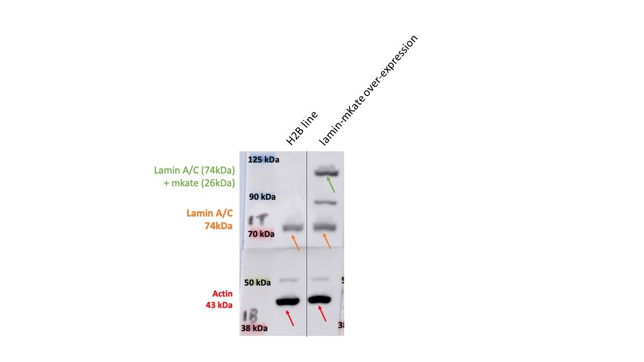
Western Blot: Rabbit Polyclonal Lamin A + C Antibody [NBP1-85550] -
Western Blot: Rabbit Polyclonal Lamin A + C Antibody [NBP1-85550] - Western blot showing over-expression of lamin A/C tagged with mKate, as well as endogenous lamin A/C, in zebrafish embryos. Actin used as loading control. Image from a verified customer review.Applications for Lamin A + C Antibody - BSA Free
Application
Recommended Usage
Immunohistochemistry
1:50 - 1:200
Immunohistochemistry-Paraffin
1:50 - 1:200
Western Blot
0.04-0.4 ug/ml
Application Notes
IHC-Paraffin, HIER pH 6 retrieval is recommended.
Reviewed Applications
Read 2 reviews rated 4.5 using NBP1-85550 in the following applications:
Formulation, Preparation, and Storage
Purification
Affinity purified
Formulation
PBS (pH 7.2) and 40% Glycerol
Format
BSA Free
Preservative
0.02% Sodium Azide
Concentration
Concentrations vary lot to lot. See vial label for concentration. If unlisted please contact technical services.
Shipping
The product is shipped with polar packs. Upon receipt, store it immediately at the temperature recommended below.
Stability & Storage
Store at 4C short term. Aliquot and store at -20C long term. Avoid freeze-thaw cycles.
Background: Lamin A + C
Alternate Names
CDCD1, CDDC, CMD1A, CMT2B1, dilated 1A (autosomal dominant), EMD2, FPLD, HGPSFPL, lamin A/C, lamin A/C-like 1, lamin-A/C, LDP1, LFP, LGMD1B, limb girdle muscular dystrophy 1B (autosomal dominant), LMN1IDC, LMNC, LMNL1, prelamin-A/C, PRO1,70 kDa lamin, progeria 1 (Hutchinson-Gilford type), renal carcinoma antigen NY-REN-32
Gene Symbol
LMNA
UniProt
Additional Lamin A + C Products
Product Documents for Lamin A + C Antibody - BSA Free
Certificate of Analysis
To download a Certificate of Analysis, please enter a lot or batch number in the search box below.
Product Specific Notices for Lamin A + C Antibody - BSA Free
This product is for research use only and is not approved for use in humans or in clinical diagnosis. Primary Antibodies are guaranteed for 1 year from date of receipt.
Customer Reviews for Lamin A + C Antibody - BSA Free (2)
4.5 out of 5
2 Customer Ratings
Have you used Lamin A + C Antibody - BSA Free?
Submit a review and receive an Amazon gift card!
$25/€18/£15/$25CAN/¥2500 Yen for a review with an image
$10/€7/£6/$10CAN/¥1110 Yen for a review without an image
Submit a review
Customer Images
Showing
1
-
2 of
2 reviews
Showing All
Filter By:
-
Application: Western BlotSample Tested: zebrafish embryosSpecies: ZebrafishVerified Customer | Posted 11/06/2023Western blot showing over-expression of lamin A/C tagged with mKate, as well as endogenous lamin A/C, in zebrafish embryos. Actin used as loading control.
 Bio-Techne ResponseThis review was submitted through the legacy Novus Innovators Program, reflecting a new species or application tested on a primary antibody.
 -
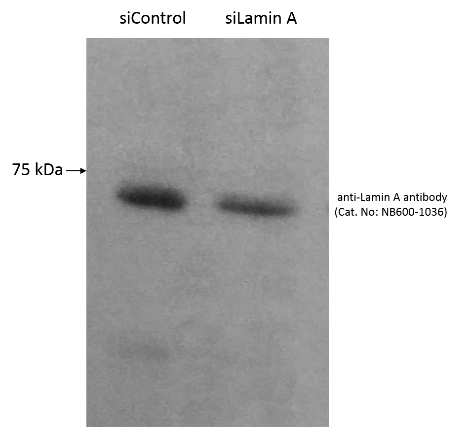
Application: Western BlotSample Tested:Species: HumanVerified Customer | Posted 05/21/2015Lamin A From HeLa Cells Using Lamin A antibody NB600-1036
There are no reviews that match your criteria.
Protocols
Find general support by application which include: protocols, troubleshooting, illustrated assays, videos and webinars.
- Antigen Retrieval Protocol (PIER)
- Antigen Retrieval for Frozen Sections Protocol
- Appropriate Fixation of IHC/ICC Samples
- Cellular Response to Hypoxia Protocols
- Chromogenic IHC Staining of Formalin-Fixed Paraffin-Embedded (FFPE) Tissue Protocol
- Chromogenic Immunohistochemistry Staining of Frozen Tissue
- ClariTSA™ Fluorophore Kits
- Detection & Visualization of Antibody Binding
- Fluorescent IHC Staining of Frozen Tissue Protocol
- Graphic Protocol for Heat-induced Epitope Retrieval
- Graphic Protocol for the Preparation and Fluorescent IHC Staining of Frozen Tissue Sections
- Graphic Protocol for the Preparation and Fluorescent IHC Staining of Paraffin-embedded Tissue Sections
- Graphic Protocol for the Preparation of Gelatin-coated Slides for Histological Tissue Sections
- IHC Sample Preparation (Frozen sections vs Paraffin)
- Immunofluorescent IHC Staining of Formalin-Fixed Paraffin-Embedded (FFPE) Tissue Protocol
- Immunohistochemistry (IHC) and Immunocytochemistry (ICC) Protocols
- Immunohistochemistry Frozen Troubleshooting
- Immunohistochemistry Paraffin Troubleshooting
- Preparing Samples for IHC/ICC Experiments
- Preventing Non-Specific Staining (Non-Specific Binding)
- Primary Antibody Selection & Optimization
- Protocol for Heat-Induced Epitope Retrieval (HIER)
- Protocol for Making a 4% Formaldehyde Solution in PBS
- Protocol for VisUCyte™ HRP Polymer Detection Reagent
- Protocol for the Preparation & Fixation of Cells on Coverslips
- Protocol for the Preparation and Chromogenic IHC Staining of Frozen Tissue Sections
- Protocol for the Preparation and Chromogenic IHC Staining of Frozen Tissue Sections - Graphic
- Protocol for the Preparation and Chromogenic IHC Staining of Paraffin-embedded Tissue Sections
- Protocol for the Preparation and Chromogenic IHC Staining of Paraffin-embedded Tissue Sections - Graphic
- Protocol for the Preparation and Fluorescent IHC Staining of Frozen Tissue Sections
- Protocol for the Preparation and Fluorescent IHC Staining of Paraffin-embedded Tissue Sections
- Protocol for the Preparation of Gelatin-coated Slides for Histological Tissue Sections
- R&D Systems Quality Control Western Blot Protocol
- TUNEL and Active Caspase-3 Detection by IHC/ICC Protocol
- The Importance of IHC/ICC Controls
- Troubleshooting Guide: Immunohistochemistry
- Troubleshooting Guide: Western Blot Figures
- Western Blot Conditions
- Western Blot Protocol
- Western Blot Protocol for Cell Lysates
- Western Blot Troubleshooting
- Western Blot Troubleshooting Guide
- View all Protocols, Troubleshooting, Illustrated assays and Webinars
Loading...