LAMP-1/CD107a Antibody - BSA Free
Novus Biologicals | Catalog # NB600-956
![Immunocytochemistry/ Immunofluorescence: LAMP-1/CD107a Antibody - BSA Free [NB600-956]](https://resources.rndsystems.com/images/products/LAMP-1-CD107a-Antibody---BSA-Free-Immunocytochemistry-Immunofluorescence-NB600-956-img0006.jpg "Immunocytochemistry/ Immunofluorescence: LAMP-1/CD107a Antibody - BSA Free [NB600-956]")
Key Product Details
Species Reactivity
Validated:
Human, Mouse, Rat
Cited:
Mouse
Applications
Validated:
Western Blot, Immunocytochemistry/ Immunofluorescence
Cited:
Immunocytochemistry/ Immunofluorescence
Label
Unconjugated
Antibody Source
Polyclonal Rabbit IgG
Format
BSA Free
Loading...
Product Specifications
Immunogen
This antibody was generated with a synthetic peptide corresponding to amino acid residues 405-416 of human LAMP1 with N-terminal added cysteine-glycine, conjugated to KLH.
Reactivity Notes
The antigen sequence is identical in rat and mouse. Rabbit reactivity reported by customer review.
Marker
Late Endosome Marker
Clonality
Polyclonal
Host
Rabbit
Isotype
IgG
Theoretical MW
120 kDa.
Disclaimer note: The observed molecular weight of the protein may vary from the listed predicted molecular weight due to post translational modifications, post translation cleavages, relative charges, and other experimental factors.
Disclaimer note: The observed molecular weight of the protein may vary from the listed predicted molecular weight due to post translational modifications, post translation cleavages, relative charges, and other experimental factors.
Scientific Data Images for LAMP-1/CD107a Antibody - BSA Free
Immunocytochemistry/ Immunofluorescence: LAMP-1/CD107a Antibody - BSA Free [NB600-956]
Immunocytochemistry/Immunofluorescence: LAMP-1/CD107a Antibody - BSA Free [NB600-956] - Human HeLa cells were fixed and permeabilized with 4% paraformaldehyde followed by 0.4% Saponin. Fixed cells were stained with 5 ug/mL Anti-LAMP1 and developed with Goat Anti-Rabbit IgG, Cy3 conjugate.![Immunocytochemistry/ Immunofluorescence: LAMP-1/CD107a Antibody - BSA Free [NB600-956]](https://resources.rndsystems.com/images/products/LAMP-1-CD107a-Antibody---BSA-Free-Immunocytochemistry-Immunofluorescence-NB600-956-img0005.jpg "Immunocytochemistry/ Immunofluorescence: LAMP-1/CD107a Antibody - BSA Free [NB600-956]")
Immunocytochemistry/ Immunofluorescence: LAMP-1/CD107a Antibody - BSA Free [NB600-956]
Immunocytochemistry/Immunofluorescence: LAMP-1/CD107a Antibody - BSA Free [NB600-956] - Rat NRK cells were fixed and permeabilized with 4% paraformaldehyde followed by 0.4% Saponin. Fixed cells were stained with 10 ug/mL Anti-LAMP1 and developed with Goat Anti-Rabbit IgG, FITC conjugate.Applications for LAMP-1/CD107a Antibody - BSA Free
Application
Recommended Usage
Immunocytochemistry/ Immunofluorescence
5-10 ug/ml
Application Notes
Useful for indirect IF using human HeLa, rat NRK, and mouse NIH3T3 cells.
Reviewed Applications
Read 1 review rated 3 using NB600-956 in the following applications:
Formulation, Preparation, and Storage
Purification
Immunogen affinity purified
Formulation
10mM PBS (pH 7.4)
Format
BSA Free
Preservative
0.09% Sodium Azide
Concentration
1.0 mg/ml
Shipping
The product is shipped with polar packs. Upon receipt, store it immediately at the temperature recommended below.
Stability & Storage
Store at 4C short term. Aliquot and store at -20C long term. Avoid freeze-thaw cycles.
Background: LAMP-1/CD107a
LAMP-1 plays an important role in autophagy-mediated ATP-release during apoptosis where lysosomes containing intracellular ATP migrate to the plasma membrane and, during exocytosis, LAMP-1 is exposed to the cell surface (5). Studies have found that knockdown of LAMP-1 blocks the ATP release from the cell (5). Furthermore, an absence of LAMP-1 and LAMP-2 leads to an accumulation of lysosomal cholesterol (6). Lysosomal membrane dysfunction or defects has also been associated with disease development (6,7). For example, one feature of pancreatitis is autophagy impairment which is caused by lysosomal dysfunction and a corresponding decrease in lysosomal-membrane associated proteins LAMP-1 and LAMP-2 (7).
References
1. Eskelinen E. L. (2006). Roles of LAMP-1 and LAMP-2 in lysosome biogenesis and autophagy. Molecular aspects of medicine, 27(5-6), 495-502. https://doi.org/10.1016/j.mam.2006.08.005
2. Cheng, X. T., Xie, Y. X., Zhou, B., Huang, N., Farfel-Becker, T., & Sheng, Z. H. (2018). Revisiting LAMP1 as a marker for degradative autophagy-lysosomal organelles in the nervous system. Autophagy, 14(8), 1472-1474. https://doi.org/10.1080/15548627.2018.1482147
3. Krzewski, K., & Coligan, J. E. (2012). Human NK cell lytic granules and regulation of their exocytosis. Frontiers in immunology, 3, 335. https://doi.org/10.3389/fimmu.2012.00335
4. Uniprot (P11279)
5. Wang, Y., Martins, I., Ma, Y., Kepp, O., Galluzzi, L., & Kroemer, G. (2013). Autophagy-dependent ATP release from dying cells via lysosomal exocytosis. Autophagy, 9(10), 1624-1625. https://doi.org/10.4161/auto.25873
6. Schwake, M., Schr0der, B., & Saftig, P. (2013). Lysosomal membrane proteins and their central role in physiology. Traffic (Copenhagen, Denmark), 14(7), 739-748. https://doi.org/10.1111/tra.12056
7. Gukovsky, I., Pandol, S. J., Mareninova, O. A., Shalbueva, N., Jia, W., & Gukovskaya, A. S. (2012). Impaired autophagy and organellar dysfunction in pancreatitis. Journal of gastroenterology and hepatology, 27 Suppl 2(Suppl 2), 27-32. https://doi.org/10.1111/j.1440-1746.2011.07004.x
Long Name
Lysosome-associated Membrane Glycoprotein 1
Alternate Names
CD107a, LAMP1
Gene Symbol
LAMP1
UniProt
Additional LAMP-1/CD107a Products
Product Documents for LAMP-1/CD107a Antibody - BSA Free
Certificate of Analysis
To download a Certificate of Analysis, please enter a lot or batch number in the search box below.
Product Specific Notices for LAMP-1/CD107a Antibody - BSA Free
This product is for research use only and is not approved for use in humans or in clinical diagnosis. Primary Antibodies are guaranteed for 1 year from date of receipt.
Citations for LAMP-1/CD107a Antibody - BSA Free
 Powered by Bioz
Customer Reviews for LAMP-1/CD107a Antibody - BSA Free (1)
3 out of 5
1 Customer Rating
Have you used LAMP-1/CD107a Antibody - BSA Free?
Submit a review and receive an Amazon gift card!
$25/€18/£15/$25CAN/¥2500 Yen for a review with an image
$10/€7/£6/$10CAN/¥1110 Yen for a review without an image
Submit a review
Customer Images
Showing
1
-
1 of
1 review
Showing All
Filter By:
-
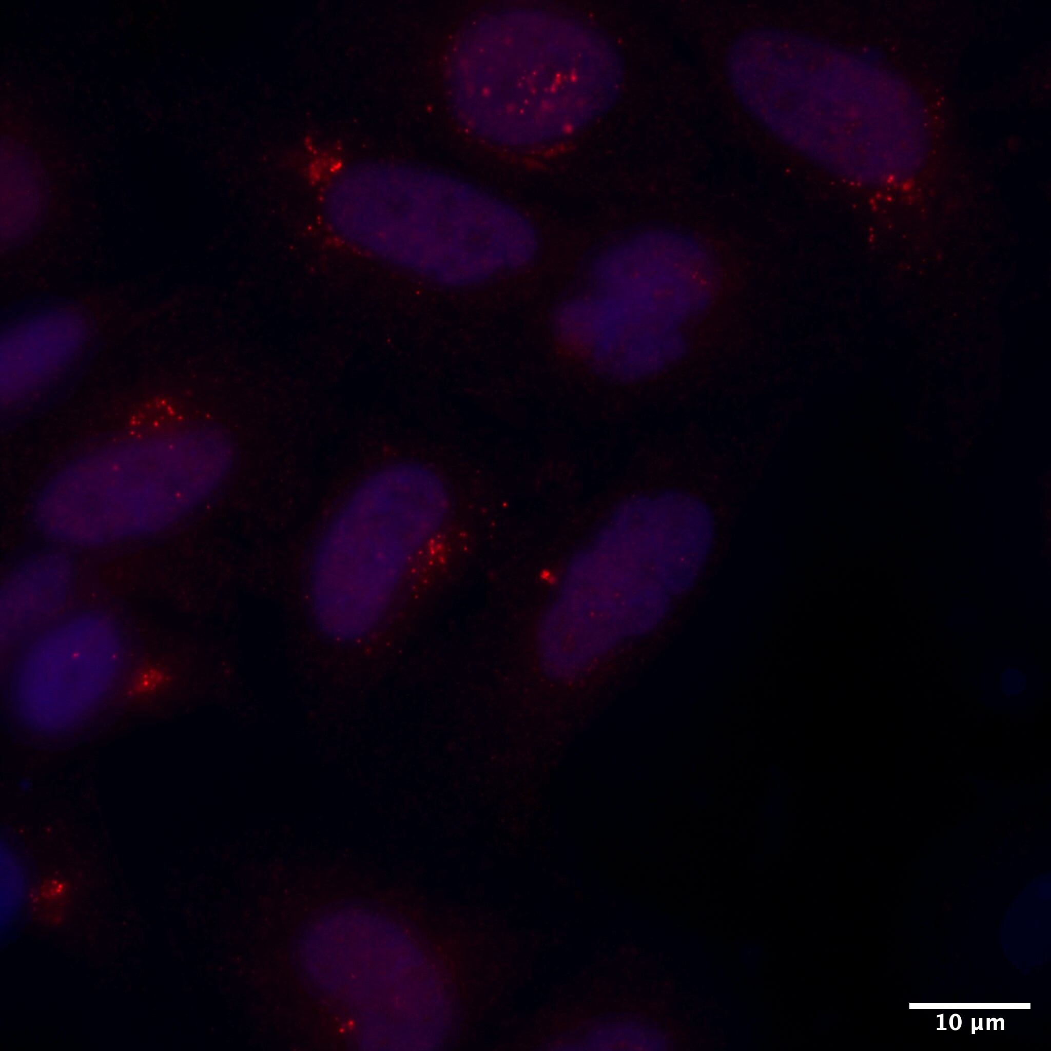
Application: ImmunocytochemistrySample Tested: hela cell and Hela cell lineSpecies: RabbitVerified Customer | Posted 01/26/2019HeLa cells were labeled with DAPI as nuclear marker (blue) and immunostained with anti-LAMP1 followed by Alexa-564 secondary antibody (Red).

There are no reviews that match your criteria.
Protocols
Find general support by application which include: protocols, troubleshooting, illustrated assays, videos and webinars.
- Appropriate Fixation of IHC/ICC Samples
- Cellular Response to Hypoxia Protocols
- ClariTSA™ Fluorophore Kits
- Detection & Visualization of Antibody Binding
- ICC Cell Smear Protocol for Suspension Cells
- ICC Immunocytochemistry Protocol Videos
- ICC for Adherent Cells
- Immunocytochemistry (ICC) Protocol
- Immunocytochemistry Troubleshooting
- Immunofluorescence of Organoids Embedded in Cultrex Basement Membrane Extract
- Immunohistochemistry (IHC) and Immunocytochemistry (ICC) Protocols
- Preparing Samples for IHC/ICC Experiments
- Preventing Non-Specific Staining (Non-Specific Binding)
- Primary Antibody Selection & Optimization
- Protocol for VisUCyte™ HRP Polymer Detection Reagent
- Protocol for the Fluorescent ICC Staining of Cell Smears - Graphic
- Protocol for the Fluorescent ICC Staining of Cultured Cells on Coverslips - Graphic
- Protocol for the Preparation and Fluorescent ICC Staining of Cells on Coverslips
- Protocol for the Preparation and Fluorescent ICC Staining of Non-adherent Cells
- Protocol for the Preparation and Fluorescent ICC Staining of Stem Cells on Coverslips
- Protocol for the Preparation of a Cell Smear for Non-adherent Cell ICC - Graphic
- R&D Systems Quality Control Western Blot Protocol
- TUNEL and Active Caspase-3 Detection by IHC/ICC Protocol
- The Importance of IHC/ICC Controls
- Troubleshooting Guide: Western Blot Figures
- Western Blot Conditions
- Western Blot Protocol
- Western Blot Protocol for Cell Lysates
- Western Blot Troubleshooting
- Western Blot Troubleshooting Guide
- View all Protocols, Troubleshooting, Illustrated assays and Webinars
Loading...
Associated Pathways
