Langerin/CD207 Antibody - BSA Free
Novus Biologicals | Catalog # NB100-56733
![Western Blot: Langerin/CD207 Antibody [NB100-56733]](https://resources.rndsystems.com/images/products/Langerin-CD207-Antibody-Western-Blot-NB100-56733-img0008.jpg "Western Blot: Langerin/CD207 Antibody [NB100-56733]")
Key Product Details
Species Reactivity
Validated:
Cited:
Predicted:
Applications
Validated:
Cited:
Label
Antibody Source
Format
Product Specifications
Immunogen
Clonality
Host
Isotype
Scientific Data Images for Langerin/CD207 Antibody - BSA Free
Western Blot: Langerin/CD207 Antibody [NB100-56733]
Western Blot: Langerin/CD207 Antibody [NB100-56733] - Analysis of langerin (CD207) in mouse lung tissue lysate using 2 ug/ml of langerin antibody. Lane 1, Without blocking peptide; Lane 2, With blocking peptide.![Immunohistochemistry-Paraffin: Langerin/CD207 Antibody [NB100-56733]](https://resources.rndsystems.com/images/products/Langerin-CD207-Antibody-Immunohistochemistry-Paraffin-NB100-56733-img0009.jpg "Immunohistochemistry-Paraffin: Langerin/CD207 Antibody [NB100-56733]")
Immunohistochemistry-Paraffin: Langerin/CD207 Antibody [NB100-56733]
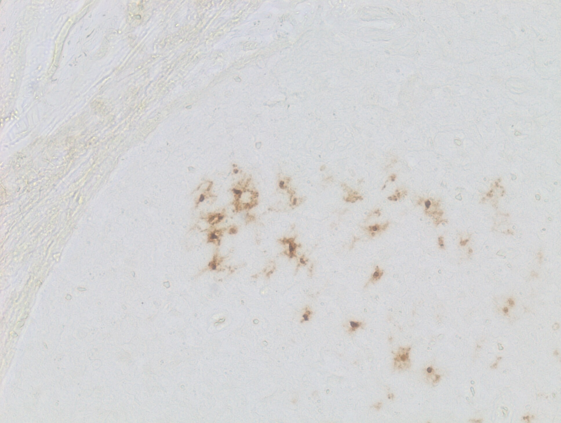
Immunohistochemistry-Paraffin: Langerin/CD207 Antibody [NB100-56733] - Human tonsil tissue. Heat mediated antigen retrieval in citrate buffer, 95C for 30 min. Image from verified customer review.![Immunohistochemistry-Paraffin: Langerin/CD207 Antibody [NB100-56733]](https://resources.rndsystems.com/images/products/Langerin-CD207-Antibody-Immunohistochemistry-Paraffin-NB100-56733-img0005.jpg "Immunohistochemistry-Paraffin: Langerin/CD207 Antibody [NB100-56733]")
Immunohistochemistry-Paraffin: Langerin/CD207 Antibody [NB100-56733]
Immunohistochemistry-Paraffin: Langerin/CD207 Antibody [NB100-56733] - Tissue microarray from normal mouse organs (NBP2-30225) using rabbit polyclonal Langerin/CD207 antibody at 5ug/ml concentration. The IHC-P protocol involved antigen retrieval with 10mM sodium citrate buffer pH 6.0 and this antibody generated specific staining of the Langerhans cells in mouse ear lobe sections.![Immunohistochemistry-Paraffin: Langerin/CD207 Antibody [NB100-56733]](https://resources.rndsystems.com/images/products/Langerin-CD207-Antibody-Immunohistochemistry-Paraffin-NB100-56733-img0006.jpg "Immunohistochemistry-Paraffin: Langerin/CD207 Antibody [NB100-56733]")
Immunohistochemistry-Paraffin: Langerin/CD207 Antibody [NB100-56733]
Immunohistochemistry-Paraffin: Langerin/CD207 Antibody [NB100-56733] - Tissue microarray from normal mouse organs (NBP2-30225) using rabbit polyclonal Langerin/CD207 antibody at 5ug/ml concentration. The IHC-P protocol involved antigen retrieval with 10mM sodium citrate buffer pH 6.0 and this antibody generated specific staining of the Langerhans cells in mouse ear lobe sections.![Immunohistochemistry-Paraffin: Langerin/CD207 Antibody [NB100-56733]](https://resources.rndsystems.com/images/products/Langerin-CD207-Antibody-Immunohistochemistry-Paraffin-NB100-56733-img0007.jpg "Immunohistochemistry-Paraffin: Langerin/CD207 Antibody [NB100-56733]")
Immunohistochemistry-Paraffin: Langerin/CD207 Antibody [NB100-56733]
Immunohistochemistry-Paraffin: Langerin/CD207 Antibody [NB100-56733] - Analysis of langerin (CD207) in mouse breast tissue using an isotype control antibody (left) and langerin (CD207) antibody (right) at 5 ug/ml.Applications for Langerin/CD207 Antibody - BSA Free
Flow (Intracellular)
Flow Cytometry
Immunohistochemistry
Immunohistochemistry-Frozen
Immunohistochemistry-Paraffin
Western Blot
Reviewed Applications
Read 2 reviews rated 5 using NB100-56733 in the following applications:
Flow Cytometry Panel Builder
Bio-Techne Knows Flow Cytometry
Save time and reduce costly mistakes by quickly finding compatible reagents using the Panel Builder Tool.
Advanced Features
- Spectra Viewer - Custom analysis of spectra from multiple fluorochromes
- Spillover Popups - Visualize the spectra of individual fluorochromes
- Antigen Density Selector - Match fluorochrome brightness with antigen density

Formulation, Preparation, and Storage
Purification
Formulation
Format
Preservative
Concentration
Shipping
Stability & Storage
Background: Langerin/CD207
Alternate Names
Gene Symbol
UniProt
Additional Langerin/CD207 Products
Product Documents for Langerin/CD207 Antibody - BSA Free
Certificate of Analysis
To download a Certificate of Analysis, please enter a lot or batch number in the search box below.
Product Specific Notices for Langerin/CD207 Antibody - BSA Free
This product is for research use only and is not approved for use in humans or in clinical diagnosis. Primary Antibodies are guaranteed for 1 year from date of receipt.
Citations for Langerin/CD207 Antibody - BSA Free
 Powered by Bioz
Customer Reviews for Langerin/CD207 Antibody - BSA Free (2)
Have you used Langerin/CD207 Antibody - BSA Free?
Submit a review and receive an Amazon gift card!
$25/€18/£15/$25CAN/¥2500 Yen for a review with an image
$10/€7/£6/$10CAN/¥1110 Yen for a review without an image
Submit a review
Customer Images
-
Application: Immunohistochemistry-ParaffinSample Tested: Tonsil tissueSpecies: HumanVerified Customer | Posted 09/25/2018Heat mediated antigen retrieval performed in citrate buffer for 30 mins at 95C

-
Application: Immunohistochemistry-ParaffinSample Tested: mouse skinSpecies: MouseVerified Customer | Posted 05/04/2016Mouse Skin stained with Langerin (CD207) antibody (NB100-56733)
There are no reviews that match your criteria.
Protocols
View specific protocols for Langerin/CD207 Antibody - BSA Free (NB100-56733):
DeParaffinization
Materials Required
- Tissue array slide - 100%, 95%, and 75% ethanol
- Xylene
Method
1. Incubate in a dry oven at 62C for 1 hour. Slides should be maintained in a vertical orientation to allow complete removal of the paraffin.
2. Dewax slides in xylene for 5 x 4 minutes.
3. Hydrate slides in 100%, 95%, and 75% ethanol for 2 x 3 minutes each.
4. Immerse slides in tap water for 5 minutes.
Suggested Antigen Retrieval Protocol
The following procedure are a suggestion only. Other protocols can be used on the array slides.
Materials Required
- Tissue array slide - Phosphate buffered saline (pH 7.6)
- Citrate buffer (0.01 M, pH 6.0) - Microwave oven (700 W)
Method 1 (Microwave)
1. Immerse slides into citrate buffer (0.01 M, pH 6.0).
2. Microwave (700 W or high) for 5 min, add citrate buffer if necessary.
3. Microwave (medium) for 5 min, add citrate buffer if necessary.
4. Microwave (low) for 5 min.
5. Immerse in cold PBS.
Method 2 (Autoclave/Pressure Cooker)
1. Immerse slides in citrate buffer.
2. Incubate in a pressure cooker for 2 min at 95C.
3. Cool to room temperature.
4. Wash slides in PBS for 3 x 5 min.
Method 3 (Enzyme treatment)
1. Incubate slides with pronase [0.05% (w/v) in PBS] or trypsin [0.05% (v/v) in PBS] or pepsin [0.05% (v/v) in 2 N HCl] at 37oC (incubation time should be adjusted according to the antibody).
2. Wash slides in PBS for 3 x 5 min.
Method 4 (Hot bath)
1. Heat citrate buffer (1mM EDTA, pH8.0 or 0.01M sodium citrate buffer, pH6.0) to about 950C.
2. Place slides in the buffer for 10-15 min.
Immunostain
Materials Required
1. Slides
2. Phosphate buffered saline (pH 7.6)
3. Hydrogen peroxide
4. Primary antibody
5. Blocking serum (normal serum)
6. Biotinylated secondary antibody
7. ABC reagent (6, 7, and 8 are included in Vectastain Elite ABC kit)
8. Diaminobenzidine
9. Meyer's hematoxylin
10.Permount
Method
1. Incubate in a dry oven at 62C for 1 hour. Slides should be maintained in a vertical orientation to allow complete removal of the paraffin.
2. Dewax slides in xylene for 5 x 4 minutes.
3. Hydrate slides in 100%, 95%, and 75% ethanol for 2 x 3 minutes each.
4. Immerse slides in tap water for 5 minutes
5. Antigen retrieval method (optional).
6. Quenching of endogenous peroxidase (optional)
a.immerse slides in 3% hydrogen peroxide solution for 6 minutes.
b.wash slides in PBS for 3 x 5 minutes.
7. Incubate slides with blocking serum (1:50) for 30 min.*
8. Blot excess serum from section, and incubate with primary Ab. Suggested incubation time may vary between antibodies:
mAb 2 hours at room temperature or overnight at 4C.
pAb: 1 ~ 1.5 hours at room temperature.
9. Wash slides in PBS for 3 x 5 minutes.
10.Incubate slides with biotin-conjugated secondary Ab for 30 min.*
11.Wash slides in PBS for 3 x 5 minutes.
12.Incubate slides with Avidin-Biotin Complexes for 30 min.*
13.Wash slides in PBS for 3 x 5 minutes.
14.Incubate slides in fresh DAB solution for 2 minutes. (We use DAB solution in Vector DAB/Ni substrate kit).**
15.Stop the reaction by washing in tap water.
16.Counterstain in Meyer's hematoxylin for 10 seconds.
17.Dehydrates slides in 75%, 80%, 95% and 100% ethanol
18.Clear slides in xylene 4 X 5 minutes.
19.Mount cover slide with Permount.
* Blocking serum, secondary antibody and avidin-biotin-peroxidase complexes are included in most of the immunostaining kit. Our lab uses the ABC kit from Vector Lab (Vectastain Elite ABC kit).
** We use DAB solution in Vector/DAB/Ni substrate kit (Vector Labs., Cat. SK-4100).
Find general support by application which include: protocols, troubleshooting, illustrated assays, videos and webinars.
- 7-Amino Actinomycin D (7-AAD) Cell Viability Flow Cytometry Protocol
- Antigen Retrieval Protocol (PIER)
- Antigen Retrieval for Frozen Sections Protocol
- Appropriate Fixation of IHC/ICC Samples
- Cellular Response to Hypoxia Protocols
- Chromogenic IHC Staining of Formalin-Fixed Paraffin-Embedded (FFPE) Tissue Protocol
- Chromogenic Immunohistochemistry Staining of Frozen Tissue
- ClariTSA™ Fluorophore Kits
- Detection & Visualization of Antibody Binding
- Extracellular Membrane Flow Cytometry Protocol
- Flow Cytometry Protocol for Cell Surface Markers
- Flow Cytometry Protocol for Staining Membrane Associated Proteins
- Flow Cytometry Staining Protocols
- Flow Cytometry Troubleshooting Guide
- Fluorescent IHC Staining of Frozen Tissue Protocol
- Graphic Protocol for Heat-induced Epitope Retrieval
- Graphic Protocol for the Preparation and Fluorescent IHC Staining of Frozen Tissue Sections
- Graphic Protocol for the Preparation and Fluorescent IHC Staining of Paraffin-embedded Tissue Sections
- Graphic Protocol for the Preparation of Gelatin-coated Slides for Histological Tissue Sections
- IHC Sample Preparation (Frozen sections vs Paraffin)
- Immunofluorescent IHC Staining of Formalin-Fixed Paraffin-Embedded (FFPE) Tissue Protocol
- Immunohistochemistry (IHC) and Immunocytochemistry (ICC) Protocols
- Immunohistochemistry Frozen Troubleshooting
- Immunohistochemistry Paraffin Troubleshooting
- Intracellular Flow Cytometry Protocol Using Alcohol (Methanol)
- Intracellular Flow Cytometry Protocol Using Detergents
- Intracellular Nuclear Staining Flow Cytometry Protocol Using Detergents
- Intracellular Staining Flow Cytometry Protocol Using Alcohol Permeabilization
- Intracellular Staining Flow Cytometry Protocol Using Detergents to Permeabilize Cells
- Preparing Samples for IHC/ICC Experiments
- Preventing Non-Specific Staining (Non-Specific Binding)
- Primary Antibody Selection & Optimization
- Propidium Iodide Cell Viability Flow Cytometry Protocol
- Protocol for Heat-Induced Epitope Retrieval (HIER)
- Protocol for Liperfluo
- Protocol for Making a 4% Formaldehyde Solution in PBS
- Protocol for VisUCyte™ HRP Polymer Detection Reagent
- Protocol for the Characterization of Human Th22 Cells
- Protocol for the Characterization of Human Th9 Cells
- Protocol for the Preparation & Fixation of Cells on Coverslips
- Protocol for the Preparation and Chromogenic IHC Staining of Frozen Tissue Sections
- Protocol for the Preparation and Chromogenic IHC Staining of Frozen Tissue Sections - Graphic
- Protocol for the Preparation and Chromogenic IHC Staining of Paraffin-embedded Tissue Sections
- Protocol for the Preparation and Chromogenic IHC Staining of Paraffin-embedded Tissue Sections - Graphic
- Protocol for the Preparation and Fluorescent IHC Staining of Frozen Tissue Sections
- Protocol for the Preparation and Fluorescent IHC Staining of Paraffin-embedded Tissue Sections
- Protocol for the Preparation of Gelatin-coated Slides for Histological Tissue Sections
- Protocol: Annexin V and PI Staining by Flow Cytometry
- Protocol: Annexin V and PI Staining for Apoptosis by Flow Cytometry
- R&D Systems Quality Control Western Blot Protocol
- TUNEL and Active Caspase-3 Detection by IHC/ICC Protocol
- The Importance of IHC/ICC Controls
- Troubleshooting Guide: Fluorokine Flow Cytometry Kits
- Troubleshooting Guide: Immunohistochemistry
- Troubleshooting Guide: Western Blot Figures
- Western Blot Conditions
- Western Blot Protocol
- Western Blot Protocol for Cell Lysates
- Western Blot Troubleshooting
- Western Blot Troubleshooting Guide
- View all Protocols, Troubleshooting, Illustrated assays and Webinars
FAQs for Langerin/CD207 Antibody - BSA Free
-
Q: I’m contacting you because I’m using often your Antibodies and I would like to conjugate some of them by myself. For that purpose I would need to have Antibodies in a buffer solution that do not contains any additive like NaAz or BSA. Could you please tell me if it is possible to order such antibodies, and if there is a minimum volume quantity to order?
A: Typically there is a minimum order quantity for custom formats of our products (1mg on average). However we are transitioning to offering more of our products in an azide/BSA free format. Please let us know which products are needed, and we can check the feasibility of offering them in the desired format.
-
Q: Would you please recommend a dilution for this application? Do you have any suggested protocol for IHC?-EndFragment-->
A:
For IHC-P we recommend a dilution range of 1:100-1:500. The lab used the following protocol
-
Q: I’m contacting you because I’m using often your Antibodies and I would like to conjugate some of them by myself. For that purpose I would need to have Antibodies in a buffer solution that do not contains any additive like NaAz or BSA. Could you please tell me if it is possible to order such antibodies, and if there is a minimum volume quantity to order?
A: Typically there is a minimum order quantity for custom formats of our products (1mg on average). However we are transitioning to offering more of our products in an azide/BSA free format. Please let us know which products are needed, and we can check the feasibility of offering them in the desired format.
-
Q: Would you please recommend a dilution for this application? Do you have any suggested protocol for IHC?-EndFragment-->
A:
For IHC-P we recommend a dilution range of 1:100-1:500. The lab used the following protocol
Associated Pathways
