LAP (TGF-beta 1) Antibody (7F6) - BSA Free
Novus Biologicals | Catalog # NBP2-22114
Key Product Details
Species Reactivity
Validated:
Cited:
Applications
Validated:
Cited:
Label
Antibody Source
Format
Product Specifications
Immunogen
Reactivity Notes
Clonality
Host
Isotype
Theoretical MW
Disclaimer note: The observed molecular weight of the protein may vary from the listed predicted molecular weight due to post translational modifications, post translation cleavages, relative charges, and other experimental factors.
Scientific Data Images for LAP (TGF-beta 1) Antibody (7F6) - BSA Free
![Western Blot: LAP (TGF-beta 1) Antibody (7F6)BSA Free [NBP2-22114]](https://resources.rndsystems.com/images/products/TGF-beta-1-Antibody-7F6-Western-Blot-NBP2-22114-img0002.jpg "Western Blot: LAP (TGF-beta 1) Antibody (7F6)BSA Free [NBP2-22114]")
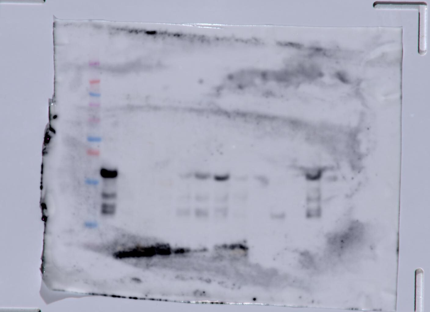
Western Blot: LAP (TGF-beta 1) Antibody (7F6)BSA Free [NBP2-22114]
Western Blot: LAP (TGF-beta 1) Antibody (7F6) [NBP2-22114] - Western blot analysis using TGF beta 1 mAb against human LAP (TGF-beta 1) recombinant protein. (Expected MW is 41 kDa)![Immunohistochemistry-Paraffin: LAP (TGF-beta 1) Antibody (7F6) - BSA Free [NBP2-22114]](https://resources.rndsystems.com/images/products/TGF-beta-1-Antibody-7F6-Immunohistochemistry-Paraffin-NBP2-22114-img0005.jpg "Immunohistochemistry-Paraffin: LAP (TGF-beta 1) Antibody (7F6) - BSA Free [NBP2-22114]")
Immunohistochemistry-Paraffin: LAP (TGF-beta 1) Antibody (7F6) - BSA Free [NBP2-22114]
Immunohistochemistry-Paraffin: LAP (TGF-beta 1) Antibody (7F6) [NBP2-22114] - Immunohistochemical analysis of paraffin-embedded lymphoid tissue tissues using LAP (TGF-beta 1) mouse mAb with DAB staining.![Flow Cytometry: LAP (TGF-beta 1) Antibody (7F6) - BSA Free [NBP2-22114]](https://resources.rndsystems.com/images/products/TGF-beta-1-Antibody-7F6-Flow-Cytometry-NBP2-22114-img0003.jpg "Flow Cytometry: LAP (TGF-beta 1) Antibody (7F6) - BSA Free [NBP2-22114]")
Flow Cytometry: LAP (TGF-beta 1) Antibody (7F6) - BSA Free [NBP2-22114]
Flow Cytometry: LAP (TGF-beta 1) Antibody (7F6) [NBP2-22114] - Flow cytometric analysis of A549 cells using LAP (TGF-beta 1) mouse mAb (green) and negative control (red).![Western Blot: LAP (TGF-beta 1) Antibody (7F6)BSA Free [NBP2-22114]](https://resources.rndsystems.com/images/products/TGF-beta-1-Antibody-7F6-Western-Blot-NBP2-22114-img0007.jpg "Western Blot: LAP (TGF-beta 1) Antibody (7F6)BSA Free [NBP2-22114]")
Western Blot: LAP (TGF-beta 1) Antibody (7F6)BSA Free [NBP2-22114]
Western Blot: LAP (TGF-beta 1) Antibody (7F6) [NBP2-22114] - Western blot analysis of human stomach tissue (A) and human small intestine tissue (B) using TGF beta 1 antibody (NBP2-22114) at 2 ug/ml.![Immunohistochemistry-Paraffin: LAP (TGF-beta 1) Antibody (7F6) - BSA Free [NBP2-22114]](https://resources.rndsystems.com/images/products/TGF-beta-1-Antibody-7F6-Immunohistochemistry-Paraffin-NBP2-22114-img0006.jpg "Immunohistochemistry-Paraffin: LAP (TGF-beta 1) Antibody (7F6) - BSA Free [NBP2-22114]")
Immunohistochemistry-Paraffin: LAP (TGF-beta 1) Antibody (7F6) - BSA Free [NBP2-22114]
Immunohistochemistry-Paraffin: LAP (TGF-beta 1) Antibody (7F6) [NBP2-22114] - Human tonsil. Image from verified customer review.![Immunohistochemistry-Paraffin: LAP (TGF-beta 1) Antibody (7F6) - BSA Free [NBP2-22114]](https://resources.rndsystems.com/images/products/TGF-beta-1-Antibody-7F6-Immunohistochemistry-Paraffin-NBP2-22114-img0004.jpg "Immunohistochemistry-Paraffin: LAP (TGF-beta 1) Antibody (7F6) - BSA Free [NBP2-22114]")
Immunohistochemistry-Paraffin: LAP (TGF-beta 1) Antibody (7F6) - BSA Free [NBP2-22114]
Immunohistochemistry-Paraffin: LAP (TGF-beta 1) Antibody (7F6) [NBP2-22114] - Immunohistochemical analysis of paraffin-embedded lung cancer tissues using LAP (TGF-beta 1) mouse mAb with DAB staining.![ELISA: LAP (TGF-beta 1) Antibody (7F6) - BSA Free [NBP2-22114]](https://resources.rndsystems.com/images/products/TGF-beta-1-Antibody-7F6-ELISA-NBP2-22114-img0001.jpg "ELISA: LAP (TGF-beta 1) Antibody (7F6) - BSA Free [NBP2-22114]")
ELISA: LAP (TGF-beta 1) Antibody (7F6) - BSA Free [NBP2-22114]
ELISA: LAP (TGF-beta 1) Antibody (7F6) [NBP2-22114] - Red: Control Antigen (100ng); Purple: Antigen (10ng); Green: Antigen (50ng); Blue: Antigen (100ng). Antibody (7F6) - BSA Free [NBP2-22114] -")
Western Blot: LAP (TGF-beta 1) Antibody (7F6) - BSA Free [NBP2-22114] -
CD47 regulates thrombospondin-1, TGF-beta 1, and collagen deposition after injury. a Whole cell lysates from freshly isolated intestinal epithelial cells from Cd47f/f and Cd47 delta IEC mice were subjected to SDS–PAGE and immunoblot for thrombospondin-1/TSP-1, TGF-beta 1, and phosphorylated SMAD2 and SMAD3. Results are representative of three independent experiments. b Representative Masson’s trichrome staining of wounds beds and chronic DSS-colitis colons from Cd47f/f and Cd47 delta IEC mice. Scale bars = 50 μm. Results representative of three independent experiments with 5–7 mice per group. Source data are provided as a Source Data file Image collected and cropped by CiteAb from the following open publication (https://pubmed.ncbi.nlm.nih.gov/31676794), licensed under a CC-BY license. Not internally tested by Novus Biologicals. Antibody (7F6) - BSA Free [NBP2-22114] -")
Western Blot: LAP (TGF-beta 1) Antibody (7F6) - BSA Free [NBP2-22114] -
Inhibition of ERK1/2 activation attenuates myocardial remodeling and inflammation in permanent MI.Trametinib- and vehicle-treated mice were studied for 7 days after permanent LAD ligation. (A) Relative mRNA expression analysis of Il6, Tnf, Ccl2, and Ccr2 from the infarcted myocardium. (B) Flow cytometry analysis of the infarcted myocardium obtained from vehicle- or trametinib-treated mice normalized to heart weight. Representative gating strategies for quantification of CD45+ leukocytes: CD45+CD90.2–B220–NK1.1–CD11b+ myelomonocytic cells, CD45+CD90.2–B220–NK1.1–CD11b+Ly6G–F4/80–Ly6Chi monocytes, and CD45+CD90.2–B220–NK1.1–CD11b+Ly6G–F4/80–Ly6Clo macrophages. (C) Protein expression analysis of p-ERK1/2 (normalized to total ERK1/2) and activated TGF-beta 1 (normalized to GAPDH) in infarcted myocardium obtained from vehicle- or trametinib-treated mice. IOD, integrated optical density. (D) Protein expression analysis of p-ERK1/2 (normalized to ERK1/2) and activated TGF-beta 1 (normalized to GAPDH) in PBMCs isolated from vehicle- or trametinib-treated mice. Ordinary 1-way ANOVA, Šidak’s multiple-comparison test; n = 4–6 animals per group. Data are shown as mean +/- SEM. *P < 0.05, **P < 0.01, ***P < 0.001, ****P < 0.0001. Image collected and cropped by CiteAb from the following open publication (https://pubmed.ncbi.nlm.nih.gov/36548062), licensed under a CC-BY license. Not internally tested by Novus Biologicals. Antibody (7F6) - BSA Free [NBP2-22114] -")
Western Blot: LAP (TGF-beta 1) Antibody (7F6) - BSA Free [NBP2-22114] -
The upregulation of TGF beta 1/IGF-1/BDNF expression in the brain of AD mice by PBMT extended to promote adult hippocampal neurogenesis. A, B Western blotting analysis (A) and quantification (B) of TGF beta 1/IGF-1/BDNF expression in APP/PS1 and 3xTg-AD mouse brain after PBMT, (n = 3–4 per group). C The concentration of TGF beta 1/IGF-1/BDNF in brain tissues were measured by enzyme linked immunosorbent assay (ELISA), (n = 3–6 per group). D Representative images of Nestin+ (neural stem cell staining) and neuronal class-III beta -tubulin (Tuj1)+ (newborn neurons staining) expression cells in APP/PS1 and 3xTg-AD mouse hippocampal dentate gyrus (DG) at the end of PBMT. Scale bars, 50 μm. E Quantitative analyses of Nestin+ and Tuj1+ area in the hippocampal DG of each group, (n = 4 per group). F Quantitative analyses of the Nestin and Tuj1 mean fluorescence (MFI) in the brain tissues of each group after PBMT. The Nestin and Tuj1 MFI were detected by flow cytometer, (n = 5–6 per group). G Tuj1 antibody was used to staining the newborn neurons, and then, the expression of alpha -amino-3-hydroxy-5-methyl-4-isoxazole-propionic acid receptors (AMPAR) and postsynaptic density protein 95 (PSD95) on Tuj1+ neurons were detected by flow cytometer. All quantifications are presented as mean +/- SEM and were analyzed by one-way ANOVA test; ***p < 0.001, **p < 0.01, *p < 0.05 versus WT group; ###p < 0.001, ##p < 0.01, #p < 0.05 versus indicated group Image collected and cropped by CiteAb from the following open publication (https://pubmed.ncbi.nlm.nih.gov/36217178), licensed under a CC-BY license. Not internally tested by Novus Biologicals. Antibody (7F6) - BSA Free [NBP2-22114] -")
Western Blot: LAP (TGF-beta 1) Antibody (7F6) - BSA Free [NBP2-22114] -
TF cytoplasmic domain phosphorylation–dependent increased TGF-beta 1 activation in clinical setting of MI.(A) Representative confocal images of phosphorylation status of TF in infarcted myocardium obtained from WT or TF delta CT mice after 7 days. Representative images and quantification of biological replicates. Kruskal-Wallis test and Dunn’s multiple-comparison test; n = 3–4 animals per group. Scale bars: 50 μm. (B) Representative immunofluorescence confocal microscopy images of CD45+ and CD45/p-TF double-positive cells in human myocardium specimens obtained from n = 5 nonischemic (NI) donor hearts and n = 7 IHF patients. Quantification of biological replicates. Mann-Whitney test. Scale bars: 50 μm. (C and D) Western blot analysis and quantification of human LV tissue obtained from n = 5 nonischemic donor hearts and n = 9 IHF patients for p-TF (normalized to total TF) and TF (C) or TGF-beta 1 (normalized to GAPDH) and p-SMAD2 (normalized to total SMAD2) (D). Mann-Whitney test. Data are shown as mean +/- SEM. *P < 0.05. Image collected and cropped by CiteAb from the following open publication (https://pubmed.ncbi.nlm.nih.gov/36548062), licensed under a CC-BY license. Not internally tested by Novus Biologicals. Antibody (7F6) - BSA Free [NBP2-22114] -")
Western Blot: LAP (TGF-beta 1) Antibody (7F6) - BSA Free [NBP2-22114] -
A profibrotic MEK1/2–TGF-beta 1 pathway is linked to PAR2-mediated ROS signaling in monocytes.(A and B) Protein expression analysis of monocytes isolated from WT mice and pretreated in vitro with trametinib (10 μM) for 1 hour (A), or isolated from PAR2–/– versus WT mice (B). Cells were stimulated with an inflammatory cytokine cocktail containing IL-6, TNF-alpha, and CCL2 at a concentration of 20 ng/mL with and without hypoxia for 4 hours. Western blotting of p-ERK1/2 (normalized to total ERK1/2) and activated TGF-beta 1 (normalized to GAPDH). Ordinary 1-way ANOVA, Šidak’s multiple-comparison test; n = 5 replicates (2–3 mice were pooled for each sample). (C–E) PAR2fl/fl and PAR2fl/fl LysMCre littermates were subjected to permanent LAD ligation and investigated after 7 days; n = 5–10 animals per group. (C) Western blot analysis of activated TGF-beta 1 (normalized to GAPDH) and p-SMAD2 (normalized to total SMAD2) in the infarcted myocardium. Representative blots and quantification of biological replicates. (D) High-frequency ultrasound echocardiography obtained from PAR2fl/fl LysMCre and PAR2fl/fl littermate control mice with measurement of LVEF (%) and LVEDV (μL). Mann-Whitney test. (E) Kaplan-Meier survival analysis of permanently LAD-ligated PAR2fl/fl LysMCre and PAR2fl/fl littermate control mice over 7 days. Log-rank (Mantel-Cox) test. (F) Sirius red staining and deconvoluted images of fibrotic area on paraffin-embedded heart sections 4 weeks after permanent LAD ligation to induce IHF. Representative images and quantification of fibrotic areas normalized to surface area. Unpaired, 2-sided t test; n = 5 animals per group. Data are shown as mean +/- SEM. *P < 0.05, **P < 0.01, ****P < 0.0001. Image collected and cropped by CiteAb from the following open publication (https://pubmed.ncbi.nlm.nih.gov/36548062), licensed under a CC-BY license. Not internally tested by Novus Biologicals. Antibody (7F6) - BSA Free [NBP2-22114] -")
Western Blot: LAP (TGF-beta 1) Antibody (7F6) - BSA Free [NBP2-22114] -
Myeloid cell–derived TF-PAR2 complex is required for TGF-beta 1 activation.(A) Confocal microscopy of myocardial cryosections obtained from n = 5 sham-operated and n = 5 LAD-ligated WT (C57BL/6J) mice at day 7. Representative images and quantification of TF+ cells costained for CD45. Unpaired, 2-sided t test. Scale bar: 50 μm. (B–D) TFfl/fl LysMCre and TFfl/fl littermates were subjected to permanent LAD ligation versus sham surgery and investigated after 7 days; n = 5–7 animals per group. (B) Protein expression analysis of p-ERK1/2 (normalized to total ERK1/2) in the infarcted myocardium. Ordinary 1-way ANOVA, Šidak’s multiple-comparison test. (C) Western blot analysis of activated TGF-beta 1 (normalized to GAPDH) and p-SMAD2 (normalized to total SMAD2) in the infarcted myocardium obtained from TFfl/fl LysMCre and TFfl/fl littermates. Representative blots and quantification of biological replicates. (D) High-frequency ultrasound echocardiography obtained from TFfl/fl LysMCre and TFfl/fl littermates. Ordinary 1-way ANOVA, Šidak’s multiple-comparison test. (E) Sirius red staining and deconvoluted images of fibrotic area on paraffin-embedded heart sections 4 weeks after permanent LAD ligation to induce IHF versus sham surgery. Representative images and quantification of fibrotic areas normalized to surface area. Ordinary 1-way ANOVA, Šidak’s multiple-comparison test; n = 5 animals per group. (F) Kaplan-Meier survival analysis of permanently LAD-ligated TFfl/fl LysMCre and TFfl/fl littermate mice over 4 weeks. Log-rank (Mantel-Cox) test; n = 10–15 animals per group. Data are shown as mean +/- SEM. *P < 0.05, **P < 0.01, ***P < 0.001. Image collected and cropped by CiteAb from the following open publication (https://pubmed.ncbi.nlm.nih.gov/36548062), licensed under a CC-BY license. Not internally tested by Novus Biologicals. Antibody (7F6) - BSA Free [NBP2-22114] -")
Western Blot: LAP (TGF-beta 1) Antibody (7F6) - BSA Free [NBP2-22114] -
Myeloid cell TF cytoplasmic domain phosphorylation mediates ERK1/2–TGF-beta 1–dependent cardiac remodeling in permanent LAD ligation.(A) Confocal microscopy of myocardial cryosections obtained from WT (C57BL/6J) and TF delta CT mice. Representative images and quantification of MFI of Ly6C+TGF beta -1+ and CD31+TGF-beta 1+ cells. Ordinary 1-way ANOVA, Šidak’s multiple-comparison test; n = 5 animals per group. Scale bars: 25 μm. (B) Mice with transplanted BM were subjected to permanent LAD ligation versus sham surgery and investigated 7 days later. Western blot analysis of NOX2 (normalized to GAPDH), p-ERK1/2 (normalized to total ERK1/2), and TGF-beta 1 (normalized to GAPDH) in infarcted myocardium obtained from chimeric mice. Ordinary 1-way ANOVA, Šidak’s multiple-comparison test; n = 5–7 animals per group. Data are shown as mean +/- SEM. *P < 0.05, **P < 0.01, ***P < 0.001. Image collected and cropped by CiteAb from the following open publication (https://pubmed.ncbi.nlm.nih.gov/36548062), licensed under a CC-BY license. Not internally tested by Novus Biologicals. Antibody (7F6) - BSA Free [NBP2-22114] -")
Western Blot: LAP (TGF-beta 1) Antibody (7F6) - BSA Free [NBP2-22114] -
A profibrotic MEK1/2–TGF-beta 1 pathway is linked to PAR2-mediated ROS signaling in monocytes.(A and B) Protein expression analysis of monocytes isolated from WT mice and pretreated in vitro with trametinib (10 μM) for 1 hour (A), or isolated from PAR2–/– versus WT mice (B). Cells were stimulated with an inflammatory cytokine cocktail containing IL-6, TNF-alpha, and CCL2 at a concentration of 20 ng/mL with and without hypoxia for 4 hours. Western blotting of p-ERK1/2 (normalized to total ERK1/2) and activated TGF-beta 1 (normalized to GAPDH). Ordinary 1-way ANOVA, Šidak’s multiple-comparison test; n = 5 replicates (2–3 mice were pooled for each sample). (C–E) PAR2fl/fl and PAR2fl/fl LysMCre littermates were subjected to permanent LAD ligation and investigated after 7 days; n = 5–10 animals per group. (C) Western blot analysis of activated TGF-beta 1 (normalized to GAPDH) and p-SMAD2 (normalized to total SMAD2) in the infarcted myocardium. Representative blots and quantification of biological replicates. (D) High-frequency ultrasound echocardiography obtained from PAR2fl/fl LysMCre and PAR2fl/fl littermate control mice with measurement of LVEF (%) and LVEDV (μL). Mann-Whitney test. (E) Kaplan-Meier survival analysis of permanently LAD-ligated PAR2fl/fl LysMCre and PAR2fl/fl littermate control mice over 7 days. Log-rank (Mantel-Cox) test. (F) Sirius red staining and deconvoluted images of fibrotic area on paraffin-embedded heart sections 4 weeks after permanent LAD ligation to induce IHF. Representative images and quantification of fibrotic areas normalized to surface area. Unpaired, 2-sided t test; n = 5 animals per group. Data are shown as mean +/- SEM. *P < 0.05, **P < 0.01, ****P < 0.0001. Image collected and cropped by CiteAb from the following open publication (https://pubmed.ncbi.nlm.nih.gov/36548062), licensed under a CC-BY license. Not internally tested by Novus Biologicals.Applications for LAP (TGF-beta 1) Antibody (7F6) - BSA Free
ELISA
Flow Cytometry
Immunohistochemistry
Immunohistochemistry-Paraffin
Western Blot
Reviewed Applications
Read 3 reviews rated 3.7 using NBP2-22114 in the following applications:
Flow Cytometry Panel Builder
Bio-Techne Knows Flow Cytometry
Save time and reduce costly mistakes by quickly finding compatible reagents using the Panel Builder Tool.
Advanced Features
- Spectra Viewer - Custom analysis of spectra from multiple fluorochromes
- Spillover Popups - Visualize the spectra of individual fluorochromes
- Antigen Density Selector - Match fluorochrome brightness with antigen density

Formulation, Preparation, and Storage
Purification
Formulation
Format
Preservative
Concentration
Shipping
Stability & Storage
Background: LAP (TGF-beta 1)
Long Name
Alternate Names
Entrez Gene IDs
Gene Symbol
UniProt
Additional LAP (TGF-beta 1) Products
Product Documents for LAP (TGF-beta 1) Antibody (7F6) - BSA Free
Certificate of Analysis
To download a Certificate of Analysis, please enter a lot or batch number in the search box below.
Product Specific Notices for LAP (TGF-beta 1) Antibody (7F6) - BSA Free
This product is for research use only and is not approved for use in humans or in clinical diagnosis. Primary Antibodies are guaranteed for 1 year from date of receipt.
Citations for LAP (TGF-beta 1) Antibody (7F6) - BSA Free
 Powered by Bioz
Customer Reviews for LAP (TGF-beta 1) Antibody (7F6) - BSA Free (3)
Have you used LAP (TGF-beta 1) Antibody (7F6) - BSA Free?
Submit a review and receive an Amazon gift card!
$25/€18/£15/$25CAN/¥2500 Yen for a review with an image
$10/€7/£6/$10CAN/¥1110 Yen for a review without an image
Submit a review
Customer Images
-(01-ml)_NBP2-22114_6771.jpg)
-
Application: Western BlotSample Tested: Mouse PancreasSpecies: MouseVerified Customer | Posted 10/02/2020Caerulein dosed mouse pancreas

-
Application: Immunohistochemistry-ParaffinSample Tested: Human kidneySpecies: HumanVerified Customer | Posted 10/28/2015
-
Application: Immunohistochemistry-ParaffinSample Tested: human tonsilSpecies: HumanVerified Customer | Posted 04/04/2014Human tonsil - TGF beta
There are no reviews that match your criteria.
Protocols
Find general support by application which include: protocols, troubleshooting, illustrated assays, videos and webinars.
- 7-Amino Actinomycin D (7-AAD) Cell Viability Flow Cytometry Protocol
- Antigen Retrieval Protocol (PIER)
- Antigen Retrieval for Frozen Sections Protocol
- Appropriate Fixation of IHC/ICC Samples
- Cellular Response to Hypoxia Protocols
- Chromogenic IHC Staining of Formalin-Fixed Paraffin-Embedded (FFPE) Tissue Protocol
- Chromogenic Immunohistochemistry Staining of Frozen Tissue
- ClariTSA™ Fluorophore Kits
- Detection & Visualization of Antibody Binding
- ELISA Sample Preparation & Collection Guide
- ELISA Troubleshooting Guide
- Extracellular Membrane Flow Cytometry Protocol
- Flow Cytometry Protocol for Cell Surface Markers
- Flow Cytometry Protocol for Staining Membrane Associated Proteins
- Flow Cytometry Staining Protocols
- Flow Cytometry Troubleshooting Guide
- Fluorescent IHC Staining of Frozen Tissue Protocol
- Graphic Protocol for Heat-induced Epitope Retrieval
- Graphic Protocol for the Preparation and Fluorescent IHC Staining of Frozen Tissue Sections
- Graphic Protocol for the Preparation and Fluorescent IHC Staining of Paraffin-embedded Tissue Sections
- Graphic Protocol for the Preparation of Gelatin-coated Slides for Histological Tissue Sections
- How to Run an R&D Systems DuoSet ELISA
- How to Run an R&D Systems Quantikine ELISA
- How to Run an R&D Systems Quantikine™ QuicKit™ ELISA
- IHC Sample Preparation (Frozen sections vs Paraffin)
- Immunofluorescent IHC Staining of Formalin-Fixed Paraffin-Embedded (FFPE) Tissue Protocol
- Immunohistochemistry (IHC) and Immunocytochemistry (ICC) Protocols
- Immunohistochemistry Frozen Troubleshooting
- Immunohistochemistry Paraffin Troubleshooting
- Intracellular Flow Cytometry Protocol Using Alcohol (Methanol)
- Intracellular Flow Cytometry Protocol Using Detergents
- Intracellular Nuclear Staining Flow Cytometry Protocol Using Detergents
- Intracellular Staining Flow Cytometry Protocol Using Alcohol Permeabilization
- Intracellular Staining Flow Cytometry Protocol Using Detergents to Permeabilize Cells
- Preparing Samples for IHC/ICC Experiments
- Preventing Non-Specific Staining (Non-Specific Binding)
- Primary Antibody Selection & Optimization
- Propidium Iodide Cell Viability Flow Cytometry Protocol
- Protocol for Heat-Induced Epitope Retrieval (HIER)
- Protocol for Liperfluo
- Protocol for Making a 4% Formaldehyde Solution in PBS
- Protocol for VisUCyte™ HRP Polymer Detection Reagent
- Protocol for the Characterization of Human Th22 Cells
- Protocol for the Characterization of Human Th9 Cells
- Protocol for the Preparation & Fixation of Cells on Coverslips
- Protocol for the Preparation and Chromogenic IHC Staining of Frozen Tissue Sections
- Protocol for the Preparation and Chromogenic IHC Staining of Frozen Tissue Sections - Graphic
- Protocol for the Preparation and Chromogenic IHC Staining of Paraffin-embedded Tissue Sections
- Protocol for the Preparation and Chromogenic IHC Staining of Paraffin-embedded Tissue Sections - Graphic
- Protocol for the Preparation and Fluorescent IHC Staining of Frozen Tissue Sections
- Protocol for the Preparation and Fluorescent IHC Staining of Paraffin-embedded Tissue Sections
- Protocol for the Preparation of Gelatin-coated Slides for Histological Tissue Sections
- Protocol: Annexin V and PI Staining by Flow Cytometry
- Protocol: Annexin V and PI Staining for Apoptosis by Flow Cytometry
- Quantikine HS ELISA Kit Assay Principle, Alkaline Phosphatase
- Quantikine HS ELISA Kit Principle, Streptavidin-HRP Polymer
- R&D Systems Quality Control Western Blot Protocol
- Sandwich ELISA (Colorimetric) – Biotin/Streptavidin Detection Protocol
- Sandwich ELISA (Colorimetric) – Direct Detection Protocol
- TUNEL and Active Caspase-3 Detection by IHC/ICC Protocol
- The Importance of IHC/ICC Controls
- Troubleshooting Guide: ELISA
- Troubleshooting Guide: Fluorokine Flow Cytometry Kits
- Troubleshooting Guide: Immunohistochemistry
- Troubleshooting Guide: Western Blot Figures
- Western Blot Conditions
- Western Blot Protocol
- Western Blot Protocol for Cell Lysates
- Western Blot Troubleshooting
- Western Blot Troubleshooting Guide
- View all Protocols, Troubleshooting, Illustrated assays and Webinars
FAQs for LAP (TGF-beta 1) Antibody (7F6) - BSA Free
-
Q: Info regarding this product NBP2-22114SS, does this product cross reacts with Latent TGF-beta 1 also?
A: Based on the immunogen sequence, we predict it would cross react.