![Immunohistochemistry: Loricrin Antibody [NBP1-33610]](https://resources.rndsystems.com/images/products/Loricrin-Antibody-Immunohistochemistry-NBP1-33610-img0018.jpg "Immunohistochemistry: Loricrin Antibody [NBP1-33610]")
Loading...
Key Product Details
Species Reactivity
Validated:
Human
Cited:
Human, Mouse
Predicted:
Rhesus Macaque (93%). Backed by our 100% Guarantee.
Applications
Validated:
Immunohistochemistry, Immunohistochemistry-Paraffin, Western Blot, Immunocytochemistry/ Immunofluorescence, Simple Western
Cited:
Immunohistochemistry-Paraffin, Western Blot, Immunocytochemistry/ Immunofluorescence, IF/IHC
Label
Unconjugated
Antibody Source
Polyclonal Rabbit IgG
Loading...
Product Specifications
Immunogen
Carrier-protein conjugated synthetic peptide encompassing a sequence within the N-terminus region of human Loricrin. The exact sequence is proprietary.
Reactivity Notes
Immunogen displays the following percentage of sequence identity for non-tested species: Bovine (87%).
Localization
Cytoplasm, Nucleus, nucleoplasm
Clonality
Polyclonal
Host
Rabbit
Isotype
IgG
Theoretical MW
26 kDa.
Disclaimer note: The observed molecular weight of the protein may vary from the listed predicted molecular weight due to post translational modifications, post translation cleavages, relative charges, and other experimental factors.
Disclaimer note: The observed molecular weight of the protein may vary from the listed predicted molecular weight due to post translational modifications, post translation cleavages, relative charges, and other experimental factors.
Scientific Data Images for Loricrin Antibody
Immunohistochemistry: Loricrin Antibody [NBP1-33610]
Immunohistochemistry: Loricrin Antibody [NBP1-33610] - Prominent differences in the expressions of innate immune and barrier molecules between sebaceous gland poor (SGR) and sebaceous gland rich (SGP) skin regions. Representative images for immunostaining and quantification of epidermal levels of LOR in SGP and SGR skin sections. Images of negative control stainings are shown in the bottom left corner of SGP immunostainings. Size bars=100um. The graphs show the mean +/- SEM of measured protein levels (*p<0.05; **p<0.01; ***p<0.001, as determined by Mann-Whitney U-test). Image collected and cropped by CiteAb from the following publication (frontiersin.org/articles/10.3389/fimmu.2018.00424/full), licensed under a CC-BY license.![Immunocytochemistry/ Immunofluorescence: Loricrin Antibody [NBP1-33610]](https://resources.rndsystems.com/images/products/Loricrin-Antibody-Immunocytochemistry-Immunofluorescence-NBP1-33610-img0014.jpg "Immunocytochemistry/ Immunofluorescence: Loricrin Antibody [NBP1-33610]")
Immunocytochemistry/ Immunofluorescence: Loricrin Antibody [NBP1-33610]
Immunocytochemistry/Immunofluorescence: Loricrin Antibody [NBP1-33610] - Analysis of methanol-fixed Hep3B, using antibody at 1:500 dilution.![Western Blot: Loricrin Antibody [NBP1-33610]](https://resources.rndsystems.com/images/products/Loricrin-Antibody-Western-Blot-NBP1-33610-img0013.jpg "Western Blot: Loricrin Antibody [NBP1-33610]")
Western Blot: Loricrin Antibody [NBP1-33610]
Western Blot: Loricrin Antibody [NBP1-33610] - Whole cell extract (30 ug) was separated by 12% SDS-PAGE, and the membrane was blotted with Loricrin antibody [N1], N-term diluted at 1:1000. The HRP-conjugated anti-rabbit IgG antibody (NBP2-19301) was used to detect the primary antibody.![Immunohistochemistry-Paraffin: Loricrin Antibody [NBP1-33610]](https://resources.rndsystems.com/images/products/Loricrin-Antibody-Immunohistochemistry-Paraffin-NBP1-33610-img0010.jpg "Immunohistochemistry-Paraffin: Loricrin Antibody [NBP1-33610]")
Immunohistochemistry-Paraffin: Loricrin Antibody [NBP1-33610]
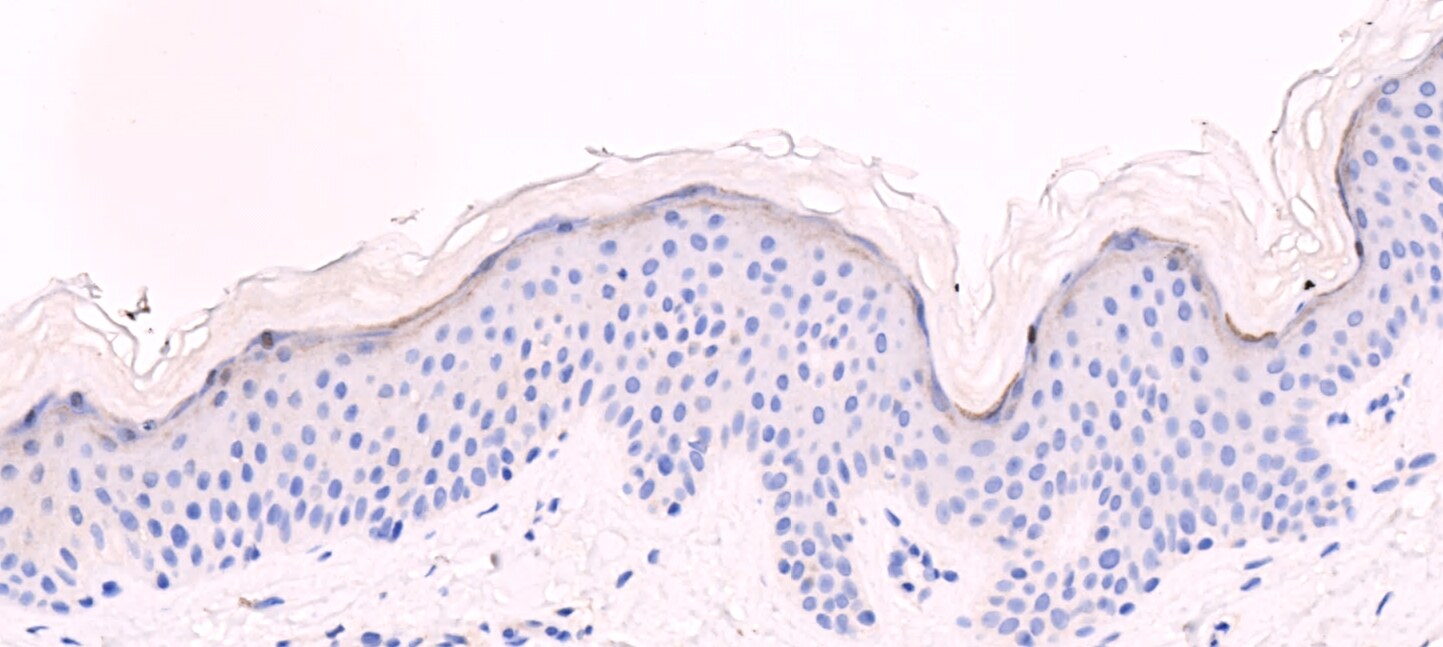
Immunohistochemistry-Paraffin: Loricrin Antibody [NBP1-33610] - HBL438 xenograft. Loricrin antibody [N1], N-term dilution: 1:500. Antigen Retrieval: Trilogy™ (EDTA based, pH 8.0) buffer, 15min.![Immunohistochemistry-Paraffin: Loricrin Antibody [NBP1-33610]](https://resources.rndsystems.com/images/products/Loricrin-Antibody-Immunohistochemistry-Paraffin-NBP1-33610-img0016.jpg "Immunohistochemistry-Paraffin: Loricrin Antibody [NBP1-33610]")
Immunohistochemistry-Paraffin: Loricrin Antibody [NBP1-33610]
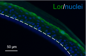
Immunohistochemistry-Paraffin: Loricrin Antibody [NBP1-33610] - Reconstructed human epidermis tissues were embedded in paraffin. After successives baths in xylene and ethanol, antigen was retrieved using warm citrate (for 30 min). Specimens were incubated overnight with the primary antibody (1:100 dilution). Blue staining corresponds to nuclei while loricrin is visible in green. IHC-P image submitted by a verified customer review.![Simple Western: Loricrin Antibody [NBP1-33610]](https://resources.rndsystems.com/images/products/Loricrin-Antibody-Simple-Western-NBP1-33610-img0015.jpg "Simple Western: Loricrin Antibody [NBP1-33610]")
Simple Western: Loricrin Antibody [NBP1-33610]
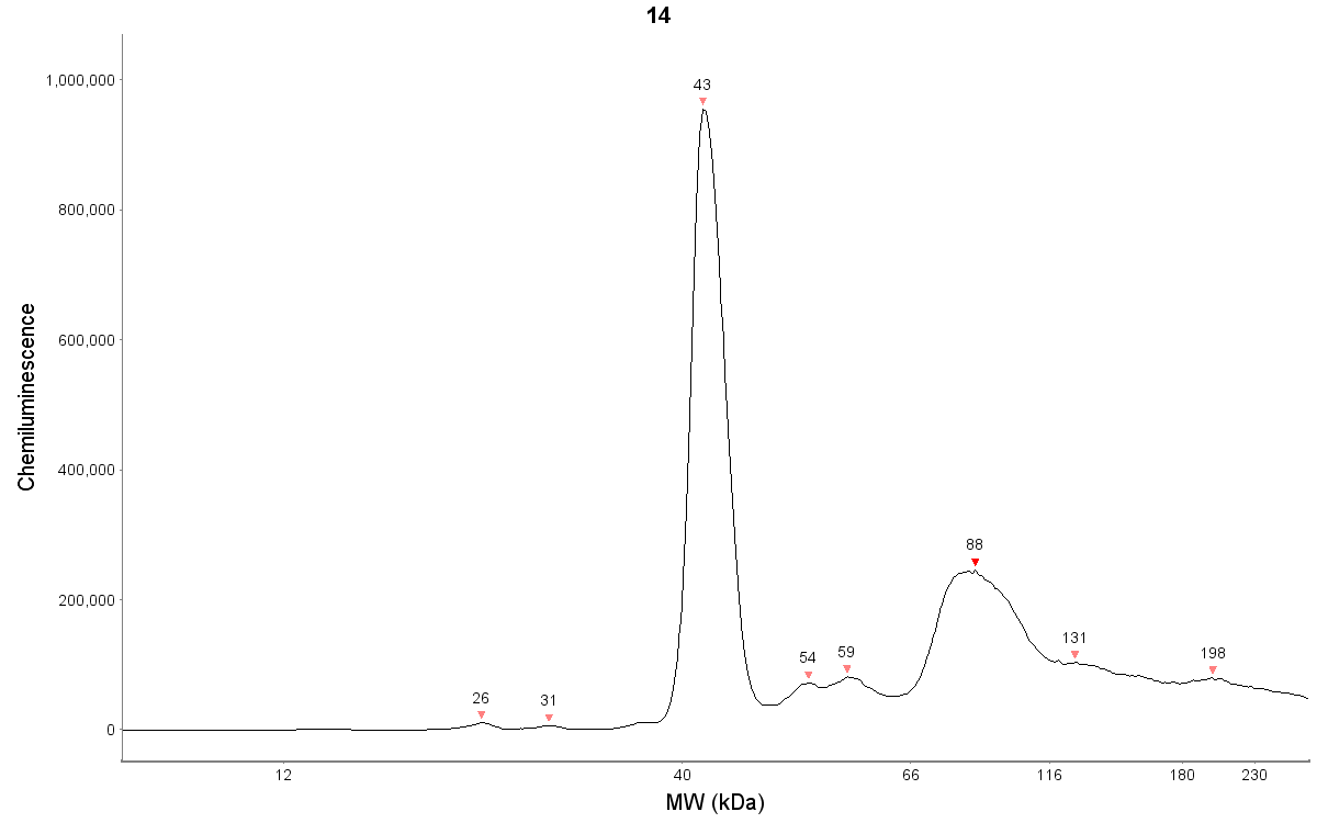
Simple Western: Loricrin Antibody [NBP1-33610] - NHEK human normal epidermal keratinocytes. Antibody dilution of 1:50. Protein concentration of 450 ug/mL. Detection is chemiluminescence. Simple Western image submitted by a verified customer review.
Western Blot: Loricrin Antibody [NBP1-33610] -
Western Blot: Loricrin Antibody [NBP1-33610] - Various whole cell extracts (30 ug) were separated by 12% SDS-PAGE, and the membrane was blotted with Loricrin antibody [N1], N-term (NBP1-33610) diluted at 1:1000. The HRP-conjugated anti-rabbit IgG antibody was used to detect the primary antibody.
Western Blot: Loricrin Antibody [NBP1-33610] -
The effect of YAP expression on skin barrier in AD mice. (A) Trans epidermal water loss (TEWL) and stratum corneum hydration (SCH) of each group. (B) YAP protein expression from immunohistochemistry with the quantification results shown in the graphs (×400), bar length = 50 μm. n = 3. (C) Filaggrin (FLG) protein expression from immunohistochemistry with the quantification results shown in the graphs (×400), bar length = 50 μm. n = 3. (D) FLG, involucrin (IVL), and loricrin (LOR) protein expression from western blot with the quantification results shown in the graphs. (E) YAP, p-YAP, mTOR, and p-mTOR protein expression from western blot with the quantification results shown in the graphs. Ctrl, control mice; AD, atopic dermatitis model mice; BLK, mice injected with empty vector lentivirus; YAP, mice injected with YAP overexpression lentivirus; AD+YAP, atopic dermatitis model mice injected with YAP overexpression lentivirus; sh-NC, mice injected with shRNA-NC lentivirus; sh-YAP, mice injected with YAP shRNA lentivirus; AD+sh-YAP, atopic dermatitis model mice injected with YAP shRNA lentivirus; AD+sh-YAP+RPM, AD+sh-YAP group with topical 0.2% rapamycin ointment. *P < 0.05, ***P < 0.001 Ctrl v.s. AD; #P < 0.05, ##P < 0.01, ###P < 0.001 AD+YAP v.s. AD; +P < 0.05, +++P < 0.001 AD+sh-YAP v.s. AD; ^^P < 0.01, ^^^P < 0.001 AD+sh-YAP+RPM v.s. AD+sh-YAP. Image collected and cropped by CiteAb from the following open publication (https://www.frontiersin.org/articles/10.3389/fimmu.2025.1681148/full), licensed under a CC-BY license. Not internally tested by Novus Biologicals.
Western Blot: Loricrin Antibody [NBP1-33610] -
The effect of beta -endorphin directly through the u-opioid receptor, and the effect of inhibition of Akt/mTOR signaling on UVB-induced disruption of epidermal homeostasis. (a) NHKs were pre-incubated with 100 nM of CTOP for 30 min before 15 mJ/cm2 UVB irradiation and the addition of 100 nM beta -endorphin. (b, c) Representative immunoblots and the quantification of the phosphorylation level of proteins in Akt/mTOR signaling pathway (b) and expression levels of differentiation markers (c) showing the reversal effect of beta -endorphin is suppressed by 100 nM CTOP treatment and the reversal effect of 500 nM rapamycin treatment in NHKs for 30 min before 15 mJ/cm2 UVB irradiation. The original blots are presented in Supplementary Fig. S6. * p < 0.05, ** p < 0.01, *** p < 0.001 compared to the designated group. n.s. means not significant. Image collected and cropped by CiteAb from the following open publication (https://www.nature.com/articles/s41598-023-49886-5), licensed under a CC-BY license. Not internally tested by Novus Biologicals.
Western Blot: Loricrin Antibody [NBP1-33610] -
Treatment with beta -endorphin salvaged UVB irradiation-induced increased proliferation and reduced the expression of epidermal differentiation markers in NHKs. (a) Representative images of 5-ethynyl-2ʹ-deoxyuridine (EdU)-positive cells in keratinocytes after UVB irradiation, followed by beta -endorphin treatment for 24 h. DAPI: 4′,6-diamidino-2-phenylindole. The histogram shows the quantification of EdU as the percentage of cells with positive staining. (b) NHKs were exposed to 15 mJ/cm2 of UVB light, followed by 100 nM beta -endorphin treatment for 48 h. RNA was isolated, and the mRNA expression of loricrin, involucrin, filaggrin, keratin 1, and keratin 10 was analyzed using RT-qPCR. Each mRNA level was normalized to that of the ribosomal gene ribosomal protein L13a (RPL13A). (c) Representative immunoblots showing differentiation markers expression levels. The original blots are presented in Supplementary Fig. S4. The protein expression levels of differentiation markers were determined via western blot analysis, and the quantification of these proteins is shown in the histogram. Data are presented as the mean +/- SEM of six independent experiments. # p < 0.05, ##p<0.01, ### p < 0.001 compared to the non-irradiated group, and * p < 0.05, ** p < 0.01, *** p < 0.001 compared to the irradiated vehicle-treated group. Image collected and cropped by CiteAb from the following open publication (https://www.nature.com/articles/s41598-023-49886-5), licensed under a CC-BY license. Not internally tested by Novus Biologicals.Applications for Loricrin Antibody
Application
Recommended Usage
Immunocytochemistry/ Immunofluorescence
1:100-1:1000
Immunohistochemistry
1:100-1:1000
Immunohistochemistry-Paraffin
1:100-1:1000
Simple Western
1:50
Western Blot
1:500-1:3000
Application Notes
See Simple Western Antibody Database for Simple Western validation: Tested in NHEK human normal epidermal keratinocytes, separated by Size, antibody dilution of 1:50
Reviewed Applications
Read 3 reviews rated 4.3 using NBP1-33610 in the following applications:
Formulation, Preparation, and Storage
Purification
Antigen Affinity-purified
Formulation
PBS, 1% BSA, 20% Glycerol
Preservative
0.025% Proclin 300
Concentration
Concentrations vary lot to lot. See vial label for concentration. If unlisted please contact technical services.
Shipping
The product is shipped with polar packs. Upon receipt, store it immediately at the temperature recommended below.
Stability & Storage
Aliquot and store at -20C or -80C. Avoid freeze-thaw cycles.
Background: Loricrin
Alternate Names
loricrin, LRN, MGC111513
Gene Symbol
LORICRIN
Additional Loricrin Products
Product Documents for Loricrin Antibody
Certificate of Analysis
To download a Certificate of Analysis, please enter a lot or batch number in the search box below.
Product Specific Notices for Loricrin Antibody
This product is for research use only and is not approved for use in humans or in clinical diagnosis. Primary Antibodies are guaranteed for 1 year from date of receipt.
Citations for Loricrin Antibody
 Powered by Bioz
Customer Reviews for Loricrin Antibody (3)
4.3 out of 5
3 Customer Ratings
Have you used Loricrin Antibody?
Submit a review and receive an Amazon gift card!
$25/€18/£15/$25CAN/¥2500 Yen for a review with an image
$10/€7/£6/$10CAN/¥1110 Yen for a review without an image
Submit a review
Customer Images
Showing
1
-
3 of
3 reviews
Showing All
Filter By:
-
Application: Immunohistochemistry-ParaffinSample Tested: Skin tissueSpecies: HumanVerified Customer | Posted 02/27/2020Reconstructed human epidermis tissues were embedded in paraffin. After successives baths in xylene and ethanol, antigen was retrieved using warm citrate (for 30 min). After a step of saturation, spécimens were incubated overnight with the primary antibody (1:100 dilution). Blue staining corresponds to nuclei while loricrin is visible in green.

-
Application: Simple WesternSample Tested: NHEK human normal epidermal keratinocytesSpecies: HumanVerified Customer | Posted 05/24/2019Antibody dilution of 1:50. Protein concentration of 450 ug/mL. Detection is chemiluminescence.Standard JESS run default settings.
-
Application: Immunohistochemistry-ParaffinSample Tested: human skinSpecies: HumanVerified Customer | Posted 12/08/2016
There are no reviews that match your criteria.
Protocols
Find general support by application which include: protocols, troubleshooting, illustrated assays, videos and webinars.
- Antigen Retrieval Protocol (PIER)
- Antigen Retrieval for Frozen Sections Protocol
- Appropriate Fixation of IHC/ICC Samples
- Cellular Response to Hypoxia Protocols
- Chromogenic IHC Staining of Formalin-Fixed Paraffin-Embedded (FFPE) Tissue Protocol
- Chromogenic Immunohistochemistry Staining of Frozen Tissue
- ClariTSA™ Fluorophore Kits
- Detection & Visualization of Antibody Binding
- Fluorescent IHC Staining of Frozen Tissue Protocol
- Graphic Protocol for Heat-induced Epitope Retrieval
- Graphic Protocol for the Preparation and Fluorescent IHC Staining of Frozen Tissue Sections
- Graphic Protocol for the Preparation and Fluorescent IHC Staining of Paraffin-embedded Tissue Sections
- Graphic Protocol for the Preparation of Gelatin-coated Slides for Histological Tissue Sections
- ICC Cell Smear Protocol for Suspension Cells
- ICC Immunocytochemistry Protocol Videos
- ICC for Adherent Cells
- IHC Sample Preparation (Frozen sections vs Paraffin)
- Immunocytochemistry (ICC) Protocol
- Immunocytochemistry Troubleshooting
- Immunofluorescence of Organoids Embedded in Cultrex Basement Membrane Extract
- Immunofluorescent IHC Staining of Formalin-Fixed Paraffin-Embedded (FFPE) Tissue Protocol
- Immunohistochemistry (IHC) and Immunocytochemistry (ICC) Protocols
- Immunohistochemistry Frozen Troubleshooting
- Immunohistochemistry Paraffin Troubleshooting
- Preparing Samples for IHC/ICC Experiments
- Preventing Non-Specific Staining (Non-Specific Binding)
- Primary Antibody Selection & Optimization
- Protocol for Heat-Induced Epitope Retrieval (HIER)
- Protocol for Making a 4% Formaldehyde Solution in PBS
- Protocol for VisUCyte™ HRP Polymer Detection Reagent
- Protocol for the Fluorescent ICC Staining of Cell Smears - Graphic
- Protocol for the Fluorescent ICC Staining of Cultured Cells on Coverslips - Graphic
- Protocol for the Preparation & Fixation of Cells on Coverslips
- Protocol for the Preparation and Chromogenic IHC Staining of Frozen Tissue Sections
- Protocol for the Preparation and Chromogenic IHC Staining of Frozen Tissue Sections - Graphic
- Protocol for the Preparation and Chromogenic IHC Staining of Paraffin-embedded Tissue Sections
- Protocol for the Preparation and Chromogenic IHC Staining of Paraffin-embedded Tissue Sections - Graphic
- Protocol for the Preparation and Fluorescent ICC Staining of Cells on Coverslips
- Protocol for the Preparation and Fluorescent ICC Staining of Non-adherent Cells
- Protocol for the Preparation and Fluorescent ICC Staining of Stem Cells on Coverslips
- Protocol for the Preparation and Fluorescent IHC Staining of Frozen Tissue Sections
- Protocol for the Preparation and Fluorescent IHC Staining of Paraffin-embedded Tissue Sections
- Protocol for the Preparation of Gelatin-coated Slides for Histological Tissue Sections
- Protocol for the Preparation of a Cell Smear for Non-adherent Cell ICC - Graphic
- R&D Systems Quality Control Western Blot Protocol
- TUNEL and Active Caspase-3 Detection by IHC/ICC Protocol
- The Importance of IHC/ICC Controls
- Troubleshooting Guide: Immunohistochemistry
- Troubleshooting Guide: Western Blot Figures
- Western Blot Conditions
- Western Blot Protocol
- Western Blot Protocol for Cell Lysates
- Western Blot Troubleshooting
- Western Blot Troubleshooting Guide
- View all Protocols, Troubleshooting, Illustrated assays and Webinars
Loading...