LOXL1 Antibody - Azide and BSA Free
Novus Biologicals | Catalog # H00004016-D01P
![Western Blot: LOXL1 Antibody [H00004016-D01P]](https://resources.rndsystems.com/images/products/LOXL1-Antibody-Western-Blot-H00004016-D01P-img0004.jpg "Western Blot: LOXL1 Antibody [H00004016-D01P]")
Loading...
Key Product Details
Species Reactivity
Validated:
Human, Mouse
Cited:
Human, Mouse
Applications
Validated:
Immunohistochemistry, Western Blot, Immunocytochemistry/ Immunofluorescence, Simple Western
Cited:
Western Blot, IF/IHC
Label
Unconjugated
Antibody Source
Polyclonal Rabbit IgG
Format
Azide and BSA Free
Loading...
Product Specifications
Immunogen
LOXL1 (NP_005567.2, 1 a.a. - 574 a.a.) full-length human protein. MALARGSRQLGALVWGACLCVLVHGQQAQPGQGSDPARWRQLIQWENNGQVYSLLNSGSEYVPAGPQRSESSSRVLLAGAPQAQQRRSHGSPRRRQAPSLPLPGRVGSDTVRGQARHPFGFGQVPDNWREVAVGDSTGMARARTSVSQQRHGGSASSVSASAFASTYRQQPSYPQQFPYPQAPFVSQYENYDPASRTYDQGFVYYRPAGGGVGAGAAAVASAGVIYPYQPRARYEEYGGGEELPEYPPQGFYPAPERPYVPPPPPPPDGLDRRYSHSLYSEGTPGFEQAYPDPGPEAAQAHGGDPRLGWYPPYANPPPEAYGPPRALEPPYLPVRSSDTPPPGGERNGAQQGRLSVGSVYRPNQNGRGLPDLVPDPNYVQASTYVQRAHLYSLRCAAEEKCLASTAYAPEATDYDVRVLLRFPQRVKNQGTADFLPNRPRHTWEWHSCHQHYHSMDEFSHYDLLDAATGKKVAEGHKASFCLEDSTCDFGNLKRYACTSHTQGLSPGCYDTYNADIDCQWIDITDVQPGNYILKVHVNPKYIVLESDFTNNVVRCNIHYTGRYVSATNCKIVQS
Reactivity Notes
Use in Mouse reported in secitific publication PMID: 32424143
Specificity
LOXL1 - lysyl oxidase-like 1,
Clonality
Polyclonal
Host
Rabbit
Isotype
IgG
Description
Quality control test: Antibody reactive against mammalian transfected lysate.
Scientific Data Images for LOXL1 Antibody - Azide and BSA Free
Western Blot: LOXL1 Antibody [H00004016-D01P]
Western Blot: LOXL1 Antibody [H00004016-D01P] - Analysis of LOXL1 expression in transfected 293T cell line by LOXL1 polyclonal antibody.Lane 1: LOXL1 transfected lysate(63.10 KDa).Lane 2: Non-transfected lysate.![Immunocytochemistry/ Immunofluorescence: LOXL1 Antibody [H00004016-D01P]](https://resources.rndsystems.com/images/products/LOXL1-Antibody-Immunocytochemistry-Immunofluorescence-H00004016-D01P-img0003.jpg "Immunocytochemistry/ Immunofluorescence: LOXL1 Antibody [H00004016-D01P]")
Immunocytochemistry/ Immunofluorescence: LOXL1 Antibody [H00004016-D01P]
Immunocytochemistry/Immunofluorescence: LOXL1 Antibody [H00004016-D01P] - Analysis of antibody to LOXL1 on HeLa cell. Antibody concentration 10 ug/ml.![Simple Western: LOXL1 Antibody [H00004016-D01P]](https://resources.rndsystems.com/images/products/LOXL1-Antibody-Simple-Western-H00004016-D01P-img0005.jpg "Simple Western: LOXL1 Antibody [H00004016-D01P]")
Simple Western: LOXL1 Antibody [H00004016-D01P]
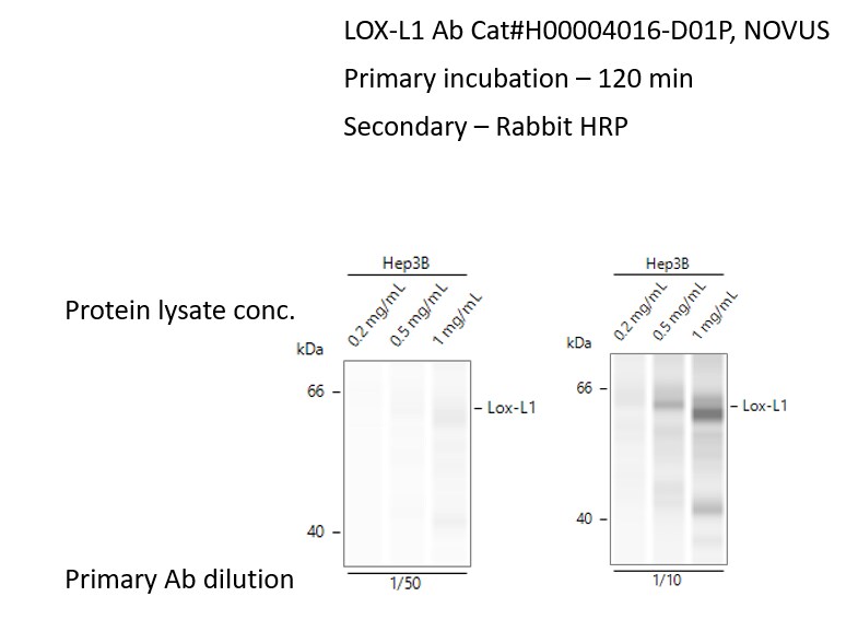
Simple Western: LOXL1 Antibody [H00004016-D01P] - Simple Western lane views show lysates of human Hep3B cell line. Specific bands were detected for LOXL1 using LOXL1 antibody at 1:50 and 1:10 diltutions. This experiment was conducted using the 12-230 kDa separation system. Image from verified customer review.
Western Blot: LOXL1 Antibody - Azide and BSA Free [H00004016-D01P] -
LOXL1 interacts with apoptosis-related modulators.a Schematic of LOXL1 interactor discovery. The potential interactors were presented at higher levels in the LOXL1 experimental group than in the IgG control group. Two replicates of IP-MS were conducted. b Scatter plots of the log2 ratios of iBAQ intensities for the proteins quantified using MS in the LOXL1 Co-IP samples from two replicates compared with the IgG control samples. Proteins displayed in red displayed a fold change of > 2 in two replicates and were determined to potentially interact with LOXL1. c Protein-protein interactions among potential LOXL1 interactors, as revealed by the STRING database. The gray lines indicate the relationships among these proteins, and the black lines indicate the direct interactions between LOXL1 and other proteins. d LOXL1 interacts with multiple proteins, especially BAG2 in U87 cells overexpressing LOXL1. e Endogenous reciprocal Co-IP assay was further performed to test the protein-protein interaction between LOXL1 and BAG2, BAG3 or HSPA1B in LN18 cells. f BAG2 was depleted by transiently transfecting specific siRNA and siNC was used as a negative control. Indicated antibodies against BAG2 and LOXL1 were used to test their protein levels. The percentages of apoptotic cells were examined. (means +/- SD, unpaired t test, two-tailed). g The scatter plot of correlation data between LOXL1 and BAG2 in glioma was provided by mining TCGA database. Image collected and cropped by CiteAb from the following open publication (https://pubmed.ncbi.nlm.nih.gov/32424143), licensed under a CC-BY license. Not internally tested by Novus Biologicals.
Western Blot: LOXL1 Antibody - Azide and BSA Free [H00004016-D01P] -
LOXL1 regulates BAG2 stability through both its enzymatic activity and direct interaction with BAG2.a, b BAG2 protein levels were increased by LOXL1. U87 cells were transiently transfected with plasmids overexpressing wild type (WT) or ED mutant LOXL1, and Vec was used as a negative control. CHX (cycloheximide, 1 μM) was used to treat cells over time (b). c GST-LOXL1, including full length (FL), 1 to 363 AAs (1-363) and 364 to the end AAs (364-END), was incubated with His-BAG2. d His-BAG2, including full length (FL), 1 to 108 AAs (1-108) and 109 to the end AAs (109-END), was incubated with GST-LOXL1. e Molecular simulations were performed to find the potential sites required for interacting with the BAG domain of BAG2. Then, measurement of LOXL1 enzymatic activity was performed. f D515A mutation reduced the direct interaction between LOXL1 and BAG2. GST-LOXL1 (including WT, N513A and D515A) was incubated with His-BAG2. g The interaction diagram of LOXL1-D515 with BAG2-K186. h U87 cells were transiently transfected with plasmids overexpressing WT, ED or D515A mutant LOXL1. i Endogenous BAG2 was determined with a specific antibody when LOXL1 D515 was mutated into A515. j The reduced interaction between LOXL1 and BAG2 decreased the ability of glioma cells to resist apoptosis. Image collected and cropped by CiteAb from the following open publication (https://pubmed.ncbi.nlm.nih.gov/32424143), licensed under a CC-BY license. Not internally tested by Novus Biologicals.
Flow Cytometry: LOXL1 Antibody - Azide and BSA Free [H00004016-D01P] -
VEGFR-Src axis signaling increases LOXL1 expression which positively correlates with BAG2 during glioma progression.a Left panel, inhibitor screen of receptor-mediated signaling pathways required for LOXL1 upregulation in U87 cells; right panel, inhibitor screen of central kinases required for LOXL1 upregulation in U87 cells. b Knocking down of SRC gene by siRNA reduced LOXL1 protein in LN18 cells. c Forced expression of LOXL1 rescued the inhibition of Src kinase activity, as determined using apoptosis assays (means +/- SD, one-way ANOVA). d Upper panel, Promo web software predicted the potential transcriptional factors that bound to the LOXL1 promoter (3000 bp upstream of the TSS); Lower panel, a ChIP assay identified that CEBPA targeted the LOXL1 promoter at 480 base pairs upstream of the TSS in LN18 cells (means +/- SD, unpaired t test, two-tailed). e Knocking down CEBPA reduced LOXL1 expression in LN18 cells. Four pairs of siRNAs were applied to target the CEBPA gene. f Real-time qPCR analyses of glioma specimens were performed. The correlation of LOXL1 expression with CEBPA is shown as R2 and p value. g Representative images of IHC staining of glioma specimens are shown. Scale bar, 100 μm. h Semi-quantitative scoring (using a scale from 0 to 300 points) was conducted, and Pearson’s correlation test was performed and evaluated using the R2 and p value. i Blood LOXL1 levels were measured using ELISA, and 21 glioma specimens were divided into two groups: high (H) or low (L) (means +/- SD, unpaired t test, two-tailed). j BAG2 levels were higher in the H group than in the L group. (means +/- SD, unpaired t test, two-tailed) k Diagram showing the mechanism by which LOXL1 exerts its antiapoptotic activity. Image collected and cropped by CiteAb from the following open publication (https://pubmed.ncbi.nlm.nih.gov/32424143), licensed under a CC-BY license. Not internally tested by Novus Biologicals.
Western Blot: LOXL1 Antibody - Azide and BSA Free [H00004016-D01P] -
LOXL1 regulates BAG2 stability through both its enzymatic activity and direct interaction with BAG2.a, b BAG2 protein levels were increased by LOXL1. U87 cells were transiently transfected with plasmids overexpressing wild type (WT) or ED mutant LOXL1, and Vec was used as a negative control. CHX (cycloheximide, 1 μM) was used to treat cells over time (b). c GST-LOXL1, including full length (FL), 1 to 363 AAs (1-363) and 364 to the end AAs (364-END), was incubated with His-BAG2. d His-BAG2, including full length (FL), 1 to 108 AAs (1-108) and 109 to the end AAs (109-END), was incubated with GST-LOXL1. e Molecular simulations were performed to find the potential sites required for interacting with the BAG domain of BAG2. Then, measurement of LOXL1 enzymatic activity was performed. f D515A mutation reduced the direct interaction between LOXL1 and BAG2. GST-LOXL1 (including WT, N513A and D515A) was incubated with His-BAG2. g The interaction diagram of LOXL1-D515 with BAG2-K186. h U87 cells were transiently transfected with plasmids overexpressing WT, ED or D515A mutant LOXL1. i Endogenous BAG2 was determined with a specific antibody when LOXL1 D515 was mutated into A515. j The reduced interaction between LOXL1 and BAG2 decreased the ability of glioma cells to resist apoptosis. Image collected and cropped by CiteAb from the following open publication (https://pubmed.ncbi.nlm.nih.gov/32424143), licensed under a CC-BY license. Not internally tested by Novus Biologicals.
Immunohistochemistry: LOXL1 Antibody - Azide and BSA Free [H00004016-D01P] -
VEGFR-Src axis signaling increases LOXL1 expression which positively correlates with BAG2 during glioma progression.a Left panel, inhibitor screen of receptor-mediated signaling pathways required for LOXL1 upregulation in U87 cells; right panel, inhibitor screen of central kinases required for LOXL1 upregulation in U87 cells. b Knocking down of SRC gene by siRNA reduced LOXL1 protein in LN18 cells. c Forced expression of LOXL1 rescued the inhibition of Src kinase activity, as determined using apoptosis assays (means +/- SD, one-way ANOVA). d Upper panel, Promo web software predicted the potential transcriptional factors that bound to the LOXL1 promoter (3000 bp upstream of the TSS); Lower panel, a ChIP assay identified that CEBPA targeted the LOXL1 promoter at 480 base pairs upstream of the TSS in LN18 cells (means +/- SD, unpaired t test, two-tailed). e Knocking down CEBPA reduced LOXL1 expression in LN18 cells. Four pairs of siRNAs were applied to target the CEBPA gene. f Real-time qPCR analyses of glioma specimens were performed. The correlation of LOXL1 expression with CEBPA is shown as R2 and p value. g Representative images of IHC staining of glioma specimens are shown. Scale bar, 100 μm. h Semi-quantitative scoring (using a scale from 0 to 300 points) was conducted, and Pearson’s correlation test was performed and evaluated using the R2 and p value. i Blood LOXL1 levels were measured using ELISA, and 21 glioma specimens were divided into two groups: high (H) or low (L) (means +/- SD, unpaired t test, two-tailed). j BAG2 levels were higher in the H group than in the L group. (means +/- SD, unpaired t test, two-tailed) k Diagram showing the mechanism by which LOXL1 exerts its antiapoptotic activity. Image collected and cropped by CiteAb from the following open publication (https://pubmed.ncbi.nlm.nih.gov/32424143), licensed under a CC-BY license. Not internally tested by Novus Biologicals.
Immunohistochemistry: LOXL1 Antibody - Azide and BSA Free [H00004016-D01P] -
Representative immunohistochemistry of LOXs in PDAC tissues and normal pancreas tissues. (A) IHC staining of LOX and quantification showed higher expression in PDAC than normal pancreas tissues. (B) IHC staining and quantification showed higher expression of LOXL1 in PDAC. (C) IHC staining and quantification showed higher expression of LOXL2 in PDAC. (D) IHC staining and quantification showed lower expression of LOXL3 in PDAC. (E) LOXL4 expression levels were similar in PDAC and normal tissues. *p < 0.01; NS, no significant difference. n = 6, repeated 5 times. Image collected and cropped by CiteAb from the following open publication (https://pubmed.ncbi.nlm.nih.gov/35433829), licensed under a CC-BY license. Not internally tested by Novus Biologicals.Applications for LOXL1 Antibody - Azide and BSA Free
Application
Recommended Usage
Western Blot
1:500
Application Notes
Antibody reactivity against Recombinant Protein with GST tag on ELISA and WB and also on transfected lysate in WB. GST tag alone is used as a negative control. Use in IHC reported in secitific publication PMID: 32424143. LOXL1 Antibody validated for Simple Western from a verified customer review.
Reviewed Applications
Read 1 review rated 5 using H00004016-D01P in the following applications:
Formulation, Preparation, and Storage
Purification
Protein A purified
Formulation
PBS (pH 7.4)
Format
Azide and BSA Free
Preservative
No Preservative
Concentration
Concentrations vary lot to lot. See vial label for concentration. If unlisted please contact technical services.
Shipping
The product is shipped with polar packs. Upon receipt, store it immediately at the temperature recommended below.
Stability & Storage
Aliquot and store at -20C or -80C. Avoid freeze-thaw cycles.
Background: LOXL1
Long Name
Lysyl Oxidase like 1
Alternate Names
LOXL, Lysyl Oxidase Homolog 1, Lysyl Oxidase like 1
Entrez Gene IDs
4016 (Human)
Gene Symbol
LOXL1
UniProt
Additional LOXL1 Products
Product Documents for LOXL1 Antibody - Azide and BSA Free
Certificate of Analysis
To download a Certificate of Analysis, please enter a lot or batch number in the search box below.
Product Specific Notices for LOXL1 Antibody - Azide and BSA Free
This product is produced by and distributed for Abnova, a company based in Taiwan.
This product is for research use only and is not approved for use in humans or in clinical diagnosis. Primary Antibodies are guaranteed for 1 year from date of receipt.
Related Research Areas
Citations for LOXL1 Antibody - Azide and BSA Free
 Powered by Bioz
Customer Reviews for LOXL1 Antibody - Azide and BSA Free (1)
5 out of 5
1 Customer Rating
Have you used LOXL1 Antibody - Azide and BSA Free?
Submit a review and receive an Amazon gift card!
$25/€18/£15/$25CAN/¥2500 Yen for a review with an image
$10/€7/£6/$10CAN/¥1110 Yen for a review without an image
Submit a review
Customer Images
Showing
1
-
1 of
1 review
Showing All
Filter By:
-
Application: Simple WesternSample Tested: hep3b cell lineSpecies: HumanVerified Customer | Posted 12/06/2022Simple protein (Jess): 12-230 kda seperation modules. Best Protein Lysate concentration: 1mg/ml Best Primary Ab dilution: 1:10Test Ab: LOX-L1 Ab Cat#H00004016-D01P, NOVUS Sample: Hep3B cell line Simple protein (Jess): 12-230 kda seperation modules. Best Protein Lysate concentration: 1mg/ml Best Primary Ab dilution: 1:10 Secondary - Rabbit HRP
 Bio-Techne ResponseThis review was submitted through the legacy Novus Innovators Program, reflecting a new species or application tested on a primary antibody.
There are no reviews that match your criteria.
Protocols
Find general support by application which include: protocols, troubleshooting, illustrated assays, videos and webinars.
- Antigen Retrieval Protocol (PIER)
- Antigen Retrieval for Frozen Sections Protocol
- Appropriate Fixation of IHC/ICC Samples
- Cellular Response to Hypoxia Protocols
- Chromogenic IHC Staining of Formalin-Fixed Paraffin-Embedded (FFPE) Tissue Protocol
- Chromogenic Immunohistochemistry Staining of Frozen Tissue
- ClariTSA™ Fluorophore Kits
- Detection & Visualization of Antibody Binding
- Fluorescent IHC Staining of Frozen Tissue Protocol
- Graphic Protocol for Heat-induced Epitope Retrieval
- Graphic Protocol for the Preparation and Fluorescent IHC Staining of Frozen Tissue Sections
- Graphic Protocol for the Preparation and Fluorescent IHC Staining of Paraffin-embedded Tissue Sections
- Graphic Protocol for the Preparation of Gelatin-coated Slides for Histological Tissue Sections
- ICC Cell Smear Protocol for Suspension Cells
- ICC Immunocytochemistry Protocol Videos
- ICC for Adherent Cells
- IHC Sample Preparation (Frozen sections vs Paraffin)
- Immunocytochemistry (ICC) Protocol
- Immunocytochemistry Troubleshooting
- Immunofluorescence of Organoids Embedded in Cultrex Basement Membrane Extract
- Immunofluorescent IHC Staining of Formalin-Fixed Paraffin-Embedded (FFPE) Tissue Protocol
- Immunohistochemistry (IHC) and Immunocytochemistry (ICC) Protocols
- Immunohistochemistry Frozen Troubleshooting
- Immunohistochemistry Paraffin Troubleshooting
- Preparing Samples for IHC/ICC Experiments
- Preventing Non-Specific Staining (Non-Specific Binding)
- Primary Antibody Selection & Optimization
- Protocol for Heat-Induced Epitope Retrieval (HIER)
- Protocol for Making a 4% Formaldehyde Solution in PBS
- Protocol for VisUCyte™ HRP Polymer Detection Reagent
- Protocol for the Fluorescent ICC Staining of Cell Smears - Graphic
- Protocol for the Fluorescent ICC Staining of Cultured Cells on Coverslips - Graphic
- Protocol for the Preparation & Fixation of Cells on Coverslips
- Protocol for the Preparation and Chromogenic IHC Staining of Frozen Tissue Sections
- Protocol for the Preparation and Chromogenic IHC Staining of Frozen Tissue Sections - Graphic
- Protocol for the Preparation and Chromogenic IHC Staining of Paraffin-embedded Tissue Sections
- Protocol for the Preparation and Chromogenic IHC Staining of Paraffin-embedded Tissue Sections - Graphic
- Protocol for the Preparation and Fluorescent ICC Staining of Cells on Coverslips
- Protocol for the Preparation and Fluorescent ICC Staining of Non-adherent Cells
- Protocol for the Preparation and Fluorescent ICC Staining of Stem Cells on Coverslips
- Protocol for the Preparation and Fluorescent IHC Staining of Frozen Tissue Sections
- Protocol for the Preparation and Fluorescent IHC Staining of Paraffin-embedded Tissue Sections
- Protocol for the Preparation of Gelatin-coated Slides for Histological Tissue Sections
- Protocol for the Preparation of a Cell Smear for Non-adherent Cell ICC - Graphic
- R&D Systems Quality Control Western Blot Protocol
- TUNEL and Active Caspase-3 Detection by IHC/ICC Protocol
- The Importance of IHC/ICC Controls
- Troubleshooting Guide: Immunohistochemistry
- Troubleshooting Guide: Western Blot Figures
- Western Blot Conditions
- Western Blot Protocol
- Western Blot Protocol for Cell Lysates
- Western Blot Troubleshooting
- Western Blot Troubleshooting Guide
- View all Protocols, Troubleshooting, Illustrated assays and Webinars
Loading...