LPAR1/LPA1/EDG-2 Antibody - BSA Free
Novus Biologicals | Catalog # NBP1-03363
![Western Blot: LPAR1/LPA1/EDG-2 AntibodyBSA Free [NBP1-03363]](https://resources.rndsystems.com/images/products/LPAR1-LPA1-EDG-2-Antibody-Western-Blot-NBP1-03363-img0004.jpg "Western Blot: LPAR1/LPA1/EDG-2 AntibodyBSA Free [NBP1-03363]")
Key Product Details
Species Reactivity
Validated:
Human, Mouse, Rat, Chicken
Cited:
Human, Mouse, Rat
Predicted:
Bovine (100%), Sheep (100%). Backed by our 100% Guarantee.
Applications
Validated:
Immunohistochemistry, Immunohistochemistry-Paraffin, Immunohistochemistry-Frozen, Western Blot, Immunocytochemistry/ Immunofluorescence, Simple Western
Cited:
Knockout Validated, Western Blot, Flow Cytometry, Immunocytochemistry/ Immunofluorescence, IF/IHC
Label
Unconjugated
Antibody Source
Polyclonal Rabbit IgG
Format
BSA Free
Loading...
Product Specifications
Immunogen
Synthetic peptide made to an internal portion of human EDG2 (within residues 200-300). [Swiss-Prot# Q92633]
Reactivity Notes
Predicted to react with sheep, and bovine based on 100% sequence homology. Positive staining in IHC-Fr/IF with 16 um cryostat sections of embryonic day 6 (E6) chicken brain was reported by a customer. Mouse reactivity reported in scientific literature (PMID: 24147068).
Localization
Cell membrane; Multi-pass membrane protein.
Clonality
Polyclonal
Host
Rabbit
Isotype
IgG
Theoretical MW
43 kDa.
Disclaimer note: The observed molecular weight of the protein may vary from the listed predicted molecular weight due to post translational modifications, post translation cleavages, relative charges, and other experimental factors.
Disclaimer note: The observed molecular weight of the protein may vary from the listed predicted molecular weight due to post translational modifications, post translation cleavages, relative charges, and other experimental factors.
Scientific Data Images for LPAR1/LPA1/EDG-2 Antibody - BSA Free
Western Blot: LPAR1/LPA1/EDG-2 AntibodyBSA Free [NBP1-03363]
Western Blot: LPAR1/LPA1/EDG-2 Antibody [NBP1-03363] - Detection of EDG2 in A549 whole cell extracts using NBP1-03363. The band at ~41 kDa position represents the target protein EDG2, whereas, the band at ~55kDa may potentially be the post-translationally modified (glycosylated, palmitoylated or lipidated) form of this protein.![Immunocytochemistry/ Immunofluorescence: LPAR1/LPA1/EDG-2 Antibody - BSA Free [NBP1-03363]](https://resources.rndsystems.com/images/products/LPAR1-LPA1-EDG-2-Antibody-Immunocytochemistry-Immunofluorescence-NBP1-03363-img0005.jpg "Immunocytochemistry/ Immunofluorescence: LPAR1/LPA1/EDG-2 Antibody - BSA Free [NBP1-03363]")
Immunocytochemistry/ Immunofluorescence: LPAR1/LPA1/EDG-2 Antibody - BSA Free [NBP1-03363]
Immunocytochemistry/Immunofluorescence: LPAR1/LPA1/EDG-2 Antibody [NBP1-03363] - A431 cells were fixed for 10 minutes using 10% formalin and then permeabilized for 5 minutes using 1X TBS + 0.5% Triton-X100. The cells were incubated with anti-EDG2/LPAR1/LPA1 (NBP1-03363) at a 1:100 dilution overnight at 4C and detected with an anti-rabbit Dylight 488 (Green) at a 1:500 dilution. Alpha tubulin was used as a co-stain at a 1:1000 dilution and detected with and anti-mouse Dylight 550 (Red) at a 1:500 dilution. Nuclei were counterstained with DAPI (Blue). Cells were imaged using a 40X objective.![Immunohistochemistry-Paraffin: LPAR1/LPA1/EDG-2 Antibody - BSA Free [NBP1-03363]](https://resources.rndsystems.com/images/products/LPAR1-LPA1-EDG-2-Antibody-Immunohistochemistry-Paraffin-NBP1-03363-img0006.jpg "Immunohistochemistry-Paraffin: LPAR1/LPA1/EDG-2 Antibody - BSA Free [NBP1-03363]")
Immunohistochemistry-Paraffin: LPAR1/LPA1/EDG-2 Antibody - BSA Free [NBP1-03363]
Immunohistochemistry-Paraffin: LPAR1/LPA1/EDG-2 Antibody [NBP1-03363] - IHC analysis of formalin fixed paraffin-embedded (FFPE) human prostate cancer using LPAR1 antibody at 1:100 on a Bond Rx autostainer (Leica Biosystems). The assay involved 20 minutes of heat induced antigen retrieval (HIER) using 10mM sodium citrate buffer (pH 6.0) and endogenous peroxidase quenching with peroxide block. The sections were incubated with primary antibody for 30 minutes and Bond Polymer Refine Detection (Leica Biosystems) with DAB was used for signal development followed by counterstaining with hematoxylin. Whole slide scanning and capturing of representative images was performed using Aperio AT2 (Leica Biosystems). Staining was obseved in the scattered ducts. Staining was performed by Histowiz.![Simple Western: LPAR1/LPA1/EDG-2 AntibodyBSA Free [NBP1-03363]](https://resources.rndsystems.com/images/products/LPAR1-LPA1-EDG-2-Antibody-Simple-Western-NBP1-03363-img0002.jpg "Simple Western: LPAR1/LPA1/EDG-2 AntibodyBSA Free [NBP1-03363]")
Simple Western: LPAR1/LPA1/EDG-2 AntibodyBSA Free [NBP1-03363]
Simple Western: LPAR1/LPA1/EDG-2 Antibody [NBP1-03363] - Simple Western lane view shows a specific band for EDG2 in 0.5 mg/ml of A431 lysate. This experiment was performed under reducing conditions using the 12-230 kDa separation system.Applications for LPAR1/LPA1/EDG-2 Antibody - BSA Free
Application
Recommended Usage
Immunocytochemistry/ Immunofluorescence
1:100-1:500
Immunohistochemistry
1:100-1:500
Immunohistochemistry-Frozen
1:100-1:500
Immunohistochemistry-Paraffin
1:100 -1:500
Simple Western
1:200
Western Blot
0.5 - 2.0 ug/ml
Application Notes
This EDG2 antibody is useful for Immunocytochemistry/Immunofluorescence, Immunohistochemistry-Frozen and Western Blot, where a band is seen approx. 43 kDa.
In Simple Western only 10 - 15 uL of the recommended dilution is used per data point.
See Simple Western Antibody Database for Simple Western validation: Tested in A431 lysate 0.5 mg/mL, separated by Size, antibody dilution of 1:200, apparent MW was 43 kDa. Separated by Size-Wes, Sally Sue/Peggy Sue.
The observed molecular weight of the protein may vary from the listed predicted molecular weight due to post translational modifications, post translation cleavages, relative charges, and other experimental factors.
Reviewed Applications
Read 1 review rated 4 using NBP1-03363 in the following applications:
Formulation, Preparation, and Storage
Purification
Immunogen affinity purified
Formulation
PBS
Format
BSA Free
Preservative
0.02% Sodium Azide
Concentration
1 mg/ml
Shipping
The product is shipped with polar packs. Upon receipt, store it immediately at the temperature recommended below.
Stability & Storage
Store at 4C short term. Aliquot and store at -20C long term. Avoid freeze-thaw cycles.
Background: LPAR1/LPA1/EDG-2
Long Name
Lysophosphatidic Acid Receptor 1
Alternate Names
EDG2, GPCR26, GPR26, LPA1, rec.1.3, vzg-1
Gene Symbol
LPAR1
UniProt
Additional LPAR1/LPA1/EDG-2 Products
Product Documents for LPAR1/LPA1/EDG-2 Antibody - BSA Free
Certificate of Analysis
To download a Certificate of Analysis, please enter a lot or batch number in the search box below.
Product Specific Notices for LPAR1/LPA1/EDG-2 Antibody - BSA Free
This product is for research use only and is not approved for use in humans or in clinical diagnosis. Primary Antibodies are guaranteed for 1 year from date of receipt.
Related Research Areas
Citations for LPAR1/LPA1/EDG-2 Antibody - BSA Free
 Powered by Bioz
Customer Reviews for LPAR1/LPA1/EDG-2 Antibody - BSA Free (1)
4 out of 5
1 Customer Rating
Have you used LPAR1/LPA1/EDG-2 Antibody - BSA Free?
Submit a review and receive an Amazon gift card!
$25/€18/£15/$25CAN/¥2500 Yen for a review with an image
$10/€7/£6/$10CAN/¥1110 Yen for a review without an image
Submit a review
Customer Images
Showing
1
-
1 of
1 review
Showing All
Filter By:
-
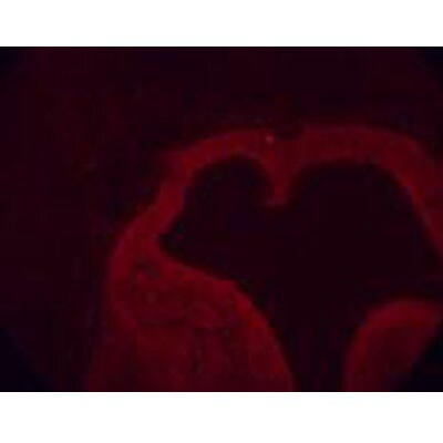
Application: ImmunofluorescenceSample Tested: 16 um cryostat sections of embryonic day 6 (E6) chicken brainSpecies: OtherVerified Customer | Posted 10/20/2011

There are no reviews that match your criteria.
Protocols
View specific protocols for LPAR1/LPA1/EDG-2 Antibody - BSA Free (NBP1-03363):
LPAR1/LPA1/EDG-2 Antibody:
Immunocytochemistry Protocol
Culture cells to appropriate density in 35 mm culture dishes or 6-well plates.
1. Remove culture medium and add 10% formalin to the dish. Fix at room temperature for 30 minutes.
2. Remove the formalin and add ice cold methanol. Incubate for 5-10 minutes.
3. Remove methanol and add washing solution (i.e. PBS). Be sure to not let the specimen dry out. Wash three times for 10 minutes.
4. To block nonspecific antibody binding incubate in 10% normal goat serum from 1 hour to overnight at room temperature.
5. Add primary antibody at appropriate dilution and incubate at room temperature from 2 hours to overnight at room temperature.
6. Remove primary antibody and replace with washing solution. Wash three times for 10 minutes.
7. Add secondary antibody at appropriate dilution. Incubate for 1 hour at room temperature.
8. Remove antibody and replace with wash solution, then wash for 10 minutes. Add Hoechst 33258 to wash solution at 1:25,0000 and incubate for 10 minutes. Wash a third time for 10 minutes.
9. Cells can be viewed directly after washing. The plates can also be stored in PBS containing Azide covered in Parafilm (TM). Cells can also be cover-slipped using Fluoromount, with appropriate sealing.
*The above information is only intended as a guide. The researcher should determine what protocol best meets their needs. Please follow safe laboratory procedures.
LPAR1/LPA1/EDG-2 Antibody:
Western Blot Protocol
1. Perform SDS-PAGE on samples to be analyzed, loading 25 ug of total protein per lane.
2. Transfer proteins to membrane according to the instructions provided by the manufacturer of the membrane and transfer apparatus.
3. Stain according to standard Ponceau S procedure (or similar product) to assess transfer success, and mark molecular weight standards where appropriate.
4. Rinse the blot.
5. Block the membrane using standard blocking buffer for at least 1 hour.
6. Wash the membrane in wash buffer three times for 10 minutes each.
7. Dilute anti-EDG-2 primary antibody in blocking buffer and incubate 1 hour at room temperature.
8. Wash the membrane in wash buffer three times for 10 minutes each.
9. Apply the diluted HRP conjugated secondary antibody in blocking buffer (as per manufacturers instructions) and incubate 1 hour at room temperature.
10. Wash the blot in wash buffer three times for 10 minutes each (this step can be repeated as required to reduce background).
11. Apply the detection reagent of choice in accordance with the manufacturers instructions.
Note: Tween-20 can be added to the blocking or antibody dilution buffer at a final concentration of 0.05-0.2%.
Find general support by application which include: protocols, troubleshooting, illustrated assays, videos and webinars.
- Antigen Retrieval Protocol (PIER)
- Antigen Retrieval for Frozen Sections Protocol
- Appropriate Fixation of IHC/ICC Samples
- Cellular Response to Hypoxia Protocols
- Chromogenic IHC Staining of Formalin-Fixed Paraffin-Embedded (FFPE) Tissue Protocol
- Chromogenic Immunohistochemistry Staining of Frozen Tissue
- ClariTSA™ Fluorophore Kits
- Detection & Visualization of Antibody Binding
- Fluorescent IHC Staining of Frozen Tissue Protocol
- Graphic Protocol for Heat-induced Epitope Retrieval
- Graphic Protocol for the Preparation and Fluorescent IHC Staining of Frozen Tissue Sections
- Graphic Protocol for the Preparation and Fluorescent IHC Staining of Paraffin-embedded Tissue Sections
- Graphic Protocol for the Preparation of Gelatin-coated Slides for Histological Tissue Sections
- ICC Cell Smear Protocol for Suspension Cells
- ICC Immunocytochemistry Protocol Videos
- ICC for Adherent Cells
- IHC Sample Preparation (Frozen sections vs Paraffin)
- Immunocytochemistry (ICC) Protocol
- Immunocytochemistry Troubleshooting
- Immunofluorescence of Organoids Embedded in Cultrex Basement Membrane Extract
- Immunofluorescent IHC Staining of Formalin-Fixed Paraffin-Embedded (FFPE) Tissue Protocol
- Immunohistochemistry (IHC) and Immunocytochemistry (ICC) Protocols
- Immunohistochemistry Frozen Troubleshooting
- Immunohistochemistry Paraffin Troubleshooting
- Preparing Samples for IHC/ICC Experiments
- Preventing Non-Specific Staining (Non-Specific Binding)
- Primary Antibody Selection & Optimization
- Protocol for Heat-Induced Epitope Retrieval (HIER)
- Protocol for Making a 4% Formaldehyde Solution in PBS
- Protocol for VisUCyte™ HRP Polymer Detection Reagent
- Protocol for the Fluorescent ICC Staining of Cell Smears - Graphic
- Protocol for the Fluorescent ICC Staining of Cultured Cells on Coverslips - Graphic
- Protocol for the Preparation & Fixation of Cells on Coverslips
- Protocol for the Preparation and Chromogenic IHC Staining of Frozen Tissue Sections
- Protocol for the Preparation and Chromogenic IHC Staining of Frozen Tissue Sections - Graphic
- Protocol for the Preparation and Chromogenic IHC Staining of Paraffin-embedded Tissue Sections
- Protocol for the Preparation and Chromogenic IHC Staining of Paraffin-embedded Tissue Sections - Graphic
- Protocol for the Preparation and Fluorescent ICC Staining of Cells on Coverslips
- Protocol for the Preparation and Fluorescent ICC Staining of Non-adherent Cells
- Protocol for the Preparation and Fluorescent ICC Staining of Stem Cells on Coverslips
- Protocol for the Preparation and Fluorescent IHC Staining of Frozen Tissue Sections
- Protocol for the Preparation and Fluorescent IHC Staining of Paraffin-embedded Tissue Sections
- Protocol for the Preparation of Gelatin-coated Slides for Histological Tissue Sections
- Protocol for the Preparation of a Cell Smear for Non-adherent Cell ICC - Graphic
- R&D Systems Quality Control Western Blot Protocol
- TUNEL and Active Caspase-3 Detection by IHC/ICC Protocol
- The Importance of IHC/ICC Controls
- Troubleshooting Guide: Immunohistochemistry
- Troubleshooting Guide: Western Blot Figures
- Western Blot Conditions
- Western Blot Protocol
- Western Blot Protocol for Cell Lysates
- Western Blot Troubleshooting
- Western Blot Troubleshooting Guide
- View all Protocols, Troubleshooting, Illustrated assays and Webinars
Loading...