LRP2 Antibody (CD7D5) - Azide and BSA Free
Novus Biologicals | Catalog # NB110-96417
![Immunohistochemistry: LRP2 Antibody (CD7D5) - Azide and BSA Free [NB110-96417]](https://resources.rndsystems.com/images/products/LRP2-Antibody-CD7D5-Immunocytochemistry-Immunofluorescence-NB110-96417-img0005.jpg "Immunohistochemistry: LRP2 Antibody (CD7D5) - Azide and BSA Free [NB110-96417]")
Key Product Details
Species Reactivity
Validated:
Cited:
Applications
Validated:
Cited:
Label
Antibody Source
Format
Product Specifications
Immunogen
Reactivity Notes
Clonality
Host
Isotype
Theoretical MW
Disclaimer note: The observed molecular weight of the protein may vary from the listed predicted molecular weight due to post translational modifications, post translation cleavages, relative charges, and other experimental factors.
Scientific Data Images for LRP2 Antibody (CD7D5) - Azide and BSA Free
Immunohistochemistry: LRP2 Antibody (CD7D5) - Azide and BSA Free [NB110-96417]
LRP2-Antibody-CD7D5-Immunocytochemistry-Immunofluorescence-NB110-96417-img0005.jpg![Immunohistochemistry-Paraffin: LRP2 Antibody (CD7D5) - Azide and BSA Free [NB110-96417]](https://resources.rndsystems.com/images/products/LRP2-Antibody-CD7D5-Immunohistochemistry-Paraffin-NB110-96417-img0002.jpg "Immunohistochemistry-Paraffin: LRP2 Antibody (CD7D5) - Azide and BSA Free [NB110-96417]")
Immunohistochemistry-Paraffin: LRP2 Antibody (CD7D5) - Azide and BSA Free [NB110-96417]
Immunohistochemistry-Paraffin: LRP2 Antibody (CD7D5) [NB110-96417] - Staining (paraffin-embedded tissue-sections fixed in formalin) of Megalin in the brush border of the proximal tubule.![Immunocytochemistry/ Immunofluorescence: LRP2 Antibody (CD7D5) - Azide and BSA Free [NB110-96417]](https://resources.rndsystems.com/images/products/LRP2-Antibody-CD7D5-Immunocytochemistry-Immunofluorescence-NB110-96417-img0003.jpg "Immunocytochemistry/ Immunofluorescence: LRP2 Antibody (CD7D5) - Azide and BSA Free [NB110-96417]")
Immunocytochemistry/ Immunofluorescence: LRP2 Antibody (CD7D5) - Azide and BSA Free [NB110-96417]
Immunocytochemistry/Immunofluorescence: LRP2 Antibody (CD7D5) [NB110-96417] - Immunofluorescence staining of Megalin in the brush border of the proximal tubule.![Immunocytochemistry/ Immunofluorescence: LRP2 Antibody (CD7D5) - Azide and BSA Free [NB110-96417]](https://resources.rndsystems.com/images/products/LRP2-Antibody-CD7D5-Immunocytochemistry-Immunofluorescence-NB110-96417-img0007.jpg "Immunocytochemistry/ Immunofluorescence: LRP2 Antibody (CD7D5) - Azide and BSA Free [NB110-96417]")
Immunocytochemistry/ Immunofluorescence: LRP2 Antibody (CD7D5) - Azide and BSA Free [NB110-96417]
Immunocytochemistry/Immunofluorescence: LRP2 Antibody (CD7D5) [NB110-96417] - Hek293 cells were fixed in 4% paraformaldehyde for 10 minutes and permeabilized in 0.05% Triton X-100 in PBS for 5 minutes. The cells were incubated with anti-LRP2 Antibody (CD7D5) NB110-96417 at 1 ug/ml overnight at 4C and detected with an anti-mouse Dylight 488 (Green) at a 1:1000 dilution for 60 minutes. Nuclei were counterstained with DAPI (Blue). Cells were imaged using a 40X objective.![Immunohistochemistry-Paraffin: LRP2 Antibody (CD7D5) - Azide and BSA Free [NB110-96417]](https://resources.rndsystems.com/images/products/LRP2-Antibody-CD7D5-Immunohistochemistry-Paraffin-NB110-96417-img0006.jpg "Immunohistochemistry-Paraffin: LRP2 Antibody (CD7D5) - Azide and BSA Free [NB110-96417]")
Immunohistochemistry-Paraffin: LRP2 Antibody (CD7D5) - Azide and BSA Free [NB110-96417]
Immunohistochemistry-Paraffin: LRP2 Antibody (CD7D5) [NB110-96417] - Analysis of a FFPE tissue section of mouse kidney using 1:200 dilution of LRP2 [CD7D5] antibody. The staining was developed using HRP labeled anti-mouse secondary antibody and DAB reagent, and nuclei of cells were counter-stained with hematoxylin.![Immunocytochemistry/ Immunofluorescence: LRP2 Antibody (CD7D5) - Azide and BSA Free [NB110-96417]](https://resources.rndsystems.com/images/products/LRP2-Antibody-CD7D5-Immunocytochemistry-Immunofluorescence-NB110-96417-img0004.jpg "Immunocytochemistry/ Immunofluorescence: LRP2 Antibody (CD7D5) - Azide and BSA Free [NB110-96417]")
Immunocytochemistry/ Immunofluorescence: LRP2 Antibody (CD7D5) - Azide and BSA Free [NB110-96417]
Immunocytochemistry/Immunofluorescence: LRP2 Antibody (CD7D5) [NB110-96417] - U2OS cells were fixed for 10 minutes using 10% formalin and then permeabilized for 5 minutes using 1X PBS + 0.1% Saponin. The cells were incubated with anti-LRP2 (CD7D5) at 20 ug/ml overnight at 4C and detected with an anti-mouse Dylight 488 (Green) at a 1:500 dilution. Nuclei were counterstained with DAPI (Blue). Cells were imaged using a 40X objective.![Immunohistochemistry-Paraffin: LRP2 Antibody (CD7D5) - Azide and BSA Free [NB110-96417]](https://resources.rndsystems.com/images/products/LRP2-Antibody-CD7D5-Immunohistochemistry-Paraffin-NB110-96417-img0001.jpg "Immunohistochemistry-Paraffin: LRP2 Antibody (CD7D5) - Azide and BSA Free [NB110-96417]")
Immunohistochemistry-Paraffin: LRP2 Antibody (CD7D5) - Azide and BSA Free [NB110-96417]
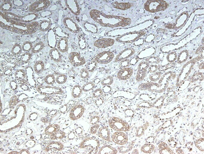
Immunohistochemistry-Paraffin: LRP2 Antibody (CD7D5) [NB110-96417] - Staining on human kidney at 1:100, overnight incubation at 4C. Image from a review by a confirmed customer. - Azide and BSA Free [NB110-96417] -")
Immunocytochemistry/ Immunofluorescence: LRP2 Antibody (CD7D5) - Azide and BSA Free [NB110-96417] -
Immunocytochemistry/ Immunofluorescence: LRP2 Antibody (CD7D5) - Azide and BSA Free [NB110-96417] - ICM & TE progenitors show loss of responsiveness to Hippo signaling manipulation at the same time as they loose responsiveness to positional changes. (D) Dox-inducible DN Lats2-IRES-mCherry transgenic embryos imaged before Dox treatment (top panel) & the same embryo was imaged following 24 hr of Dox live (middle panel) & fixed/stained for lineage markers (bottom panel). A representative embryo is shown for each stage. Live mCherry is shown as an extended focus image, IF stainings shown as single plane images. mCherry positive ICMs in mosaic transgenic embryos circled with a dotted line. Arrow points to a rare ICM cell in a 64 cell stage-induced embryo with weak Cdx2 expression, which also co-expressed an ICM marker. Scale bar: 25 µm. n indicates number of transgenic embryos analyzed. Image collected & cropped by CiteAb from the following publication (https://elifesciences.org/articles/22906), licensed under a CC-BY license. Not internally tested by Novus Biologicals. - Azide and BSA Free [NB110-96417] -")
Immunocytochemistry/ Immunofluorescence: LRP2 Antibody (CD7D5) - Azide and BSA Free [NB110-96417] -
Immunocytochemistry/ Immunofluorescence: LRP2 Antibody (CD7D5) - Azide and BSA Free [NB110-96417] - Impairment of autophagy in Atg7-deficient renal proximal tubular cells. (A) Decrease of Atg7 & increase of LC3-I in 2-month-old Atg7flox/flox;KAP-Cre+ mouse kidney. Atg7 & LC3 Western blot (LC3-I/unconjugated LC3 & LC3-II/lipidated LC3) using whole kidney lysate of Atg7flox/flox;KAP-Cre+ mice. As a control, Atg7flox/flox mouse kidney was employed. The intensity of each band of Atg7 & beta -actin was estimated by densitometry. The ratio of Atg7 to beta -actin was calculated. The amount of Atg7-positive signals in the Atg7flox/flox;KAP-Cre+ mice kidney was about 63% lower than that in the Atg7flox/flox mice kidney (n = 3). Note that LC3-I was increased in the Atg7flox/flox;KAP-Cre+ mouse kidney. (B) The massive accumulation of p62 in kidneys of 2-month old Atg7flox/flox;KAP-Cre+ mouse. The cortico-medullary region of each kidney in 2 month-old Atg7flox/flox;KAP-Cre+ mouse & Atg7flox/flox mouse was recognized with anti-p62 antibody (red). As a marker of renal proximal tubular cells, megalin (green) was employed. Nuclei were stained with 4′,6-diamidino-2-phenylindole (DAPI; blue). (C) Quantification of p62 positive area of 2-month-old Atg7flox/flox;KAP-Cre+ (n = 5) & Atg7flox/flox kidney (n = 4, *** p < 0.01). Data in graphs are expressed as the mean ± SEM. Statistical analyses were performed using a student’s t-test. (D) Age-dependent accumulation of p62 (red) in the Atg7flox/flox;KAP-Cre+ mouse kidney in 1-, 2-, 6-, & 9-month-old Atg7flox/flox;KAP-Cre+ (lower) & Atg7flox/flox (upper) mouse kidney. Image collected & cropped by CiteAb from the following publication (https://pubmed.ncbi.nlm.nih.gov/31881660), licensed under a CC-BY license. Not internally tested by Novus Biologicals.Applications for LRP2 Antibody (CD7D5) - Azide and BSA Free
Immunocytochemistry/ Immunofluorescence
Immunohistochemistry
Immunohistochemistry-Frozen
Immunohistochemistry-Paraffin
Reviewed Applications
Read 1 review rated 4 using NB110-96417 in the following applications:
Formulation, Preparation, and Storage
Purification
Formulation
Format
Preservative
Concentration
Shipping
Stability & Storage
Background: Megalin/LRP2
Long Name
Alternate Names
Gene Symbol
UniProt
Additional Megalin/LRP2 Products
Product Documents for LRP2 Antibody (CD7D5) - Azide and BSA Free
Certificate of Analysis
To download a Certificate of Analysis, please enter a lot or batch number in the search box below.
Product Specific Notices for LRP2 Antibody (CD7D5) - Azide and BSA Free
This product is for research use only and is not approved for use in humans or in clinical diagnosis. Primary Antibodies are guaranteed for 1 year from date of receipt.
Related Research Areas
Citations for LRP2 Antibody (CD7D5) - Azide and BSA Free
 Powered by Bioz
Customer Reviews for LRP2 Antibody (CD7D5) - Azide and BSA Free (1)
Have you used LRP2 Antibody (CD7D5) - Azide and BSA Free?
Submit a review and receive an Amazon gift card!
$25/€18/£15/$25CAN/¥2500 Yen for a review with an image
$10/€7/£6/$10CAN/¥1110 Yen for a review without an image
Submit a review
Customer Images
-
Application: Immunohistochemistry-ParaffinSample Tested: Human kidneySpecies: HumanVerified Customer | Posted 10/24/2013

There are no reviews that match your criteria.
Protocols
Find general support by application which include: protocols, troubleshooting, illustrated assays, videos and webinars.
- Antigen Retrieval Protocol (PIER)
- Antigen Retrieval for Frozen Sections Protocol
- Appropriate Fixation of IHC/ICC Samples
- Cellular Response to Hypoxia Protocols
- Chromogenic IHC Staining of Formalin-Fixed Paraffin-Embedded (FFPE) Tissue Protocol
- Chromogenic Immunohistochemistry Staining of Frozen Tissue
- ClariTSA™ Fluorophore Kits
- Detection & Visualization of Antibody Binding
- Fluorescent IHC Staining of Frozen Tissue Protocol
- Graphic Protocol for Heat-induced Epitope Retrieval
- Graphic Protocol for the Preparation and Fluorescent IHC Staining of Frozen Tissue Sections
- Graphic Protocol for the Preparation and Fluorescent IHC Staining of Paraffin-embedded Tissue Sections
- Graphic Protocol for the Preparation of Gelatin-coated Slides for Histological Tissue Sections
- ICC Cell Smear Protocol for Suspension Cells
- ICC Immunocytochemistry Protocol Videos
- ICC for Adherent Cells
- IHC Sample Preparation (Frozen sections vs Paraffin)
- Immunocytochemistry (ICC) Protocol
- Immunocytochemistry Troubleshooting
- Immunofluorescence of Organoids Embedded in Cultrex Basement Membrane Extract
- Immunofluorescent IHC Staining of Formalin-Fixed Paraffin-Embedded (FFPE) Tissue Protocol
- Immunohistochemistry (IHC) and Immunocytochemistry (ICC) Protocols
- Immunohistochemistry Frozen Troubleshooting
- Immunohistochemistry Paraffin Troubleshooting
- Preparing Samples for IHC/ICC Experiments
- Preventing Non-Specific Staining (Non-Specific Binding)
- Primary Antibody Selection & Optimization
- Protocol for Heat-Induced Epitope Retrieval (HIER)
- Protocol for Making a 4% Formaldehyde Solution in PBS
- Protocol for VisUCyte™ HRP Polymer Detection Reagent
- Protocol for the Fluorescent ICC Staining of Cell Smears - Graphic
- Protocol for the Fluorescent ICC Staining of Cultured Cells on Coverslips - Graphic
- Protocol for the Preparation & Fixation of Cells on Coverslips
- Protocol for the Preparation and Chromogenic IHC Staining of Frozen Tissue Sections
- Protocol for the Preparation and Chromogenic IHC Staining of Frozen Tissue Sections - Graphic
- Protocol for the Preparation and Chromogenic IHC Staining of Paraffin-embedded Tissue Sections
- Protocol for the Preparation and Chromogenic IHC Staining of Paraffin-embedded Tissue Sections - Graphic
- Protocol for the Preparation and Fluorescent ICC Staining of Cells on Coverslips
- Protocol for the Preparation and Fluorescent ICC Staining of Non-adherent Cells
- Protocol for the Preparation and Fluorescent ICC Staining of Stem Cells on Coverslips
- Protocol for the Preparation and Fluorescent IHC Staining of Frozen Tissue Sections
- Protocol for the Preparation and Fluorescent IHC Staining of Paraffin-embedded Tissue Sections
- Protocol for the Preparation of Gelatin-coated Slides for Histological Tissue Sections
- Protocol for the Preparation of a Cell Smear for Non-adherent Cell ICC - Graphic
- TUNEL and Active Caspase-3 Detection by IHC/ICC Protocol
- The Importance of IHC/ICC Controls
- Troubleshooting Guide: Immunohistochemistry
- View all Protocols, Troubleshooting, Illustrated assays and Webinars
FAQs for LRP2 Antibody (CD7D5) - Azide and BSA Free
-
Q: Could you please forward me the above reference or an IHC protocol for this product?
A:
A: Unfortunately we do not have a product specific protocol for this particular antibody. The recommended dilution range for IHC staining is indicated on the datasheet to be between 1:10-1:2000, and should be optimized based on how highly expressed the protein is in your samples. Our standard IHC protocol will also serve as a useful guide to running your assay. Please see these links to product specific references PubMed 21896500 (https://pubmed.ncbi.nlm.nih.gov/21896500/) and PubMed 23262319 (https://pubmed.ncbi.nlm.nih.gov/23262319/) for additional protocol details.