![Western Blot: LRRFIP1 Antibody [NBP1-71835]](https://resources.rndsystems.com/images/products/LRRFIP1-Antibody-Western-Blot-NBP1-71835-img0002.jpg "Western Blot: LRRFIP1 Antibody [NBP1-71835]")
Loading...
Key Product Details
Validated by
Independent Antibodies, Biological Validation
Species Reactivity
Human
Applications
Western Blot, Immunocytochemistry/ Immunofluorescence, Immunoprecipitation
Label
Unconjugated
Antibody Source
Polyclonal Rabbit IgG
Loading...
Product Specifications
Immunogen
The immunogen for this product maps to a region between residue 500 and 550 of human Leucine Rich Repeat (in FLII) Interacting Protein 1 using the numbering given in entry NP_001131024.1 (GeneID 9208).
Clonality
Polyclonal
Host
Rabbit
Isotype
IgG
Scientific Data Images for LRRFIP1 Antibody
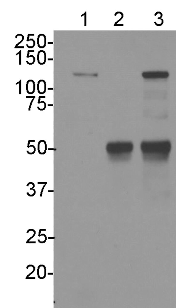
Western Blot: LRRFIP1 Antibody [NBP1-71835]
Western Blot: LRRFIP1 Antibody [NBP1-71835] - Whole cell lysate from HeLa (5, 15 and 50 ug for WB; 1 mg for IP, 20% of IP loaded) and 293T (50 ug) cells. Affinity purified rabbit anti-LRRFIP1 antibody used for WB at 0.04 ug/mL (A) and 0.4 ug/mL (B) and used for IP at 6 ug/mg lysate. LRRFIP1 was also immunoprecipitated by rabbit anti-LRRFIP1 antibodies NBP1-71834 and NBP1-71836, which recognize other epitopes.![Immunocytochemistry/ Immunofluorescence: LRRFIP1 Antibody [NBP1-71835]](https://resources.rndsystems.com/images/products/LRRFIP1-Antibody-Immunocytochemistry-Immunofluorescence-NBP1-71835-img0003.jpg "Immunocytochemistry/ Immunofluorescence: LRRFIP1 Antibody [NBP1-71835]")
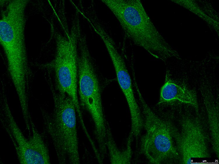
Immunocytochemistry/ Immunofluorescence: LRRFIP1 Antibody [NBP1-71835]
Immunocytochemistry/Immunofluorescence: LRRFIP1 Antibody [NBP1-71835] - LRRFIP1 in human fibroblasts (Green). 1:250 dilution in 1X PBS/1% BSA/0.3% Triton X-100. 16 hours, 4C. Image submitted by a verified customer review.![Immunoprecipitation: LRRFIP1 Antibody [NBP1-71835]](https://resources.rndsystems.com/images/products/LRRFIP1-Antibody-Immunoprecipitation-NBP1-71835-img0005.jpg "Immunoprecipitation: LRRFIP1 Antibody [NBP1-71835]")
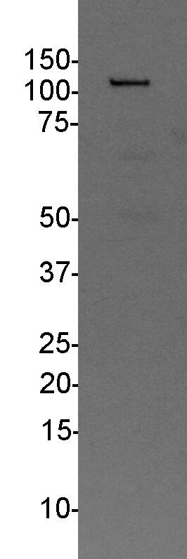
Immunoprecipitation: LRRFIP1 Antibody [NBP1-71835]
Immunoprecipitation: LRRFIP1 Antibody [NBP1-71835] - LRRFIP1 IP in B-cell whole cell lysate. 2 ug antibody : 500 ug whole cell lysate. Same antibody used at 1:2000 for Western blot. Image submitted by a verified customer review.Applications for LRRFIP1 Antibody
Application
Recommended Usage
Immunocytochemistry/ Immunofluorescence
From verified customer review.
Immunoprecipitation
2 - 5 ug/mg lysate
Western Blot
1:2000-1:10000
Reviewed Applications
Read 3 reviews rated 4.3 using NBP1-71835 in the following applications:
Formulation, Preparation, and Storage
Purification
Immunogen affinity purified
Formulation
TBS, 0.1% BSA
Preservative
0.09% Sodium Azide
Concentration
0.2 mg/ml
Shipping
The product is shipped with polar packs. Upon receipt, store it immediately at the temperature recommended below.
Stability & Storage
Store at 4C. Do not freeze.
Background: LRRFIP1
Alternate Names
FLAP-1, FLIIAP1, GC-binding factor 2, GCF2, GCF-2, HUFI-1, leucine rich repeat (in FLII) interacting protein 1, leucine-rich repeat flightless-interacting protein 1, LRR FLII-interacting protein 1, MGC10947, MGC119738, MGC119739, NEDD8-conjugating enzyme, TRIPTAR RNA-interacting protein
Gene Symbol
LRRFIP1
UniProt
Additional LRRFIP1 Products
Product Documents for LRRFIP1 Antibody
Certificate of Analysis
To download a Certificate of Analysis, please enter a lot or batch number in the search box below.
Product Specific Notices for LRRFIP1 Antibody
This product is for research use only and is not approved for use in humans or in clinical diagnosis. Primary Antibodies are guaranteed for 1 year from date of receipt.
Customer Reviews for LRRFIP1 Antibody (3)
4.3 out of 5
3 Customer Ratings
Have you used LRRFIP1 Antibody?
Submit a review and receive an Amazon gift card!
$25/€18/£15/$25CAN/¥2500 Yen for a review with an image
$10/€7/£6/$10CAN/¥1110 Yen for a review without an image
Submit a review
Customer Images
Showing
1
-
3 of
3 reviews
Showing All
Filter By:
-
Application: ImmunoprecipitationSample Tested: B-cell whole cell lysateSpecies: HumanVerified Customer | Posted 11/20/2018LRRFIP1 IP in B-cells2ug antibody : 500ug whole cell lysate. Same antibody used 1:2000 for western blot.

-
Application: ImmunocytochemistrySample Tested: fibroblastsSpecies: HumanVerified Customer | Posted 01/11/2018LRRFIP1 in human fibroblasts1:250 dilution in 1X PBS/1% BSA/0.3% Triton X-100. 16 hours, 4C.
-
Application: Western BlotSample Tested: human lymphocytes, whole cell lysateSpecies: HumanVerified Customer | Posted 12/12/201725ug. 1:2000 dilution in milk, 16H 4C.25ug. 1:2000 dilution in milk, 16H 4C.
There are no reviews that match your criteria.
Protocols
Find general support by application which include: protocols, troubleshooting, illustrated assays, videos and webinars.
- Appropriate Fixation of IHC/ICC Samples
- Cellular Response to Hypoxia Protocols
- ClariTSA™ Fluorophore Kits
- Detection & Visualization of Antibody Binding
- ICC Cell Smear Protocol for Suspension Cells
- ICC Immunocytochemistry Protocol Videos
- ICC for Adherent Cells
- Immunocytochemistry (ICC) Protocol
- Immunocytochemistry Troubleshooting
- Immunofluorescence of Organoids Embedded in Cultrex Basement Membrane Extract
- Immunohistochemistry (IHC) and Immunocytochemistry (ICC) Protocols
- Immunoprecipitation Protocol
- Preparing Samples for IHC/ICC Experiments
- Preventing Non-Specific Staining (Non-Specific Binding)
- Primary Antibody Selection & Optimization
- Protocol for VisUCyte™ HRP Polymer Detection Reagent
- Protocol for the Fluorescent ICC Staining of Cell Smears - Graphic
- Protocol for the Fluorescent ICC Staining of Cultured Cells on Coverslips - Graphic
- Protocol for the Preparation and Fluorescent ICC Staining of Cells on Coverslips
- Protocol for the Preparation and Fluorescent ICC Staining of Non-adherent Cells
- Protocol for the Preparation and Fluorescent ICC Staining of Stem Cells on Coverslips
- Protocol for the Preparation of a Cell Smear for Non-adherent Cell ICC - Graphic
- R&D Systems Quality Control Western Blot Protocol
- TUNEL and Active Caspase-3 Detection by IHC/ICC Protocol
- The Importance of IHC/ICC Controls
- Troubleshooting Guide: Western Blot Figures
- Western Blot Conditions
- Western Blot Protocol
- Western Blot Protocol for Cell Lysates
- Western Blot Troubleshooting
- Western Blot Troubleshooting Guide
- View all Protocols, Troubleshooting, Illustrated assays and Webinars
Loading...