Luciferase Antibody (Luci 21 1-107) - BSA Free
Novus Biologicals | Catalog # NB600-307
Key Product Details
Species Reactivity
Validated:
Firefly
Cited:
Mouse, Firefly
Applications
Validated:
Immunohistochemistry, Immunohistochemistry-Paraffin, Immunohistochemistry-Frozen, Western Blot, Flow Cytometry, Immunocytochemistry/ Immunofluorescence
Cited:
Immunohistochemistry-Paraffin, Western Blot, Immunocytochemistry/ Immunofluorescence, IF/IHC
Label
Unconjugated
Antibody Source
Monoclonal Mouse IgG1 kappa Clone # Luci 21 1-107
Format
BSA Free
Loading...
Product Specifications
Immunogen
This Luciferase Antibody (Luci 21 1-107) was developed against luciferase protein from Photinus pyralis (North American firefly). [UniProt# P08659].
Reactivity Notes
Photinus pyralis (North American firefly).
Localization
Peroxisome
Specificity
This Luciferase Antibody (Luci 21 1-107) is specific for Luciferase, recognizing a peptide consisting of the first 258 amino acids. Further epitope mapping has not been done at this time.
Clonality
Monoclonal
Host
Mouse
Isotype
IgG1 kappa
Scientific Data Images for Luciferase Antibody (Luci 21 1-107) - BSA Free
![Western Blot: Luciferase Antibody (Luci 21 1-107)BSA Free [NB600-307]](https://resources.rndsystems.com/images/products/Luciferase-Antibody-Luci-21-1-107-Western-Blot-NB600-307-img0006.jpg "Western Blot: Luciferase Antibody (Luci 21 1-107)BSA Free [NB600-307]")
Western Blot: Luciferase Antibody (Luci 21 1-107)BSA Free [NB600-307]
Luciferase-Antibody-Luci-21-1-107-Western-Blot-NB600-307-img0006.jpg![Immunohistochemistry: Luciferase Antibody (Luci 21 1-107) - BSA Free [NB600-307]](https://resources.rndsystems.com/images/products/Luciferase-Antibody-Luci-21-1-107-Immunohistochemistry-NB600-307-img0004.jpg "Immunohistochemistry: Luciferase Antibody (Luci 21 1-107) - BSA Free [NB600-307]")
Immunohistochemistry: Luciferase Antibody (Luci 21 1-107) - BSA Free [NB600-307]
Immunohistochemistry: Luciferase Antibody (Luci 21 1-107) [NB600-307] - Detection of Luciferase expression in CD34+ cells by Immunohistochemistry. Cytospin slides prepared from transduced CD34+ cells after 3 days of culture were stained with monoclonal anti-Luciferase antibody. Luciferase-positive cells have green cytoplasm; nuclei stained with DAPI are blue. Nontransduced, cultured CD34+ cells were used as a negative control. Original magnification, 40X. Wang, X. et al., Dynamic tracking of human hematopoietic stem cell. Blood. 102(10): 3478-3482, 2003.![Western Blot: Luciferase Antibody (Luci 21 1-107)BSA Free [NB600-307]](https://resources.rndsystems.com/images/products/Luciferase-Antibody-Luci-21-1-107-Western-Blot-NB600-307-img0005.jpg "Western Blot: Luciferase Antibody (Luci 21 1-107)BSA Free [NB600-307]")
Western Blot: Luciferase Antibody (Luci 21 1-107)BSA Free [NB600-307]
Western Blot: Luciferase Antibody (Luci 21 1-107) [NB600-307] - WB analysis of NB600-307 on Luciferase Protein (NB810-74573) - BSA Free [NB600-307] -")
Western Blot: Luciferase Antibody (Luci 21 1-107) - BSA Free [NB600-307] -
Western Blot: Luciferase Antibody (Luci 21 1-107) - BSA Free [NB600-307] - TTFields application leads to increased autophagic flux.a Ultra-structural STEM electron microscopy analysis of U-87 MG (upper panel) & A172 (lower panel) cells treated with TTFields for 48 h. Autophagosomes (blue arrows) & autolysosomes (green arrows) are indicated. b U-87 MG & A172 cells were either left untreated or were treated with TTFields for 24–72 h. CQ (20 µM) was added 4 h before cells were collected. Samples were immunoblotted for LC3 & GAPDH. Upper panel: representative blots. Lower panel: densitometric quantification of immunoblot signal, showing an average of at least three independent experiments (0.01 < *P < 0.05, Student’s t-test). c U-87 MG & A172 cells were either left untreated or were treated with TTFields for 48 h. CQ (20 µM) was added at the last 3 h of treatment & cells were fixed & stained with anti-LC3 Ab (green) & DAPI (blue). Upper panel: representative images. Original magnifications: × 40. Lower panel: quantification of LC3 intensity, presented as average intensity per cell (**P < 0.01, Student’s t-test). d U-87 MG cells were either left untreated or were treated with TTFields (48 h) or with vinblastin 25 nM (0.5 h). CQ (20 µM) was added at the last 3 h of treatment & cells were fixed & stained with anti-LC3 (green), LAMP1 (red), & DAPI (blue) (upper panel). Arrowheads indicate the strongest colocalization staining in each cell. Intensity histograms of LAMP1 & LC3 fluorescent signal calculated from the region of interest indicated by the white bar (lower panel). Representative images are shown Image collected & cropped by CiteAb from the following publication (https://pubmed.ncbi.nlm.nih.gov/30341282), licensed under a CC-BY license. Not internally tested by Novus Biologicals. - BSA Free [NB600-307] -")
Western Blot: Luciferase Antibody (Luci 21 1-107) - BSA Free [NB600-307] -
Western Blot: Luciferase Antibody (Luci 21 1-107) - BSA Free [NB600-307] - Induction of autophagy by TTFields is AMPK dependent.a U-87 MG & A172 cells either left untreated or treated w/ TTFields for indicated time points. Immunoblot analysis of GFP78 protein. Numeric values represent fold increase in GRP78 signal, normalized to loading control (GAPDH), relative to untreated control. b Quantification of intracellular ATP levels in U-87 MG cells either left untreated or treated w/ TTFields for 72 h.Results presented as average ATP concentration (nmol/2 × 106 cells) from 3 independent experiments (*P < 0.01, Student’s t-test). c U-87 MG & A172 cells either left untreated or treated w/ TTFields for indicated time points. Immunoblot analysis of pAMPK & pULK1proteins. GAPDH used as loading control. (5D-F) U-87 MG cells transfected w/ AMPK-targeting siRNA (siAMPK) or w/ siRNA sham vector (siVector), & incubated for 48 h w/ or w/out TTFields application. CQ 20 µM added for last 4 h of treatment where indicated. d (left panel) Immunoblot analysis of LC3 & AMPK. Numeric values represent fold-change in LC3-II signal, normalized to GAPDH signal, relative to respective control. d (right panel) CQ-treated cells fixed & stained for LC3 (green) & DAPI (blue), original magnifications: × 40. e Cell count of siAMPK- or siVector-expressing cells after TTFields treatment. (0.01 < *P < 0.05, Student’s t-test, n = 3). f siVector- & siAMPK-transfected U-87 MG cells either left untreated or treated w/ TTFields for 48 h. Cells then fixed & stained for cleaved caspase-3 (green) & DAPI (blue) (left panel). Images from each treatment analyzed manually & fraction of cleaved caspase-3-positive cells calculated for at least 200 cells from each group (right panel) (**P < 0.01, Student’s t-test, n = 2) Image collected & cropped by CiteAb from following publication (https://pubmed.ncbi.nlm.nih.gov/30341282), licensed under a CC-BY license. Not internally tested by Novus Biologicals. - BSA Free [NB600-307] -")
Western Blot: Luciferase Antibody (Luci 21 1-107) - BSA Free [NB600-307] -
Western Blot: Luciferase Antibody (Luci 21 1-107) - BSA Free [NB600-307] - TTFields induce autophagy in glioma cell lines.a U-87 MG & A172 cells were either left untreated or treated with TTFields at the last 24 h, 48 h, or 72 h of culturing. All cultures were plated on the same time, incubated overnight to allow cell attachment, & collected 72 h afterwards. Cells were collected, lysed, & samples were analyzed using immunoblotting for LC3 & GAPDH. Upper panel: representative blots. Lower panel: densitometric quantification of immunoblot signal, showing an average of at least three independent experiments (0.01 < *P < 0.05, **P < 0.01, Student’s t-test). b Paraffin-embedded sections from sham- or TTFields-treated rats were stained with anti-LC3 Ab (green) & DAPI (blue). Representative images are presented. c Quantification of LC3 intensity, presented as fold increase from corresponding control (*P < 0.05, Student’s t-test) Image collected & cropped by CiteAb from the following publication (https://pubmed.ncbi.nlm.nih.gov/30341282), licensed under a CC-BY license. Not internally tested by Novus Biologicals. - BSA Free [NB600-307] -")
Immunocytochemistry/ Immunofluorescence: Luciferase Antibody (Luci 21 1-107) - BSA Free [NB600-307] -
Immunocytochemistry/ Immunofluorescence: Luciferase Antibody (Luci 21 1-107) - BSA Free [NB600-307] - Immunofluorescence imaging of luciferase protein as a reporter of hybrid formation. Entire primary tumor & lung sections were imaged via tile scanning, & each image of the scan was carefully analyzed to confirm or refute positive staining for luciferase. The luciferase signal was considered a positive signal if it was above background levels associated with negative controls & corresponded to the cytoplasm of a cell with a nucleus. Rare luciferase-positive cells were detected in the primary tumors. Most red signal was not in the cytoplasm of cells associated with nuclei and, therefore, considered nonspecific [insets (a), (c)]. The lungs containing metastases on the other hand [(b), (d)] contained a large number of bona fide luciferase-positive cells corresponding to fusion products. Scale bars on slide scans = 100 μm. Scale bars on 40× inset = 25 μm. Image collected & cropped by CiteAb from the following publication (https://pubmed.ncbi.nlm.nih.gov/31069316), licensed under a CC-BY license. Not internally tested by Novus Biologicals. - BSA Free [NB600-307] -")
Western Blot: Luciferase Antibody (Luci 21 1-107) - BSA Free [NB600-307] -
Western Blot: Luciferase Antibody (Luci 21 1-107) - BSA Free [NB600-307] - TTFields induce autophagy in glioma cell lines.a U-87 MG & A172 cells were either left untreated or treated with TTFields at the last 24 h, 48 h, or 72 h of culturing. All cultures were plated on the same time, incubated overnight to allow cell attachment, & collected 72 h afterwards. Cells were collected, lysed, & samples were analyzed using immunoblotting for LC3 & GAPDH. Upper panel: representative blots. Lower panel: densitometric quantification of immunoblot signal, showing an average of at least three independent experiments (0.01 < *P < 0.05, **P < 0.01, Student’s t-test). b Paraffin-embedded sections from sham- or TTFields-treated rats were stained with anti-LC3 Ab (green) & DAPI (blue). Representative images are presented. c Quantification of LC3 intensity, presented as fold increase from corresponding control (*P < 0.05, Student’s t-test) Image collected & cropped by CiteAb from the following publication (https://pubmed.ncbi.nlm.nih.gov/30341282), licensed under a CC-BY license. Not internally tested by Novus Biologicals. - BSA Free [NB600-307] -")
Immunocytochemistry/ Immunofluorescence: Luciferase Antibody (Luci 21 1-107) - BSA Free [NB600-307] -
Immunocytochemistry/ Immunofluorescence: Luciferase Antibody (Luci 21 1-107) - BSA Free [NB600-307] - TTFields application leads to increased autophagic flux.a Ultra-structural STEM electron microscopy analysis of U-87 MG (upper panel) & A172 (lower panel) cells treated with TTFields for 48 h. Autophagosomes (blue arrows) & autolysosomes (green arrows) are indicated. b U-87 MG & A172 cells were either left untreated or were treated with TTFields for 24–72 h. CQ (20 µM) was added 4 h before cells were collected. Samples were immunoblotted for LC3 & GAPDH. Upper panel: representative blots. Lower panel: densitometric quantification of immunoblot signal, showing an average of at least three independent experiments (0.01 < *P < 0.05, Student’s t-test). c U-87 MG & A172 cells were either left untreated or were treated with TTFields for 48 h. CQ (20 µM) was added at the last 3 h of treatment & cells were fixed & stained with anti-LC3 Ab (green) & DAPI (blue). Upper panel: representative images. Original magnifications: × 40. Lower panel: quantification of LC3 intensity, presented as average intensity per cell (**P < 0.01, Student’s t-test). d U-87 MG cells were either left untreated or were treated with TTFields (48 h) or with vinblastin 25 nM (0.5 h). CQ (20 µM) was added at the last 3 h of treatment & cells were fixed & stained with anti-LC3 (green), LAMP1 (red), & DAPI (blue) (upper panel). Arrowheads indicate the strongest colocalization staining in each cell. Intensity histograms of LAMP1 & LC3 fluorescent signal calculated from the region of interest indicated by the white bar (lower panel). Representative images are shown Image collected & cropped by CiteAb from the following publication (https://pubmed.ncbi.nlm.nih.gov/30341282), licensed under a CC-BY license. Not internally tested by Novus Biologicals. - BSA Free [NB600-307] -")
Immunocytochemistry/ Immunofluorescence: Luciferase Antibody (Luci 21 1-107) - BSA Free [NB600-307] -
Immunocytochemistry/ Immunofluorescence: Luciferase Antibody (Luci 21 1-107) - BSA Free [NB600-307] - TTFields application leads to increased autophagic flux.a Ultra-structural STEM electron microscopy analysis of U-87 MG (upper panel) & A172 (lower panel) cells treated with TTFields for 48 h. Autophagosomes (blue arrows) & autolysosomes (green arrows) are indicated. b U-87 MG & A172 cells were either left untreated or were treated with TTFields for 24–72 h. CQ (20 µM) was added 4 h before cells were collected. Samples were immunoblotted for LC3 & GAPDH. Upper panel: representative blots. Lower panel: densitometric quantification of immunoblot signal, showing an average of at least three independent experiments (0.01 < *P < 0.05, Student’s t-test). c U-87 MG & A172 cells were either left untreated or were treated with TTFields for 48 h. CQ (20 µM) was added at the last 3 h of treatment & cells were fixed & stained with anti-LC3 Ab (green) & DAPI (blue). Upper panel: representative images. Original magnifications: × 40. Lower panel: quantification of LC3 intensity, presented as average intensity per cell (**P < 0.01, Student’s t-test). d U-87 MG cells were either left untreated or were treated with TTFields (48 h) or with vinblastin 25 nM (0.5 h). CQ (20 µM) was added at the last 3 h of treatment & cells were fixed & stained with anti-LC3 (green), LAMP1 (red), & DAPI (blue) (upper panel). Arrowheads indicate the strongest colocalization staining in each cell. Intensity histograms of LAMP1 & LC3 fluorescent signal calculated from the region of interest indicated by the white bar (lower panel). Representative images are shown Image collected & cropped by CiteAb from the following publication (https://pubmed.ncbi.nlm.nih.gov/30341282), licensed under a CC-BY license. Not internally tested by Novus Biologicals.Applications for Luciferase Antibody (Luci 21 1-107) - BSA Free
Application
Recommended Usage
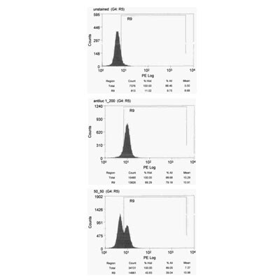
Flow Cytometry
1:200. Use reported by customer review
Immunocytochemistry/ Immunofluorescence
1:100-1:1000
Immunohistochemistry
1:100-1:1000
Immunohistochemistry-Frozen
reported in scientific literature (PMID 31069316)
Immunohistochemistry-Paraffin
1:100-1:1000
Western Blot
1:1000
Application Notes
Western blot has been tested with Drosophila embryos. Purified Luciferase protein and Luciferase expressed in Drosophila adult co-migrate on Western blots with a band seen at ~61 kDa, representing Luciferase.
Reviewed Applications
Read 2 reviews rated 4.5 using NB600-307 in the following applications:
Flow Cytometry Panel Builder
Bio-Techne Knows Flow Cytometry
Save time and reduce costly mistakes by quickly finding compatible reagents using the Panel Builder Tool.
Advanced Features
- Spectra Viewer - Custom analysis of spectra from multiple fluorochromes
- Spillover Popups - Visualize the spectra of individual fluorochromes
- Antigen Density Selector - Match fluorochrome brightness with antigen density

Formulation, Preparation, and Storage
Purification
Protein A or G purified
Formulation
PBS
Format
BSA Free
Preservative
0.02% Sodium Azide
Concentration
1.0 mg/ml
Shipping
The product is shipped with polar packs. Upon receipt, store it immediately at the temperature recommended below.
Stability & Storage
Aliquot and store at -20C or -80C. Avoid freeze-thaw cycles.
Background: Luciferase
The luciferase assay is fast and sensitive, differentiating itself from the CAT (chloramphenicol acetyltransferase) assay because it does not require a radioactive substrate.
References
1. Eun, H. (1996). Marker/Reporter enzymes. Enzymology Primer for Recombinant DNA Technology, 567-645. doi:10.1016/b978-012243740-3/50011-9
2. McNabb, D. S., Reed, R., & Marciniak, R. A. (2005). Dual luciferase assay system for rapid assessment of gene expression in Saccharomyces cerevisiae. Eukaryotic Cell, 4(9), 1539-1549. doi:10.1128/ec.4.9.1539-1549.2005
3. Fraga, H. (2008). Firefly luminescence: A historical perspective and recent developments. Photochemical & Photobiological Sciences, 7(2), 146-158. doi:10.1039/b719181b
4. Younes, A., Lukyanenko, Y. O., Lyashkov, A. E., Lakatta, E. G., & Sollott, S. J. (2011). A bioluminescence method for direct measurement of phosphodiesterase activity. Analytical Biochemistry, 417(1), 36-40. doi:10.1016/j.ab.2011.05.036
Alternate Names
LuC, luciferin 4 monooxygenase, Luciferin 4-monooxygenase
UniProt
Additional Luciferase Products
Product Documents for Luciferase Antibody (Luci 21 1-107) - BSA Free
Certificate of Analysis
To download a Certificate of Analysis, please enter a lot or batch number in the search box below.
Product Specific Notices for Luciferase Antibody (Luci 21 1-107) - BSA Free
This product is for research use only and is not approved for use in humans or in clinical diagnosis. Primary Antibodies are guaranteed for 1 year from date of receipt.
Citations for Luciferase Antibody (Luci 21 1-107) - BSA Free
 Powered by Bioz
Customer Reviews for Luciferase Antibody (Luci 21 1-107) - BSA Free (2)
4.5 out of 5
2 Customer Ratings
Have you used Luciferase Antibody (Luci 21 1-107) - BSA Free?
Submit a review and receive an Amazon gift card!
$25/€18/£15/$25CAN/¥2500 Yen for a review with an image
$10/€7/£6/$10CAN/¥1110 Yen for a review without an image
Submit a review
Customer Images
Showing
1
-
2 of
2 reviews
Showing All
Filter By:
-
Application: Flow CytometryVerified Customer | Posted 12/05/2012

-
Application: Western BlotSample Tested: cardiomyocytes cell lysate, Sample Amount: 15ugSpecies: RatVerified Customer | Posted 04/22/2010
There are no reviews that match your criteria.
Protocols
Find general support by application which include: protocols, troubleshooting, illustrated assays, videos and webinars.
- 7-Amino Actinomycin D (7-AAD) Cell Viability Flow Cytometry Protocol
- Antigen Retrieval Protocol (PIER)
- Antigen Retrieval for Frozen Sections Protocol
- Appropriate Fixation of IHC/ICC Samples
- Cellular Response to Hypoxia Protocols
- Chromogenic IHC Staining of Formalin-Fixed Paraffin-Embedded (FFPE) Tissue Protocol
- Chromogenic Immunohistochemistry Staining of Frozen Tissue
- ClariTSA™ Fluorophore Kits
- Detection & Visualization of Antibody Binding
- Extracellular Membrane Flow Cytometry Protocol
- Flow Cytometry Protocol for Cell Surface Markers
- Flow Cytometry Protocol for Staining Membrane Associated Proteins
- Flow Cytometry Staining Protocols
- Flow Cytometry Troubleshooting Guide
- Fluorescent IHC Staining of Frozen Tissue Protocol
- Graphic Protocol for Heat-induced Epitope Retrieval
- Graphic Protocol for the Preparation and Fluorescent IHC Staining of Frozen Tissue Sections
- Graphic Protocol for the Preparation and Fluorescent IHC Staining of Paraffin-embedded Tissue Sections
- Graphic Protocol for the Preparation of Gelatin-coated Slides for Histological Tissue Sections
- ICC Cell Smear Protocol for Suspension Cells
- ICC Immunocytochemistry Protocol Videos
- ICC for Adherent Cells
- IHC Sample Preparation (Frozen sections vs Paraffin)
- Immunocytochemistry (ICC) Protocol
- Immunocytochemistry Troubleshooting
- Immunofluorescence of Organoids Embedded in Cultrex Basement Membrane Extract
- Immunofluorescent IHC Staining of Formalin-Fixed Paraffin-Embedded (FFPE) Tissue Protocol
- Immunohistochemistry (IHC) and Immunocytochemistry (ICC) Protocols
- Immunohistochemistry Frozen Troubleshooting
- Immunohistochemistry Paraffin Troubleshooting
- Intracellular Flow Cytometry Protocol Using Alcohol (Methanol)
- Intracellular Flow Cytometry Protocol Using Detergents
- Intracellular Nuclear Staining Flow Cytometry Protocol Using Detergents
- Intracellular Staining Flow Cytometry Protocol Using Alcohol Permeabilization
- Intracellular Staining Flow Cytometry Protocol Using Detergents to Permeabilize Cells
- Preparing Samples for IHC/ICC Experiments
- Preventing Non-Specific Staining (Non-Specific Binding)
- Primary Antibody Selection & Optimization
- Propidium Iodide Cell Viability Flow Cytometry Protocol
- Protocol for Heat-Induced Epitope Retrieval (HIER)
- Protocol for Liperfluo
- Protocol for Making a 4% Formaldehyde Solution in PBS
- Protocol for VisUCyte™ HRP Polymer Detection Reagent
- Protocol for the Characterization of Human Th22 Cells
- Protocol for the Characterization of Human Th9 Cells
- Protocol for the Fluorescent ICC Staining of Cell Smears - Graphic
- Protocol for the Fluorescent ICC Staining of Cultured Cells on Coverslips - Graphic
- Protocol for the Preparation & Fixation of Cells on Coverslips
- Protocol for the Preparation and Chromogenic IHC Staining of Frozen Tissue Sections
- Protocol for the Preparation and Chromogenic IHC Staining of Frozen Tissue Sections - Graphic
- Protocol for the Preparation and Chromogenic IHC Staining of Paraffin-embedded Tissue Sections
- Protocol for the Preparation and Chromogenic IHC Staining of Paraffin-embedded Tissue Sections - Graphic
- Protocol for the Preparation and Fluorescent ICC Staining of Cells on Coverslips
- Protocol for the Preparation and Fluorescent ICC Staining of Non-adherent Cells
- Protocol for the Preparation and Fluorescent ICC Staining of Stem Cells on Coverslips
- Protocol for the Preparation and Fluorescent IHC Staining of Frozen Tissue Sections
- Protocol for the Preparation and Fluorescent IHC Staining of Paraffin-embedded Tissue Sections
- Protocol for the Preparation of Gelatin-coated Slides for Histological Tissue Sections
- Protocol for the Preparation of a Cell Smear for Non-adherent Cell ICC - Graphic
- Protocol: Annexin V and PI Staining by Flow Cytometry
- Protocol: Annexin V and PI Staining for Apoptosis by Flow Cytometry
- R&D Systems Quality Control Western Blot Protocol
- TUNEL and Active Caspase-3 Detection by IHC/ICC Protocol
- The Importance of IHC/ICC Controls
- Troubleshooting Guide: Fluorokine Flow Cytometry Kits
- Troubleshooting Guide: Immunohistochemistry
- Troubleshooting Guide: Western Blot Figures
- Western Blot Conditions
- Western Blot Protocol
- Western Blot Protocol for Cell Lysates
- Western Blot Troubleshooting
- Western Blot Troubleshooting Guide
- View all Protocols, Troubleshooting, Illustrated assays and Webinars
FAQs for Luciferase Antibody (Luci 21 1-107) - BSA Free
Showing
1
-
1 of
1 FAQ
Showing All
-
Q: Hello. Do you have a specific protocol for use of this antibody in FACS analysis? Luciferase Antibody (Luci 21 1-107) 0.1ml. It is uncojugated, so I am wondering if there is a specific protocol for labeling, etc. Thank you
A:
The flow protocol used with this antibody NB600-307 was our general flow protocol. Since this antibody is unconjugated, you would want to use a conjugated mouse IgG secondary with this product in order to detect the antibody. We do also have this specific clone available in a directly conjugated PE form (NB600-307PE). If you are interested, I can inquire about a custom conjugate of another fluor or dye if you like. The other option would be for you to directly conjugate the antibody yourself. We do sell several conjugation kits that are very easy to use.
Loading...