![Immunohistochemistry: LYVE-1 Antibody [NB600-1005]](https://resources.rndsystems.com/images/products/LYVE-1-Antibody-Immunohistochemistry-NB600-1005-img0011.jpg "Immunohistochemistry: LYVE-1 Antibody [NB600-1005]")
Loading...
Key Product Details
Species Reactivity
Human
Applications
Immunohistochemistry, Immunohistochemistry-Paraffin, Immunohistochemistry-Frozen, Western Blot, Flow Cytometry, CyTOF-ready
Label
Unconjugated
Antibody Source
Polyclonal Rabbit IgG
Format
Azide and BSA Free
Loading...
Product Specifications
Immunogen
Recombinant human LYVE-1
Localization
Plasma membrane
Marker
Lymphatic Vessel Marker
Clonality
Polyclonal
Host
Rabbit
Isotype
IgG
Theoretical MW
35 kDa.
Disclaimer note: The observed molecular weight of the protein may vary from the listed predicted molecular weight due to post translational modifications, post translation cleavages, relative charges, and other experimental factors.
Disclaimer note: The observed molecular weight of the protein may vary from the listed predicted molecular weight due to post translational modifications, post translation cleavages, relative charges, and other experimental factors.
Scientific Data Images for LYVE-1 Antibody - Azide and BSA Free
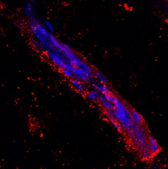
Immunohistochemistry: LYVE-1 Antibody [NB600-1005]
Immunohistochemistry: LYVE-1 Antibody [NB600-1005] - LYVE1 Antibody [NB600-1005] - Staining of human skin showing positivity in lymphatic vessels. Image provided by Patricia Redondo.![Flow Cytometry: LYVE-1 Antibody [NB600-1005]](https://resources.rndsystems.com/images/products/LYVE-1-Antibody-Flow-Cytometry-NB600-1005-img0010.jpg "Flow Cytometry: LYVE-1 Antibody [NB600-1005]")
Flow Cytometry: LYVE-1 Antibody [NB600-1005]
Flow Cytometry: LYVE-1 Antibody [NB600-1005] - Analysis with primary human dermal microvascular endothelial cells (HDMVEC).![Western Blot: LYVE-1 Antibody [NB600-1005]](https://resources.rndsystems.com/images/products/LYVE-1-Antibody-Western-Blot-NB600-1005-img0009.jpg "Western Blot: LYVE-1 Antibody [NB600-1005]")
Western Blot: LYVE-1 Antibody [NB600-1005]
Western Blot: LYVE-1 Antibody [NB600-1005] - Analysis of recombinant human sLYVE-1 and mouse sLYVE-1 using an anti-human LYVE-1 polyclonal antibody directed against the extracellular domain of human LYVE-1. There is more or less no cross reactivity withmouse LYVE-1.Applications for LYVE-1 Antibody - Azide and BSA Free
Application
Recommended Usage
Flow Cytometry
2-30 ug/ml
Immunohistochemistry
1:10-1:500
Immunohistochemistry-Frozen
6-30 ug/ml
Immunohistochemistry-Paraffin
1:10-1:500
Western Blot
2-5 ug/ml
Application Notes
IHC: Originally, it has not been tested on formalin-fixed tissue sections. However, recently one of our customers has used it successfully on this type of application (see review). Predicted molecular weight: 39 kDa. Not tested in other applications. Optimal dilutions/concentrations should be determined by the end user.
Reviewed Applications
Read 1 review rated 5 using NB600-1005 in the following applications:
Flow Cytometry Panel Builder
Bio-Techne Knows Flow Cytometry
Save time and reduce costly mistakes by quickly finding compatible reagents using the Panel Builder Tool.
Advanced Features
- Spectra Viewer - Custom analysis of spectra from multiple fluorochromes
- Spillover Popups - Visualize the spectra of individual fluorochromes
- Antigen Density Selector - Match fluorochrome brightness with antigen density

Formulation, Preparation, and Storage
Purification
Protein A purified
Reconstitution
Centrifuge vial prior to opening. Reconstitute in sterile water to a concentration of 0.1-1.0 mg/ml. Please note the sample size of this product will be provided in reconstituted liquid form.
Formulation
PBS
Format
Azide and BSA Free
Preservative
No Preservative
Concentration
LYOPH mg/ml
Shipping
The product is shipped with polar packs. Upon receipt, store it immediately at the temperature recommended below.
Stability & Storage
Store at 4C short term. Aliquot and store at -20C long term. Avoid freeze-thaw cycles.
Calculators
Background: LYVE-1
LYVE-1 has been an important marker in studies of embryonic and tumor lymphangiogenesis, as many cancers are characterized by early metastasis to the lymph nodes (1-3, 5). One study of five different vascular tumors in infants used immunohistochemical analysis and found positive LYVE-1 expression in infantile hemangioma, tufted angioma, and kaposiform hemangioendothelioma (5). LYVE-1 along with other markers such as GLUT-1, CD31, CD34, Prox-1, and WT-1 can be used to help provide immunohistologic profiles of various tumors and, when used in conjunction with clinical and histopathologic approaches, may offer better overall diagnosis and disease treatment (5).
References
1. Jackson D. G. (2019). Hyaluronan in the lymphatics: The key role of the hyaluronan receptor LYVE-1 in leucocyte trafficking. Matrix Biology : Journal of the International Society for Matrix Biology. https://doi.org/10.1016/j.matbio.2018.02.001
2. Jackson D. G. (2004). Biology of the lymphatic marker LYVE-1 and applications in research into lymphatic trafficking and lymphangiogenesis. APMIS : acta pathologica, microbiologica, et immunologica Scandinavica. https://doi.org/10.1111/j.1600-0463.2004.apm11207-0811.x
3. Jackson D. G. (2003). The lymphatics revisited: new perspectives from the hyaluronan receptor LYVE-1. Trends in Cardiovascular Medicine. https://doi.org/10.1016/s1050-1738(02)00189-5
4. Unitprot (Q9Y5Y7)
5. Johnson, E. F., Davis, D. M., Tollefson, M. M., Fritchie, K., & Gibson, L. E. (2018). Vascular Tumors in Infants: Case Report and Review of Clinical, Histopathologic, and Immunohistochemical Characteristics of Infantile Hemangioma, Pyogenic Granuloma, Noninvoluting Congenital Hemangioma, Tufted Angioma, and Kaposiform Hemangioendothelioma. The American Journal of Dermatopathology. https://doi.org/10.1097/DAD.0000000000000983
Long Name
Lymphatic Vessel Endothelial Hyaluronan Receptor 1
Alternate Names
LYVE1, XLKD1
Entrez Gene IDs
10894 (Human)
Gene Symbol
LYVE1
Additional LYVE-1 Products
Product Documents for LYVE-1 Antibody - Azide and BSA Free
Certificate of Analysis
To download a Certificate of Analysis, please enter a lot or batch number in the search box below.
Product Specific Notices for LYVE-1 Antibody - Azide and BSA Free
This product is for research use only and is not approved for use in humans or in clinical diagnosis. Primary Antibodies are guaranteed for 1 year from date of receipt.
Related Research Areas
Customer Reviews for LYVE-1 Antibody - Azide and BSA Free (1)
5 out of 5
1 Customer Rating
Have you used LYVE-1 Antibody - Azide and BSA Free?
Submit a review and receive an Amazon gift card!
$25/€18/£15/$25CAN/¥2500 Yen for a review with an image
$10/€7/£6/$10CAN/¥1110 Yen for a review without an image
Submit a review
Customer Images
Showing
1
-
1 of
1 review
Showing All
Filter By:
-
Application: ImmunohistochemistrySample Tested: skin (lymphatic vessel)Species: HumanVerified Customer | Posted 06/13/2013

There are no reviews that match your criteria.
Protocols
Find general support by application which include: protocols, troubleshooting, illustrated assays, videos and webinars.
- 7-Amino Actinomycin D (7-AAD) Cell Viability Flow Cytometry Protocol
- Antigen Retrieval Protocol (PIER)
- Antigen Retrieval for Frozen Sections Protocol
- Appropriate Fixation of IHC/ICC Samples
- Cellular Response to Hypoxia Protocols
- Chromogenic IHC Staining of Formalin-Fixed Paraffin-Embedded (FFPE) Tissue Protocol
- Chromogenic Immunohistochemistry Staining of Frozen Tissue
- ClariTSA™ Fluorophore Kits
- Detection & Visualization of Antibody Binding
- Extracellular Membrane Flow Cytometry Protocol
- Flow Cytometry Protocol for Cell Surface Markers
- Flow Cytometry Protocol for Staining Membrane Associated Proteins
- Flow Cytometry Staining Protocols
- Flow Cytometry Troubleshooting Guide
- Fluorescent IHC Staining of Frozen Tissue Protocol
- Graphic Protocol for Heat-induced Epitope Retrieval
- Graphic Protocol for the Preparation and Fluorescent IHC Staining of Frozen Tissue Sections
- Graphic Protocol for the Preparation and Fluorescent IHC Staining of Paraffin-embedded Tissue Sections
- Graphic Protocol for the Preparation of Gelatin-coated Slides for Histological Tissue Sections
- IHC Sample Preparation (Frozen sections vs Paraffin)
- Immunofluorescent IHC Staining of Formalin-Fixed Paraffin-Embedded (FFPE) Tissue Protocol
- Immunohistochemistry (IHC) and Immunocytochemistry (ICC) Protocols
- Immunohistochemistry Frozen Troubleshooting
- Immunohistochemistry Paraffin Troubleshooting
- Intracellular Flow Cytometry Protocol Using Alcohol (Methanol)
- Intracellular Flow Cytometry Protocol Using Detergents
- Intracellular Nuclear Staining Flow Cytometry Protocol Using Detergents
- Intracellular Staining Flow Cytometry Protocol Using Alcohol Permeabilization
- Intracellular Staining Flow Cytometry Protocol Using Detergents to Permeabilize Cells
- Preparing Samples for IHC/ICC Experiments
- Preventing Non-Specific Staining (Non-Specific Binding)
- Primary Antibody Selection & Optimization
- Propidium Iodide Cell Viability Flow Cytometry Protocol
- Protocol for Heat-Induced Epitope Retrieval (HIER)
- Protocol for Liperfluo
- Protocol for Making a 4% Formaldehyde Solution in PBS
- Protocol for VisUCyte™ HRP Polymer Detection Reagent
- Protocol for the Characterization of Human Th22 Cells
- Protocol for the Characterization of Human Th9 Cells
- Protocol for the Preparation & Fixation of Cells on Coverslips
- Protocol for the Preparation and Chromogenic IHC Staining of Frozen Tissue Sections
- Protocol for the Preparation and Chromogenic IHC Staining of Frozen Tissue Sections - Graphic
- Protocol for the Preparation and Chromogenic IHC Staining of Paraffin-embedded Tissue Sections
- Protocol for the Preparation and Chromogenic IHC Staining of Paraffin-embedded Tissue Sections - Graphic
- Protocol for the Preparation and Fluorescent IHC Staining of Frozen Tissue Sections
- Protocol for the Preparation and Fluorescent IHC Staining of Paraffin-embedded Tissue Sections
- Protocol for the Preparation of Gelatin-coated Slides for Histological Tissue Sections
- Protocol: Annexin V and PI Staining by Flow Cytometry
- Protocol: Annexin V and PI Staining for Apoptosis by Flow Cytometry
- R&D Systems Quality Control Western Blot Protocol
- TUNEL and Active Caspase-3 Detection by IHC/ICC Protocol
- The Importance of IHC/ICC Controls
- Troubleshooting Guide: Fluorokine Flow Cytometry Kits
- Troubleshooting Guide: Immunohistochemistry
- Troubleshooting Guide: Western Blot Figures
- Western Blot Conditions
- Western Blot Protocol
- Western Blot Protocol for Cell Lysates
- Western Blot Troubleshooting
- Western Blot Troubleshooting Guide
- View all Protocols, Troubleshooting, Illustrated assays and Webinars
Loading...