M-CSFR/CD115 Antibody (AFS98) - BSA Free
Novus Biologicals | Catalog # NBP1-43363
![Immunohistochemistry-Paraffin: M-CSFR/CD115 Antibody (AFS98) - BSA Free [NBP1-43363]](https://resources.rndsystems.com/images/products/M-CSFR-CD115-Antibody-AFS98-Immunohistochemistry-Paraffin-NBP1-43363-img0005.jpg "Immunohistochemistry-Paraffin: M-CSFR/CD115 Antibody (AFS98) - BSA Free [NBP1-43363]")
Key Product Details
Species Reactivity
Validated:
Mouse
Cited:
Mouse
Applications
Validated:
Immunohistochemistry, Immunohistochemistry-Paraffin, Immunohistochemistry-Frozen, Western Blot, Block/Neutralize, Flow Cytometry
Cited:
Block/Neutralize
Label
Unconjugated
Antibody Source
Monoclonal Rat IgG2a Kappa Clone # AFS98
Format
BSA Free
Loading...
Product Specifications
Immunogen
The immunogen for this antibody was CD115.
Clonality
Monoclonal
Host
Rat
Isotype
IgG2a Kappa
Scientific Data Images for M-CSFR/CD115 Antibody (AFS98) - BSA Free
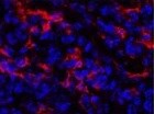
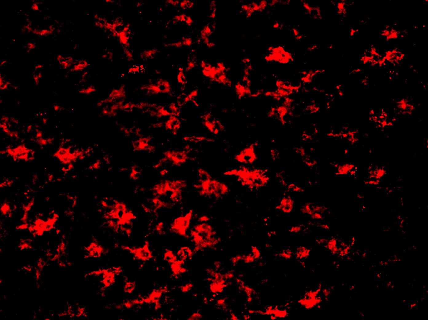
Immunohistochemistry-Paraffin: M-CSFR/CD115 Antibody (AFS98) - BSA Free [NBP1-43363]
Immunohistochemistry-Paraffin: M-CSFR/CD115 Antibody (AFS98) [NBP1-43363] - Mouse tumor tissue stained with M-CSFR/CD115 Antibody (AFS98). Tissue sections were deparaffinized and incubated with the antibody O/N at 4C. IHC-P image submitted by a verified customer review.![Flow Cytometry: M-CSFR/CD115 Antibody (AFS98) - BSA Free [NBP1-43363]](https://resources.rndsystems.com/images/products/M-CSF-R-CD115-Antibody-AFS98-Flow-Cytometry-NBP1-43363-img0003.jpg "Flow Cytometry: M-CSFR/CD115 Antibody (AFS98) - BSA Free [NBP1-43363]")
Flow Cytometry: M-CSFR/CD115 Antibody (AFS98) - BSA Free [NBP1-43363]
Flow Cytometry: M-CSF R/CD115 Antibody (AFS98) [NBP1-43363] - Analysis using the Biotin conjugate of NBP1-43363. Staining of thioglycolate-induced peritoneal exudate cells with Anti-Mouse CD115 (c-fms) PE. Appropriate isotype controls were used (open histogram).![Immunohistochemistry-Frozen: M-CSFR/CD115 Antibody (AFS98) - BSA Free [NBP1-43363]](https://resources.rndsystems.com/images/products/M-CSFR-CD115-Antibody-AFS98-Immunohistochemistry-Frozen-NBP1-43363-img0004.jpg "Immunohistochemistry-Frozen: M-CSFR/CD115 Antibody (AFS98) - BSA Free [NBP1-43363]")
Immunohistochemistry-Frozen: M-CSFR/CD115 Antibody (AFS98) - BSA Free [NBP1-43363]
Immunohistochemistry-Frozen: M-CSFR/CD115 Antibody (AFS98) [NBP1-43363] - Mouse brain section. Antibody at 1:200. IHC-Fr image submitted by a verified customer review.![Flow Cytometry: M-CSFR/CD115 Antibody (AFS98) - BSA Free [NBP1-43363]](https://resources.rndsystems.com/images/products/M-CSF-R-CD115-Antibody-AFS98-Flow-Cytometry-NBP1-43363-img0002.jpg "Flow Cytometry: M-CSFR/CD115 Antibody (AFS98) - BSA Free [NBP1-43363]")
Flow Cytometry: M-CSFR/CD115 Antibody (AFS98) - BSA Free [NBP1-43363]
Flow Cytometry: M-CSF R/CD115 Antibody (AFS98) [NBP1-43363] - Staining of thioglycolate-induced peritoneal exudate cells with Anti-Mouse CD115 (c-fms) PE. Appropriate isotype controls were used (open histogram). Cells in the large scatter population were used for analysis.![Flow Cytometry: M-CSFR/CD115 Antibody (AFS98) - BSA Free [NBP1-43363]](https://resources.rndsystems.com/images/products/M-CSF-R-CD115-Antibody-AFS98-Flow-Cytometry-NBP1-43363-img0001.jpg "Flow Cytometry: M-CSFR/CD115 Antibody (AFS98) - BSA Free [NBP1-43363]")
Flow Cytometry: M-CSFR/CD115 Antibody (AFS98) - BSA Free [NBP1-43363]
Flow Cytometry: M-CSF R/CD115 Antibody (AFS98) [NBP1-43363] - Staining of thioglycolate-induced peritoneal exudate cells (PECs) with 0.25 ug of Rat IgG2a kappa Isotype Control Purified (NBP1-43321) (open histogram) or 0.25 ug of Anti-Mouse CD115 (c-fms) Purified (filled histogram) followed by Anti-Rat IgG Biotin and Streptavidin PE. Total viable cells were used for analysis.Applications for M-CSFR/CD115 Antibody (AFS98) - BSA Free
Application
Recommended Usage
Flow Cytometry
< 1 ug/10^6 cells
Immunohistochemistry
1:10 - 1:500
Immunohistochemistry-Frozen
1:10 - 1:500
Western Blot
1:100 - 1:2000
Application Notes
The AFS98 antibody has been tested by blocking of fluorochrome conjugated AFS98 in flow cytometric analysis of peritoneal exudate cells. This can be used at less than or equal to 1 ug per test. Cell number should be determined empirically but can range from 10^5to 10^8cells/test. Use in Blocking/Neutralizing reported in scientific literature (PMID: 24056963). This M-CSFR/CD115 Antibody (AFS98) is validated for IHC-P from a verified customer review.
Reviewed Applications
Read 2 reviews rated 4.5 using NBP1-43363 in the following applications:
Flow Cytometry Panel Builder
Bio-Techne Knows Flow Cytometry
Save time and reduce costly mistakes by quickly finding compatible reagents using the Panel Builder Tool.
Advanced Features
- Spectra Viewer - Custom analysis of spectra from multiple fluorochromes
- Spillover Popups - Visualize the spectra of individual fluorochromes
- Antigen Density Selector - Match fluorochrome brightness with antigen density

Formulation, Preparation, and Storage
Purification
Protein A or G purified
Formulation
PBS (pH 7.2)
Format
BSA Free
Preservative
0.09% Sodium Azide
Concentration
0.5 mg/ml
Shipping
The product is shipped with polar packs. Upon receipt, store it immediately at the temperature recommended below.
Stability & Storage
Store at 4C. Do not freeze.
Background: M-CSF R/CD115
Long Name
Macrophage Colony Stimulating Factor Receptor
Alternate Names
c-fms, CD115, CSF1R, M-CSFR, MCSFR
Entrez Gene IDs
1436 (Human)
Gene Symbol
CSF1R
UniProt
Additional M-CSF R/CD115 Products
Product Documents for M-CSFR/CD115 Antibody (AFS98) - BSA Free
Certificate of Analysis
To download a Certificate of Analysis, please enter a lot or batch number in the search box below.
Product Specific Notices for M-CSFR/CD115 Antibody (AFS98) - BSA Free
This product is for research use only and is not approved for use in humans or in clinical diagnosis. Primary Antibodies are guaranteed for 1 year from date of receipt.
Citations for M-CSFR/CD115 Antibody (AFS98) - BSA Free
 Powered by Bioz
Customer Reviews for M-CSFR/CD115 Antibody (AFS98) - BSA Free (2)
4.5 out of 5
2 Customer Ratings
Have you used M-CSFR/CD115 Antibody (AFS98) - BSA Free?
Submit a review and receive an Amazon gift card!
$25/€18/£15/$25CAN/¥2500 Yen for a review with an image
$10/€7/£6/$10CAN/¥1110 Yen for a review without an image
Submit a review
Customer Images
Showing
1
-
2 of
2 reviews
Showing All
Filter By:
-
Application: ImmunofluorescenceSample Tested: Tumor tissueSpecies: MouseVerified Customer | Posted 11/09/2021staining in mouse tumor tissueTissue sections were deparaffinized. Incubated O/N in 4 C with the 1ry antibody.

-
Application: Immunohistochemistry-FrozenSample Tested: Mouse brainSpecies: MouseVerified Customer | Posted 09/18/2019Dilution 1:200
There are no reviews that match your criteria.
Protocols
Find general support by application which include: protocols, troubleshooting, illustrated assays, videos and webinars.
- 7-Amino Actinomycin D (7-AAD) Cell Viability Flow Cytometry Protocol
- Antigen Retrieval Protocol (PIER)
- Antigen Retrieval for Frozen Sections Protocol
- Appropriate Fixation of IHC/ICC Samples
- Cellular Response to Hypoxia Protocols
- Chromogenic IHC Staining of Formalin-Fixed Paraffin-Embedded (FFPE) Tissue Protocol
- Chromogenic Immunohistochemistry Staining of Frozen Tissue
- ClariTSA™ Fluorophore Kits
- Detection & Visualization of Antibody Binding
- Extracellular Membrane Flow Cytometry Protocol
- Flow Cytometry Protocol for Cell Surface Markers
- Flow Cytometry Protocol for Staining Membrane Associated Proteins
- Flow Cytometry Staining Protocols
- Flow Cytometry Troubleshooting Guide
- Fluorescent IHC Staining of Frozen Tissue Protocol
- Graphic Protocol for Heat-induced Epitope Retrieval
- Graphic Protocol for the Preparation and Fluorescent IHC Staining of Frozen Tissue Sections
- Graphic Protocol for the Preparation and Fluorescent IHC Staining of Paraffin-embedded Tissue Sections
- Graphic Protocol for the Preparation of Gelatin-coated Slides for Histological Tissue Sections
- IHC Sample Preparation (Frozen sections vs Paraffin)
- Immunofluorescent IHC Staining of Formalin-Fixed Paraffin-Embedded (FFPE) Tissue Protocol
- Immunohistochemistry (IHC) and Immunocytochemistry (ICC) Protocols
- Immunohistochemistry Frozen Troubleshooting
- Immunohistochemistry Paraffin Troubleshooting
- Intracellular Flow Cytometry Protocol Using Alcohol (Methanol)
- Intracellular Flow Cytometry Protocol Using Detergents
- Intracellular Nuclear Staining Flow Cytometry Protocol Using Detergents
- Intracellular Staining Flow Cytometry Protocol Using Alcohol Permeabilization
- Intracellular Staining Flow Cytometry Protocol Using Detergents to Permeabilize Cells
- Preparing Samples for IHC/ICC Experiments
- Preventing Non-Specific Staining (Non-Specific Binding)
- Primary Antibody Selection & Optimization
- Propidium Iodide Cell Viability Flow Cytometry Protocol
- Protocol for Heat-Induced Epitope Retrieval (HIER)
- Protocol for Liperfluo
- Protocol for Making a 4% Formaldehyde Solution in PBS
- Protocol for VisUCyte™ HRP Polymer Detection Reagent
- Protocol for the Characterization of Human Th22 Cells
- Protocol for the Characterization of Human Th9 Cells
- Protocol for the Preparation & Fixation of Cells on Coverslips
- Protocol for the Preparation and Chromogenic IHC Staining of Frozen Tissue Sections
- Protocol for the Preparation and Chromogenic IHC Staining of Frozen Tissue Sections - Graphic
- Protocol for the Preparation and Chromogenic IHC Staining of Paraffin-embedded Tissue Sections
- Protocol for the Preparation and Chromogenic IHC Staining of Paraffin-embedded Tissue Sections - Graphic
- Protocol for the Preparation and Fluorescent IHC Staining of Frozen Tissue Sections
- Protocol for the Preparation and Fluorescent IHC Staining of Paraffin-embedded Tissue Sections
- Protocol for the Preparation of Gelatin-coated Slides for Histological Tissue Sections
- Protocol: Annexin V and PI Staining by Flow Cytometry
- Protocol: Annexin V and PI Staining for Apoptosis by Flow Cytometry
- R&D Systems Quality Control Western Blot Protocol
- TUNEL and Active Caspase-3 Detection by IHC/ICC Protocol
- The Importance of IHC/ICC Controls
- Troubleshooting Guide: Fluorokine Flow Cytometry Kits
- Troubleshooting Guide: Immunohistochemistry
- Troubleshooting Guide: Western Blot Figures
- Western Blot Conditions
- Western Blot Protocol
- Western Blot Protocol for Cell Lysates
- Western Blot Troubleshooting
- Western Blot Troubleshooting Guide
- View all Protocols, Troubleshooting, Illustrated assays and Webinars
Loading...
Associated Pathways
