MafA Antibody - BSA Free
Novus Biologicals | Catalog # NB400-137
![Western Blot: MafA Antibody [NB400-137]](https://resources.rndsystems.com/images/products/MafA-Antibody-Western-Blot-NB400-137-img0008.jpg "Western Blot: MafA Antibody [NB400-137]")
Key Product Details
Species Reactivity
Validated:
Human, Mouse
Cited:
Human, Mouse
Applications
Validated:
Immunohistochemistry, Immunohistochemistry-Paraffin, Immunohistochemistry-Frozen, Western Blot, Immunocytochemistry/ Immunofluorescence, Chromatin Immunoprecipitation (ChIP), Gel Super Shift Assays
Cited:
Immunohistochemistry-Paraffin, Immunohistochemistry-Frozen, Western Blot, Immunocytochemistry/ Immunofluorescence, Chemotaxis
Label
Unconjugated
Antibody Source
Polyclonal Rabbit IgG
Format
BSA Free
Loading...
Product Specifications
Immunogen
The immunogen recognized by this antibody maps to a region between residue 300 and the C-terminus (residue 359) of mouse v-maf musculoaponeurotic fibrosarcoma oncogene homolog A using the numbering given in entry NP_919331.1 (GeneID 378435).
Reactivity Notes
Human reactivity reported in the scientific literature (PMID: 22665223).
Clonality
Polyclonal
Host
Rabbit
Isotype
IgG
Scientific Data Images for MafA Antibody - BSA Free
Western Blot: MafA Antibody [NB400-137]
Western Blot: MafA Antibody [NB400-137] - Nuclear extract (6 ug) from HeLa cells transfected with MafA, MafB or cMaf expression constructs. Affinity purified anti-MafA antibody (NB400-137), anti-MafB antibody (NB600-266) or anti-c-Maf antibody (NB600-267). Each antibody was used at 0.5 ug/ml.![Immunohistochemistry-Paraffin: MafA Antibody [NB400-137]](https://resources.rndsystems.com/images/products/MafA-Antibody-Immunohistochemistry-Paraffin-NB400-137-img0009.jpg "Immunohistochemistry-Paraffin: MafA Antibody [NB400-137]")
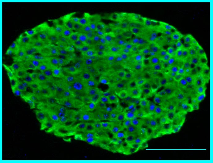
Immunohistochemistry-Paraffin: MafA Antibody [NB400-137]
Immunohistochemistry-Paraffin: MafA Antibody [NB400-137] - Immunohistochemical staining of MafA (blue) and insulin (green) in mouse pancreas. Image from verified customer review.![Western Blot: MafA Antibody [NB400-137]](https://resources.rndsystems.com/images/products/MafA-Antibody-Western-Blot-NB400-137-img0007.jpg "Western Blot: MafA Antibody [NB400-137]")
Western Blot: MafA Antibody [NB400-137]
Western Blot: MafA Antibody [NB400-137] - Binding of MafA to the enhancer region of the endogenous insulin gene. Samples: Immunoprecipitated cross-linked DNA from betaTC-3 cells was analyzed by PCR. As controls, reactions were run with no DNA, with input chromatin, with DNA obtained after precipitation with rabbit IgG or without antibody (lanes 1 through 5, respectively). Antibody: Affinity purified rabbit anti-MafA antibody (NB 400-137).![Gel Super Shift Assays: MafA Antibody [NB400-137]](https://resources.rndsystems.com/images/products/MafA-Antibody-Gel-Super-Shift-Assays-NB400-137-img0005.jpg "Gel Super Shift Assays: MafA Antibody [NB400-137]")
Gel Super Shift Assays: MafA Antibody [NB400-137]
Gel Super Shift Assays: MafA Antibody [NB400-137] - Electrophoretic Mobility Shift of MafA, MafB and c-Maf. Samples: Nuclear extract (6 ug) from HeLa cells transfected with MafA, MafB or c-Maf expression constructs. Antibodies: Affinity purified anti-MafA antibody NB400-137, anti-MafB antibody NB600-266 or anti-c-Maf antibody NB600-267.Applications for MafA Antibody - BSA Free
Application
Recommended Usage
Gel Super Shift Assays
1 to 5 ug/20 ul reaction
Immunocytochemistry/ Immunofluorescence
1:10-1:500
Immunohistochemistry
1:10-1:500
Immunohistochemistry-Frozen
1:10-1:500
Immunohistochemistry-Paraffin
1:1000 to 1:3000
Western Blot
1:1000-1:10000
Application Notes
A band ~36-40 kDa is seen in WB analysis. Use in IHC-P was reported in the scientific literature (PMID: 22665223). In some cases, the antibody may be diluted further than indicated. Optimal working dilutions should be determined experimentally by the investigator. Prepare working dilution immediately before use. Use in ICC/IF and in IHC-Frozen reported in scientific literature (PMID 23967072). Chip reported in scientific literature (PMID: 26314560).
Reviewed Applications
Read 1 review rated 5 using NB400-137 in the following applications:
Formulation, Preparation, and Storage
Purification
Immunogen affinity purified
Formulation
Tris-Citrate/Phosphate (pH 7.0 - 8.0)
Format
BSA Free
Preservative
0.09% Sodium Azide
Concentration
1.0 mg/ml
Shipping
The product is shipped with polar packs. Upon receipt, store it immediately at the temperature recommended below.
Stability & Storage
Store at 4C. Do not freeze.
Background: MafA
Alternate Names
hMafA, Pancreatic beta-cell-specific transcriptional activator, RIPE3b1, transcription factor MafA, Transcription factor RIPE3b1, V-maf musculoaponeurotic fibrosarcoma oncogene homolog A, v-maf musculoaponeurotic fibrosarcoma oncogene homolog A (avian)
Entrez Gene IDs
378435 (Mouse)
Gene Symbol
MAFA
UniProt
Additional MafA Products
Product Documents for MafA Antibody - BSA Free
Certificate of Analysis
To download a Certificate of Analysis, please enter a lot or batch number in the search box below.
Product Specific Notices for MafA Antibody - BSA Free
This product is for research use only and is not approved for use in humans or in clinical diagnosis. Primary Antibodies are guaranteed for 1 year from date of receipt.
Citations for MafA Antibody - BSA Free
 Powered by Bioz
Customer Reviews for MafA Antibody - BSA Free (1)
5 out of 5
1 Customer Rating
Have you used MafA Antibody - BSA Free?
Submit a review and receive an Amazon gift card!
$25/€18/£15/$25CAN/¥2500 Yen for a review with an image
$10/€7/£6/$10CAN/¥1110 Yen for a review without an image
Submit a review
Customer Images
Showing
1
-
1 of
1 review
Showing All
Filter By:
-
Application: Immunohistochemistry-ParaffinSample Tested: Mouse PancreasSpecies: MouseVerified Customer | Posted 09/21/2015MafA-insulin

There are no reviews that match your criteria.
Protocols
Find general support by application which include: protocols, troubleshooting, illustrated assays, videos and webinars.
- Antigen Retrieval Protocol (PIER)
- Antigen Retrieval for Frozen Sections Protocol
- Appropriate Fixation of IHC/ICC Samples
- Cellular Response to Hypoxia Protocols
- ChIP Protocol Video
- Chromatin Immunoprecipitation (ChIP) Protocol
- Chromatin Immunoprecipitation Protocol
- Chromogenic IHC Staining of Formalin-Fixed Paraffin-Embedded (FFPE) Tissue Protocol
- Chromogenic Immunohistochemistry Staining of Frozen Tissue
- ClariTSA™ Fluorophore Kits
- Detection & Visualization of Antibody Binding
- Fluorescent IHC Staining of Frozen Tissue Protocol
- Graphic Protocol for Heat-induced Epitope Retrieval
- Graphic Protocol for the Preparation and Fluorescent IHC Staining of Frozen Tissue Sections
- Graphic Protocol for the Preparation and Fluorescent IHC Staining of Paraffin-embedded Tissue Sections
- Graphic Protocol for the Preparation of Gelatin-coated Slides for Histological Tissue Sections
- ICC Cell Smear Protocol for Suspension Cells
- ICC Immunocytochemistry Protocol Videos
- ICC for Adherent Cells
- IHC Sample Preparation (Frozen sections vs Paraffin)
- Immunocytochemistry (ICC) Protocol
- Immunocytochemistry Troubleshooting
- Immunofluorescence of Organoids Embedded in Cultrex Basement Membrane Extract
- Immunofluorescent IHC Staining of Formalin-Fixed Paraffin-Embedded (FFPE) Tissue Protocol
- Immunohistochemistry (IHC) and Immunocytochemistry (ICC) Protocols
- Immunohistochemistry Frozen Troubleshooting
- Immunohistochemistry Paraffin Troubleshooting
- Preparing Samples for IHC/ICC Experiments
- Preventing Non-Specific Staining (Non-Specific Binding)
- Primary Antibody Selection & Optimization
- Protocol for Heat-Induced Epitope Retrieval (HIER)
- Protocol for Making a 4% Formaldehyde Solution in PBS
- Protocol for VisUCyte™ HRP Polymer Detection Reagent
- Protocol for the Fluorescent ICC Staining of Cell Smears - Graphic
- Protocol for the Fluorescent ICC Staining of Cultured Cells on Coverslips - Graphic
- Protocol for the Preparation & Fixation of Cells on Coverslips
- Protocol for the Preparation and Chromogenic IHC Staining of Frozen Tissue Sections
- Protocol for the Preparation and Chromogenic IHC Staining of Frozen Tissue Sections - Graphic
- Protocol for the Preparation and Chromogenic IHC Staining of Paraffin-embedded Tissue Sections
- Protocol for the Preparation and Chromogenic IHC Staining of Paraffin-embedded Tissue Sections - Graphic
- Protocol for the Preparation and Fluorescent ICC Staining of Cells on Coverslips
- Protocol for the Preparation and Fluorescent ICC Staining of Non-adherent Cells
- Protocol for the Preparation and Fluorescent ICC Staining of Stem Cells on Coverslips
- Protocol for the Preparation and Fluorescent IHC Staining of Frozen Tissue Sections
- Protocol for the Preparation and Fluorescent IHC Staining of Paraffin-embedded Tissue Sections
- Protocol for the Preparation of Gelatin-coated Slides for Histological Tissue Sections
- Protocol for the Preparation of a Cell Smear for Non-adherent Cell ICC - Graphic
- R&D Systems Quality Control Western Blot Protocol
- TUNEL and Active Caspase-3 Detection by IHC/ICC Protocol
- The Importance of IHC/ICC Controls
- Troubleshooting Guide: Immunohistochemistry
- Troubleshooting Guide: Western Blot Figures
- Western Blot Conditions
- Western Blot Protocol
- Western Blot Protocol for Cell Lysates
- Western Blot Troubleshooting
- Western Blot Troubleshooting Guide
- View all Protocols, Troubleshooting, Illustrated assays and Webinars
Loading...