![Immunocytochemistry/ Immunofluorescence: MafA Antibody [NBP1-00121]](https://resources.rndsystems.com/images/products/MafA-Antibody-Immunocytochemistry-Immunofluorescence-NBP1-00121-img0003.jpg "Immunocytochemistry/ Immunofluorescence: MafA Antibody [NBP1-00121]")
Loading...
Key Product Details
Species Reactivity
Validated:
Human, Mouse
Cited:
Human, Mouse, Rat
Applications
Validated:
Immunohistochemistry, Immunohistochemistry-Paraffin, Immunohistochemistry-Frozen, Western Blot, Immunocytochemistry/ Immunofluorescence
Cited:
Immunohistochemistry-Frozen, Western Blot, Immunocytochemistry/ Immunofluorescence, IF/IHC
Label
Unconjugated
Antibody Source
Polyclonal Rabbit IgG
Loading...
Product Specifications
Immunogen
The immunogen recognized by this antibody maps to a region between residue 300 and the C-terminus (residue 359) of mouse v-maf musculoaponeurotic fibrosarcoma oncogene homolog A using the numbering given in entry NP_919331.1 (GeneID 378435).
Reactivity Notes
Human reactivity reported in scientific literature (PMID: 26495868).
Clonality
Polyclonal
Host
Rabbit
Isotype
IgG
Scientific Data Images for MafA Antibody
Immunocytochemistry/ Immunofluorescence: MafA Antibody [NBP1-00121]
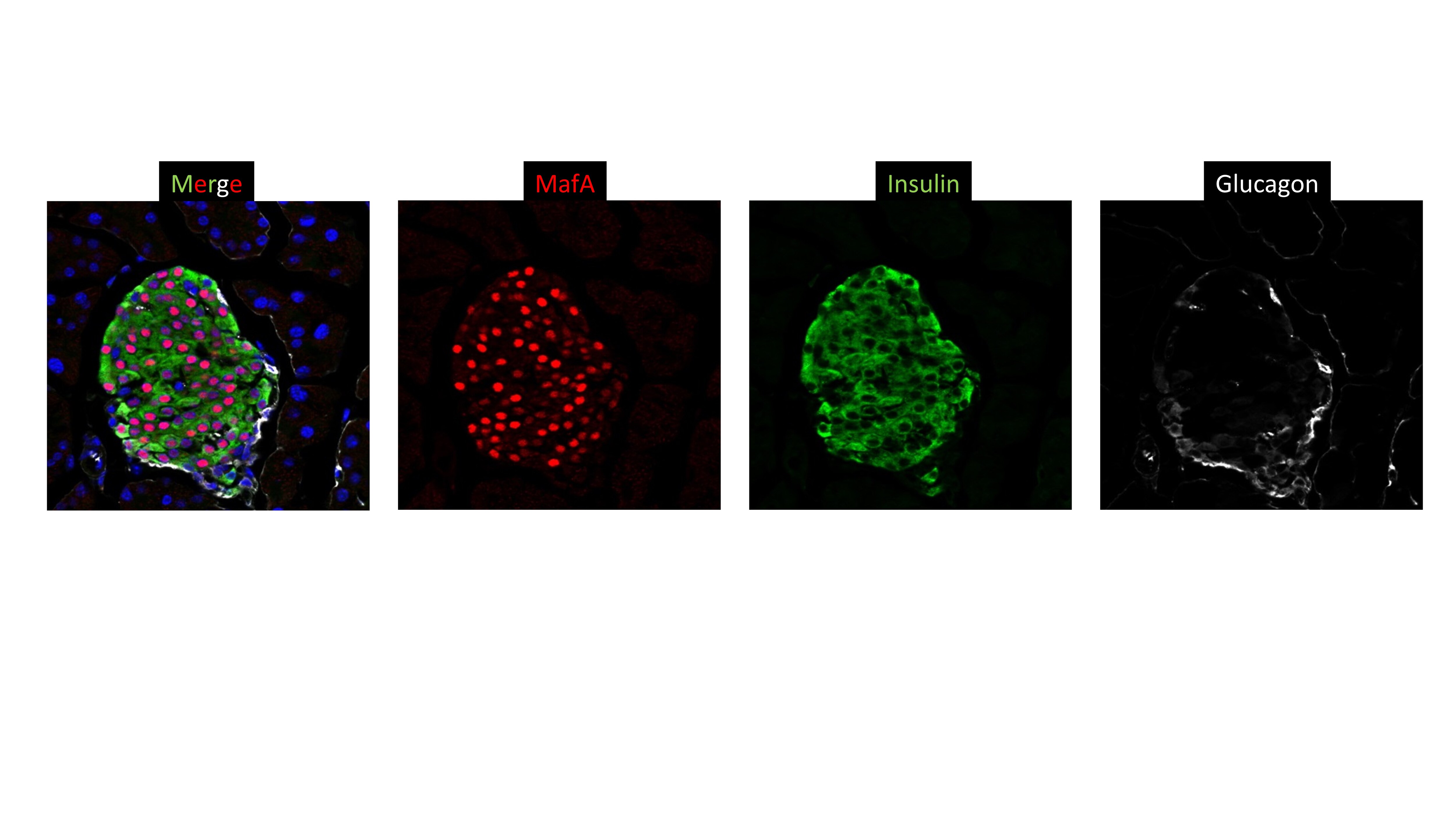
Immunocytochemistry/Immunofluorescence: MafA Antibody [NBP1-00121] - Analysis of MafA in 8-week old mouse pancretic cryosection using anti-MafA antibody (red). Image from verified customer review.![Immunohistochemistry-Paraffin: MafA Antibody [NBP1-00121]](https://resources.rndsystems.com/images/products/MafA-Antibody-Immunohistochemistry-Paraffin-NBP1-00121-img0001.jpg "Immunohistochemistry-Paraffin: MafA Antibody [NBP1-00121]")
Immunohistochemistry-Paraffin: MafA Antibody [NBP1-00121]
Immunohistochemistry-Paraffin: MafA Antibody [NBP1-00121] - Section of mouse pancreatic islet. Antibody: Affinity purified rabbit anti-MafA (Cat. No. NBP1-00121) used at a dilution of 1:250.
Western Blot: MafA Antibody [NBP1-00121] -
Critical beta -cell factors are reduced upon cytokine treatment in mouse and human islets. A-F. Relative mRNA quantification of various beta -cell transcriptional regulator genes (MafA, Pdx1, Nkx6.1, Ldb1, Isl1, and SSBP3, respectively) from primary mouse islets after 4 h culturing with single cytokines (Tnf alpha, Ifn gamma, IL-1 beta ) or a cocktail of each, as compared to PBS vehicle controls (set to 1-fold). 36B4 was used as the housekeeping gene; n = 3–4 for each treatment group. G. Left: LDB1, MAFA, NKX6.1, and ISL1 Western blot with Ins-1 cell extracts after 4 h or overnight treatment with single cytokines, or a cocktail, as compared to control. Actin was included as loading control. Right: Densitometry quantification of 4 h or overnight treated LDB1, MAFA, NKX6.1, or ISL1 Western blot protein levels, normalized to Actin. H. Human islets were treated with a DMSO vehicle or a cytokine cocktail,26,27 then mRNA measured. 18S was used as the housekeeping gene; n = 3. I. Quantification of beta -cell mRNAs from primary mouse islets after siRNA-mediated Ldb1 knockdown using Gapdh as the housekeeping gene; n = 3. *, P <.05; **, P <.01; ***, P <.001; ****, P <.0001 based on one-way ANOVA or Student’s t-test. Image collected and cropped by CiteAb from the following open publication (https://pubmed.ncbi.nlm.nih.gov/34968409), licensed under a CC-BY license. Not internally tested by Novus Biologicals.Applications for MafA Antibody
Application
Recommended Usage
Immunocytochemistry/ Immunofluorescence
Reported in scientific literature (PMID: 28705881), verified customer review
Immunohistochemistry
1:100 - 1:500
Immunohistochemistry-Frozen
Reported in scientific literature (PMID: 31201281)
Immunohistochemistry-Paraffin
1:100-1:500
Western Blot
Reported in scientific literature (PMID: 26495868)
Application Notes
Epitope exposure with citrate bufferexposure is recommended. Epitope exposure with citrate buffer will enhance staining. Likely to work with frozen sections. In some cases, the antibody may be diluted further than indicated.
Reviewed Applications
Read 1 review rated 5 using NBP1-00121 in the following applications:
Formulation, Preparation, and Storage
Purification
Immunogen affinity purified
Formulation
TBS and 0.1% BSA
Preservative
0.09% Sodium Azide
Concentration
0.05 mg/ml
Shipping
The product is shipped with polar packs. Upon receipt, store it immediately at the temperature recommended below.
Stability & Storage
Store at 4C. Do not freeze.
Background: MafA
Alternate Names
hMafA, Pancreatic beta-cell-specific transcriptional activator, RIPE3b1, transcription factor MafA, Transcription factor RIPE3b1, V-maf musculoaponeurotic fibrosarcoma oncogene homolog A, v-maf musculoaponeurotic fibrosarcoma oncogene homolog A (avian)
Entrez Gene IDs
378435 (Mouse)
Gene Symbol
MAFA
UniProt
Additional MafA Products
Product Documents for MafA Antibody
Certificate of Analysis
To download a Certificate of Analysis, please enter a lot or batch number in the search box below.
Product Specific Notices for MafA Antibody
This product is for research use only and is not approved for use in humans or in clinical diagnosis. Primary Antibodies are guaranteed for 1 year from date of receipt.
Citations for MafA Antibody
 Powered by Bioz
Customer Reviews for MafA Antibody (1)
5 out of 5
1 Customer Rating
Have you used MafA Antibody?
Submit a review and receive an Amazon gift card!
$25/€18/£15/$25CAN/¥2500 Yen for a review with an image
$10/€7/£6/$10CAN/¥1110 Yen for a review without an image
Submit a review
Customer Images
Showing
1
-
1 of
1 review
Showing All
Filter By:
-
Application: ImmunofluorescenceSample Tested: Mouse PancreasSpecies: MouseVerified Customer | Posted 09/11/2015MafA (NBP1-001121; 1:1000) /Insulin/Glucagon staining in 8 Week old pancretic cryosection

There are no reviews that match your criteria.
Protocols
Find general support by application which include: protocols, troubleshooting, illustrated assays, videos and webinars.
- Antigen Retrieval Protocol (PIER)
- Antigen Retrieval for Frozen Sections Protocol
- Appropriate Fixation of IHC/ICC Samples
- Cellular Response to Hypoxia Protocols
- Chromogenic IHC Staining of Formalin-Fixed Paraffin-Embedded (FFPE) Tissue Protocol
- Chromogenic Immunohistochemistry Staining of Frozen Tissue
- ClariTSA™ Fluorophore Kits
- Detection & Visualization of Antibody Binding
- Fluorescent IHC Staining of Frozen Tissue Protocol
- Graphic Protocol for Heat-induced Epitope Retrieval
- Graphic Protocol for the Preparation and Fluorescent IHC Staining of Frozen Tissue Sections
- Graphic Protocol for the Preparation and Fluorescent IHC Staining of Paraffin-embedded Tissue Sections
- Graphic Protocol for the Preparation of Gelatin-coated Slides for Histological Tissue Sections
- ICC Cell Smear Protocol for Suspension Cells
- ICC Immunocytochemistry Protocol Videos
- ICC for Adherent Cells
- IHC Sample Preparation (Frozen sections vs Paraffin)
- Immunocytochemistry (ICC) Protocol
- Immunocytochemistry Troubleshooting
- Immunofluorescence of Organoids Embedded in Cultrex Basement Membrane Extract
- Immunofluorescent IHC Staining of Formalin-Fixed Paraffin-Embedded (FFPE) Tissue Protocol
- Immunohistochemistry (IHC) and Immunocytochemistry (ICC) Protocols
- Immunohistochemistry Frozen Troubleshooting
- Immunohistochemistry Paraffin Troubleshooting
- Preparing Samples for IHC/ICC Experiments
- Preventing Non-Specific Staining (Non-Specific Binding)
- Primary Antibody Selection & Optimization
- Protocol for Heat-Induced Epitope Retrieval (HIER)
- Protocol for Making a 4% Formaldehyde Solution in PBS
- Protocol for VisUCyte™ HRP Polymer Detection Reagent
- Protocol for the Fluorescent ICC Staining of Cell Smears - Graphic
- Protocol for the Fluorescent ICC Staining of Cultured Cells on Coverslips - Graphic
- Protocol for the Preparation & Fixation of Cells on Coverslips
- Protocol for the Preparation and Chromogenic IHC Staining of Frozen Tissue Sections
- Protocol for the Preparation and Chromogenic IHC Staining of Frozen Tissue Sections - Graphic
- Protocol for the Preparation and Chromogenic IHC Staining of Paraffin-embedded Tissue Sections
- Protocol for the Preparation and Chromogenic IHC Staining of Paraffin-embedded Tissue Sections - Graphic
- Protocol for the Preparation and Fluorescent ICC Staining of Cells on Coverslips
- Protocol for the Preparation and Fluorescent ICC Staining of Non-adherent Cells
- Protocol for the Preparation and Fluorescent ICC Staining of Stem Cells on Coverslips
- Protocol for the Preparation and Fluorescent IHC Staining of Frozen Tissue Sections
- Protocol for the Preparation and Fluorescent IHC Staining of Paraffin-embedded Tissue Sections
- Protocol for the Preparation of Gelatin-coated Slides for Histological Tissue Sections
- Protocol for the Preparation of a Cell Smear for Non-adherent Cell ICC - Graphic
- R&D Systems Quality Control Western Blot Protocol
- TUNEL and Active Caspase-3 Detection by IHC/ICC Protocol
- The Importance of IHC/ICC Controls
- Troubleshooting Guide: Immunohistochemistry
- Troubleshooting Guide: Western Blot Figures
- Western Blot Conditions
- Western Blot Protocol
- Western Blot Protocol for Cell Lysates
- Western Blot Troubleshooting
- Western Blot Troubleshooting Guide
- View all Protocols, Troubleshooting, Illustrated assays and Webinars
Loading...