MAP1 Antibody (4A1) - Azide and BSA Free
Novus Biologicals | Catalog # H00064112-M02
![Western Blot: MAP1 Antibody (4A1) [H00064112-M02]](https://resources.rndsystems.com/images/products/MAP1-Antibody-4A1-Western-Blot-H00064112-M02-img0004.jpg "Western Blot: MAP1 Antibody (4A1) [H00064112-M02]")
Loading...
Key Product Details
Species Reactivity
Validated:
Human
Cited:
Human
Applications
Validated:
Western Blot, ELISA, Sandwich ELISA, Immunocytochemistry/ Immunofluorescence
Cited:
Western Blot
Label
Unconjugated
Antibody Source
Monoclonal Mouse IgG1 kappa Clone # 4A1
Format
Azide and BSA Free
Loading...
Product Specifications
Immunogen
MOAP1 (AAH15044.1, 1 a.a. ~ 351 a.a) full-length recombinant protein with GST tag. MW of the GST tag alone is 26 KDa. MTLRLLEDWCRGMDMNPRKALLIAGISQSCSVAEIEEALQAGLAPLGEYRLLGRMFRRDENRKVALVGLTAETSHALVPKEIPGKGGIWRVIFKPPDPDNTFLSRLNEFLAGEGMTVGELSRALGHENGSLDPEQGMIPEMWAPMLAQALEALQPALQCLKYKKLRVFSGRESPEPGEEEFGRWMFHTTQMIKAWQVPDVEKRRRLLESLRGPALDVIRVLKINNPLITVDECLQALEEVFGVTDNPRELQVKYLTTYQKDEEKLSAYVLRLEPLLQKLVQRGAIERDAVNQARLDQVIAGAVHKTIRRELNLPEDGPAPGFLQLLVLIKDYEAAEEEEALLQAILEGNFT
Reactivity Notes
This product is reactive against Human.
Clonality
Monoclonal
Host
Mouse
Isotype
IgG1 kappa
Scientific Data Images for MAP1 Antibody (4A1) - Azide and BSA Free
Western Blot: MAP1 Antibody (4A1) [H00064112-M02]
Western Blot: MAP1 Antibody (4A1) [H00064112-M02] - Analysis of MOAP1 expression in transfected 293T cell line by MOAP1 monoclonal antibody (M02), clone 4A1.Lane 1: MOAP1 transfected lysate(39.5 KDa).Lane 2: Non-transfected lysate.![Immunocytochemistry/ Immunofluorescence: MAP1 Antibody (4A1) [H00064112-M02]](https://resources.rndsystems.com/images/products/MAP1-Antibody-4A1-Immunocytochemistry-Immunofluorescence-H00064112-M02-img0002.jpg "Immunocytochemistry/ Immunofluorescence: MAP1 Antibody (4A1) [H00064112-M02]")
Immunocytochemistry/ Immunofluorescence: MAP1 Antibody (4A1) [H00064112-M02]
Immunocytochemistry/Immunofluorescence: MAP1 Antibody (4A1) [H00064112-M02] - Analysis of monoclonal antibody to MOAP1 on HeLa cell. Antibody concentration 10 ug/ml.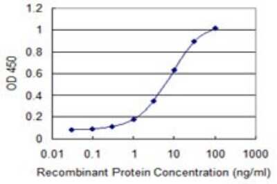
Sandwich ELISA: MAP1 Antibody (4A1) [H00064112-M02] - Detection limit for recombinant GST tagged MOAP1 is 0.3 ng/ml as a capture antibody.
 - Azide and BSA Free [H00064112-M02] -")
Western Blot: MAP1 Antibody (4A1) - Azide and BSA Free [H00064112-M02] -
Comparison of downregulation of apoptosis/MOAP1 by siRNA of MOAP1 and hcmv-miR-UL70-3p: (a) Through DAPI imaging, images (63×) showing the nuclear morphology. (b) Measurement of Caspase 3/7 activity: The Caspase 3/7 activities were measured through Caspase Glo 3/7 assay in four different groups of cells, as mentioned above, these were recorded as raw luminescence units (RLU) (+/-SEM; ****p < 0.0001). (c) MOAP1 mRNA downregulation: The relative expression levels of MOAP1 mRNA after the transfection of either hcmv-miR-UL70-3p and siRNA of MOAP1 were measured through qRT-PCR. Results were expressed as the fold change (2− delta delta Ct) (+/- SEM; **, p < 0.01; ****, p < 0.0001). (d) MOAP1 protein downregulation: The MOAP1 protein downregulation after transfection with either siRNA of MOAP1 and hcmv-miR-UL70-3p were analyzed through Western blot. The relative MOAP1 protein quantification was performed through ImageJ software after normalizing with beta -actin. Experiments were performed in triplicates (Supplementary Figure S2b), and the data from three different experiments were used for statistical analysis (+/-SEM; *, p < 0.05; **, p < 0.01, ns = non-significant). Image collected and cropped by CiteAb from the following open publication (https://pubmed.ncbi.nlm.nih.gov/35008453), licensed under a CC-BY license. Not internally tested by Novus Biologicals.Applications for MAP1 Antibody (4A1) - Azide and BSA Free
Application
Recommended Usage
ELISA
Optimal dilutions of this antibody should be experimentally determined.
Immunocytochemistry/ Immunofluorescence
Optimal dilutions of this antibody should be experimentally determined.
Sandwich ELISA
Optimal dilutions of this antibody should be experimentally determined.
Western Blot
Optimal dilutions of this antibody should be experimentally determined.
Application Notes
This antibody is useful for ELISA, Western Blot
Formulation, Preparation, and Storage
Purification
IgG purified
Formulation
In 1x PBS, pH 7.4
Format
Azide and BSA Free
Preservative
No Preservative
Concentration
Concentrations vary lot to lot. See vial label for concentration. If unlisted please contact technical services.
Shipping
The product is shipped with polar packs. Upon receipt, store it immediately at the temperature recommended below.
Stability & Storage
Aliquot and store at -20C or -80C. Avoid freeze-thaw cycles.
Background: MAP1
Alternate Names
MAP1, MAP-1PNMA4Paraneoplastic antigen Ma4, modulator of apoptosis 1, paraneoplastic antigen like 4
Gene Symbol
MOAP1
UniProt
Additional MAP1 Products
Product Documents for MAP1 Antibody (4A1) - Azide and BSA Free
Certificate of Analysis
To download a Certificate of Analysis, please enter a lot or batch number in the search box below.
Product Specific Notices for MAP1 Antibody (4A1) - Azide and BSA Free
This product is produced by and distributed for Abnova, a company based in Taiwan.
This product is for research use only and is not approved for use in humans or in clinical diagnosis. Primary Antibodies are guaranteed for 1 year from date of receipt.
Citations for MAP1 Antibody (4A1) - Azide and BSA Free
 Powered by Bioz
Customer Reviews for MAP1 Antibody (4A1) - Azide and BSA Free
There are currently no reviews for this product. Be the first to review MAP1 Antibody (4A1) - Azide and BSA Free and earn rewards!
Have you used MAP1 Antibody (4A1) - Azide and BSA Free?
Submit a review and receive an Amazon gift card!
$25/€18/£15/$25CAN/¥2500 Yen for a review with an image
$10/€7/£6/$10CAN/¥1110 Yen for a review without an image
Submit a review
Protocols
Find general support by application which include: protocols, troubleshooting, illustrated assays, videos and webinars.
- Appropriate Fixation of IHC/ICC Samples
- Cellular Response to Hypoxia Protocols
- ClariTSA™ Fluorophore Kits
- Detection & Visualization of Antibody Binding
- ELISA Sample Preparation & Collection Guide
- ELISA Troubleshooting Guide
- How to Run an R&D Systems DuoSet ELISA
- How to Run an R&D Systems Quantikine ELISA
- How to Run an R&D Systems Quantikine™ QuicKit™ ELISA
- ICC Cell Smear Protocol for Suspension Cells
- ICC Immunocytochemistry Protocol Videos
- ICC for Adherent Cells
- Immunocytochemistry (ICC) Protocol
- Immunocytochemistry Troubleshooting
- Immunofluorescence of Organoids Embedded in Cultrex Basement Membrane Extract
- Immunohistochemistry (IHC) and Immunocytochemistry (ICC) Protocols
- Preparing Samples for IHC/ICC Experiments
- Preventing Non-Specific Staining (Non-Specific Binding)
- Primary Antibody Selection & Optimization
- Protocol for VisUCyte™ HRP Polymer Detection Reagent
- Protocol for the Fluorescent ICC Staining of Cell Smears - Graphic
- Protocol for the Fluorescent ICC Staining of Cultured Cells on Coverslips - Graphic
- Protocol for the Preparation and Fluorescent ICC Staining of Cells on Coverslips
- Protocol for the Preparation and Fluorescent ICC Staining of Non-adherent Cells
- Protocol for the Preparation and Fluorescent ICC Staining of Stem Cells on Coverslips
- Protocol for the Preparation of a Cell Smear for Non-adherent Cell ICC - Graphic
- Quantikine HS ELISA Kit Assay Principle, Alkaline Phosphatase
- Quantikine HS ELISA Kit Principle, Streptavidin-HRP Polymer
- R&D Systems Quality Control Western Blot Protocol
- Sandwich ELISA (Colorimetric) – Biotin/Streptavidin Detection Protocol
- Sandwich ELISA (Colorimetric) – Direct Detection Protocol
- TUNEL and Active Caspase-3 Detection by IHC/ICC Protocol
- The Importance of IHC/ICC Controls
- Troubleshooting Guide: ELISA
- Troubleshooting Guide: Western Blot Figures
- Western Blot Conditions
- Western Blot Protocol
- Western Blot Protocol for Cell Lysates
- Western Blot Troubleshooting
- Western Blot Troubleshooting Guide
- View all Protocols, Troubleshooting, Illustrated assays and Webinars
Loading...