Mas Antibody - BSA Free
Novus Biologicals | Catalog # NBP1-78444
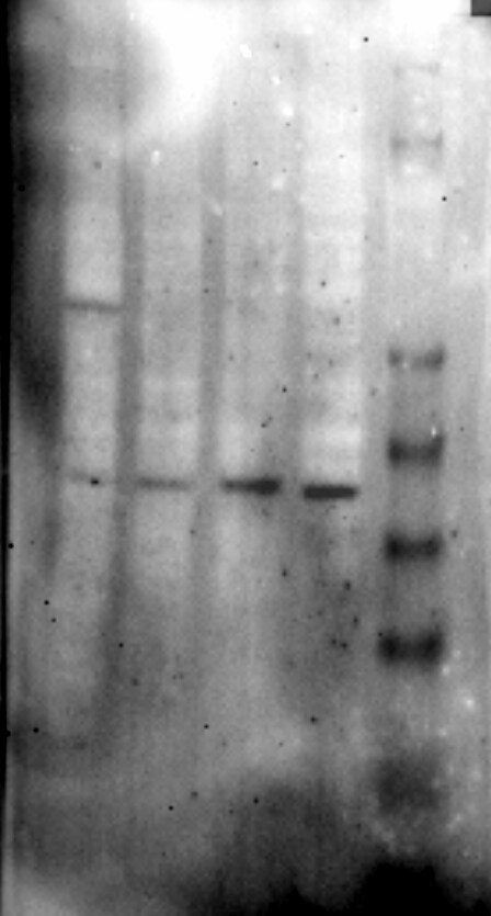
![Western Blot: Mas AntibodyBSA Free [NBP1-78444]](https://resources.rndsystems.com/images/products/Mas-Antibody-Western-Blot-NBP1-78444-img0008.jpg "Western Blot: Mas AntibodyBSA Free [NBP1-78444]")
Key Product Details
Species Reactivity
Validated:
Cited:
Applications
Validated:
Cited:
Label
Antibody Source
Format
Product Specifications
Immunogen
Reactivity Notes
Localization
Clonality
Host
Isotype
Scientific Data Images for Mas Antibody - BSA Free
Western Blot: Mas AntibodyBSA Free [NBP1-78444]
Western Blot: Mas Antibody [NBP1-78444] - Analysis of MAS1 in: (1) Ntera 2, (2) A431, (3) HepG2, (4) MCF7, (5) 3T3, and (6) Cos7.![Immunocytochemistry/ Immunofluorescence: Mas Antibody - BSA Free [NBP1-78444]](https://resources.rndsystems.com/images/products/Mas-Antibody-Immunocytochemistry-Immunofluorescence-NBP1-78444-img0006.jpg "Immunocytochemistry/ Immunofluorescence: Mas Antibody - BSA Free [NBP1-78444]")
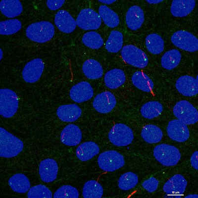
Immunocytochemistry/ Immunofluorescence: Mas Antibody - BSA Free [NBP1-78444]
Immunocytochemistry/Immunofluorescence: Mas Antibody [NBP1-78444] - Tested at 1:25 in HepG2 cells with FITC (green). Nuclei were counterstained with DAPI (blue).![Immunohistochemistry: Mas Antibody - BSA Free [NBP1-78444]](https://resources.rndsystems.com/images/products/Mas-Antibody-Immunohistochemistry-NBP1-78444-img0007.jpg "Immunohistochemistry: Mas Antibody - BSA Free [NBP1-78444]")
Immunohistochemistry: Mas Antibody - BSA Free [NBP1-78444]
Immunohistochemistry: Mas Antibody [NBP1-78444] - Analysis of MAS1 in mouse kidney using DAB with hematoxylin counterstain.![Simple Western: Mas AntibodyBSA Free [NBP1-78444]](https://resources.rndsystems.com/images/products/Mas-Antibody-Simple-Western-NBP1-78444-img0009.jpg "Simple Western: Mas AntibodyBSA Free [NBP1-78444]")
Simple Western: Mas AntibodyBSA Free [NBP1-78444]
Simple Western: Mas Antibody [NBP1-78444] - Lane view shows a specific band for MAS1 in 0.5 mg/ml of HepG2 lysate. This experiment was performed under reducing conditions using the 12-230 kDa separation system.
Western Blot: Mas Antibody - BSA Free [NBP1-78444] -
MasR activation restores LPS‐induced overactivation of ACE/AngII/AT1 axis and contains neuroprotective properties. (a–j) The impacts of MasR agonist, AVE, and the ACE2 activator, DIZE, on brain RAS following repeated LPS treatment. (a) Representative western blots. ACE protein expression (b) and activity (c). (d) AngII concentration. (e) AT1 protein expression. ACE2 protein expression (f) and activity (g). (h) Ang(1–7) concentration. MasR protein expression (i) and immunofluorescence staining (j). (m) Representative images of TUNEL and Nissl staining. (k, l) mRNA expression of biomarkers of microglial M1 phenotype (k) and M2 phenotype (l). Scale bar = 50 μm. Data are means +/- SD (n = 7). **p < 0.01 compared to control group. +p < 0.05, ++p < 0.01 compared to LPS group Image collected and cropped by CiteAb from the following open publication (https://pubmed.ncbi.nlm.nih.gov/34529881), licensed under a CC-BY license. Not internally tested by Novus Biologicals.
Western Blot: Mas Antibody - BSA Free [NBP1-78444] -
LPS exposure shifts the balance of brain RAS. (a) The impacts of acute LPS (AcLPS) or repeated LPS (ReLPS) exposure on microglial activation (IBA‐1 staining) and ROS generation (DHE staining) in the brain cortex of C57BL/6 mice. (b) Biomarkers of microglial M1 phenotype mRNA expression. (c) Biomarkers of microglial M2 phenotype mRNA expression. (d–m) Alterations of ACE/AngII/AT1 and ACE2/Ang(1–7)/MasR pathways following LPS treatment. (d) Representative western blots. ACE protein expression (e) and activity (f). (g) AngII concentration. (h) AT1 protein expression. (i) AT2 protein expression. ACE2 protein expression (j) and activity (k). (L) Ang(1–7) concentration. (m) MasR protein expression. Scale bar = 50 μm. Data are means +/- SD (n = 7–9). *p < 0.05, **p < 0.01 compared to control group Image collected and cropped by CiteAb from the following open publication (https://pubmed.ncbi.nlm.nih.gov/34529881), licensed under a CC-BY license. Not internally tested by Novus Biologicals.Applications for Mas Antibody - BSA Free
Immunocytochemistry/ Immunofluorescence
Immunohistochemistry
Immunohistochemistry-Paraffin
Simple Western
Western Blot
See Simple Western Antibody Database for Simple Western validation: Tested in HepG2 lysate 0.5 mg/mL, separated by Size, antibody dilution of 1:100, apparent MW was 47 kDa. Separated by Size-Wes, Sally Sue/Peggy Sue.
Reviewed Applications
Read 2 reviews rated 5 using NBP1-78444 in the following applications:
Formulation, Preparation, and Storage
Purification
Formulation
Format
Preservative
Concentration
Shipping
Stability & Storage
Background: Mas
Additional Mas Products
Product Documents for Mas Antibody - BSA Free
Certificate of Analysis
To download a Certificate of Analysis, please enter a lot or batch number in the search box below.
Product Specific Notices for Mas Antibody - BSA Free
This product is for research use only and is not approved for use in humans or in clinical diagnosis. Primary Antibodies are guaranteed for 1 year from date of receipt.
Related Research Areas
Citations for Mas Antibody - BSA Free
 Powered by Bioz
Customer Reviews for Mas Antibody - BSA Free (2)
Have you used Mas Antibody - BSA Free?
Submit a review and receive an Amazon gift card!
$25/€18/£15/$25CAN/¥2500 Yen for a review with an image
$10/€7/£6/$10CAN/¥1110 Yen for a review without an image
Submit a review
Customer Images
-
Application: Western BlotSample Tested: IMCD3, hTertRPE1, HDF, SHSY5Y whole cell lysatesSpecies: HumanVerified Customer | Posted 10/30/2013

-
Application: ImmunofluorescenceSample Tested: Mouse inner medullary collecting duct (IMCD3) cell lineSpecies: MouseVerified Customer | Posted 10/14/2013
There are no reviews that match your criteria.
Protocols
View specific protocols for Mas Antibody - BSA Free (NBP1-78444):
Immunocytochemistry Protocol
Culture cells to appropriate density in 35 mm culture dishes or 6-well plates.
1. Remove culture medium and add 10% formalin to the dish. Fix at room temperature for 30 minutes.
2. Remove the formalin and add ice cold methanol. Incubate for 5-10 minutes.
3. Remove methanol and add washing solution (i.e. PBS). Be sure to not let the specimen dry out. Wash three times for 10 minutes.
4. To block nonspecific antibody binding incubate in 10% normal goat serum from 1 hour to overnight at room temperature.
5. Add primary antibody at appropriate dilution and incubate at room temperature from 2 hours to overnight at room temperature.
6. Remove primary antibody and replace with washing solution. Wash three times for 10 minutes.
7. Add secondary antibody at appropriate dilution. Incubate for 1 hour at room temperature.
8. Remove antibody and replace with wash solution, then wash for 10 minutes. Add Hoechst 33258 to wash solution at 1:25,0000 and incubate for 10 minutes. Wash a third time for 10 minutes.
9. Cells can be viewed directly after washing. The plates can also be stored in PBS containing Azide covered in Parafilm (TM). Cells can also be cover-slipped using Fluoromount, with appropriate sealing.
*The above information is only intended as a guide. The researcher should determine what protocol best meets their needs. Please follow safe laboratory procedures.
Immunohistochemistry-Paraffin Embedded Sections
Antigen Unmasking:
Bring slides to a boil in 10 mM sodium citrate buffer (pH 6.0) then maintain at a sub-boiling temperature for 10 minutes. Cool slides on bench-top for 30 minutes.
Staining:
1. Wash sections in deionized water three times for 5 minutes each.
2. Wash sections in wash buffer for 5 minutes.
3. Block each section with 100-400 ul blocking solution for 1 hour at room temperature.
4. Remove blocking solution and add 100-400 ul diluted primary antibody. Incubate overnight at 4C.
5. Remove antibody solution and wash sections in wash buffer three times for 5 minutes each.
6. Add 100-400 ul biotinylated diluted secondary antibody. Incubate 30 minutes at room temperature.
7. Remove secondary antibody solution and wash sections three times with wash buffer for 5 minutes each.
8. Add 100-400 ul Streptavidin-HRP reagent to each section and incubate for 30 minutes at room temperature.
9. Wash sections three times in wash buffer for 5 minutes each.
10. Add 100-400 ul DAB substrate to each section and monitor staining closely.
11. As soon as the sections develop, immerse slides in deionized water.
12. Counterstain sections in hematoxylin.
13. Wash sections in deionized water two times for 5 minutes each.
14. Dehydrate sections.
15. Mount coverslips.
*The above information is only intended as a guide. The researcher should determine what protocol best meets their needs. Please follow safe laboratory procedures.
Western Blot Protocol
1. Perform SDS-PAGE on samples to be analyzed, loading 40 ug of total protein per lane.
2. Transfer proteins to membrane according to the instructions provided by the manufacturer of the membrane and transfer apparatus.
3. Stain according to standard Ponceau S procedure (or similar product) to assess transfer success, and mark molecular weight standards where appropriate.
4. Rinse the blot.
5. Block the membrane using standard blocking buffer for at least 1 hour.
6. Wash the membrane in wash buffer three times for 10 minutes each.
7. Dilute primary antibody in blocking buffer and incubate 1 hour at room temperature.
8. Wash the membrane in wash buffer three times for 10 minutes each.
9. Apply the diluted HRP conjugated secondary antibody in blocking buffer (as per manufacturers instructions) and incubate 1 hour at room temperature.
10. Wash the blot in wash buffer three times for 10 minutes each (this step can be repeated as required to reduce background).
11. Apply the detection reagent of choice in accordance with the manufacturers instructions.
Note: Tween-20 can be added to the blocking or antibody dilution buffer at a final concentration of 0.05-0.2%.
*The above information is only intended as a guide. The researcher should determine what protocol best meets their needs. Please follow safe laboratory procedures.
Find general support by application which include: protocols, troubleshooting, illustrated assays, videos and webinars.
- Antigen Retrieval Protocol (PIER)
- Antigen Retrieval for Frozen Sections Protocol
- Appropriate Fixation of IHC/ICC Samples
- Cellular Response to Hypoxia Protocols
- Chromogenic IHC Staining of Formalin-Fixed Paraffin-Embedded (FFPE) Tissue Protocol
- Chromogenic Immunohistochemistry Staining of Frozen Tissue
- ClariTSA™ Fluorophore Kits
- Detection & Visualization of Antibody Binding
- Fluorescent IHC Staining of Frozen Tissue Protocol
- Graphic Protocol for Heat-induced Epitope Retrieval
- Graphic Protocol for the Preparation and Fluorescent IHC Staining of Frozen Tissue Sections
- Graphic Protocol for the Preparation and Fluorescent IHC Staining of Paraffin-embedded Tissue Sections
- Graphic Protocol for the Preparation of Gelatin-coated Slides for Histological Tissue Sections
- ICC Cell Smear Protocol for Suspension Cells
- ICC Immunocytochemistry Protocol Videos
- ICC for Adherent Cells
- IHC Sample Preparation (Frozen sections vs Paraffin)
- Immunocytochemistry (ICC) Protocol
- Immunocytochemistry Troubleshooting
- Immunofluorescence of Organoids Embedded in Cultrex Basement Membrane Extract
- Immunofluorescent IHC Staining of Formalin-Fixed Paraffin-Embedded (FFPE) Tissue Protocol
- Immunohistochemistry (IHC) and Immunocytochemistry (ICC) Protocols
- Immunohistochemistry Frozen Troubleshooting
- Immunohistochemistry Paraffin Troubleshooting
- Preparing Samples for IHC/ICC Experiments
- Preventing Non-Specific Staining (Non-Specific Binding)
- Primary Antibody Selection & Optimization
- Protocol for Heat-Induced Epitope Retrieval (HIER)
- Protocol for Making a 4% Formaldehyde Solution in PBS
- Protocol for VisUCyte™ HRP Polymer Detection Reagent
- Protocol for the Fluorescent ICC Staining of Cell Smears - Graphic
- Protocol for the Fluorescent ICC Staining of Cultured Cells on Coverslips - Graphic
- Protocol for the Preparation & Fixation of Cells on Coverslips
- Protocol for the Preparation and Chromogenic IHC Staining of Frozen Tissue Sections
- Protocol for the Preparation and Chromogenic IHC Staining of Frozen Tissue Sections - Graphic
- Protocol for the Preparation and Chromogenic IHC Staining of Paraffin-embedded Tissue Sections
- Protocol for the Preparation and Chromogenic IHC Staining of Paraffin-embedded Tissue Sections - Graphic
- Protocol for the Preparation and Fluorescent ICC Staining of Cells on Coverslips
- Protocol for the Preparation and Fluorescent ICC Staining of Non-adherent Cells
- Protocol for the Preparation and Fluorescent ICC Staining of Stem Cells on Coverslips
- Protocol for the Preparation and Fluorescent IHC Staining of Frozen Tissue Sections
- Protocol for the Preparation and Fluorescent IHC Staining of Paraffin-embedded Tissue Sections
- Protocol for the Preparation of Gelatin-coated Slides for Histological Tissue Sections
- Protocol for the Preparation of a Cell Smear for Non-adherent Cell ICC - Graphic
- R&D Systems Quality Control Western Blot Protocol
- TUNEL and Active Caspase-3 Detection by IHC/ICC Protocol
- The Importance of IHC/ICC Controls
- Troubleshooting Guide: Immunohistochemistry
- Troubleshooting Guide: Western Blot Figures
- Western Blot Conditions
- Western Blot Protocol
- Western Blot Protocol for Cell Lysates
- Western Blot Troubleshooting
- Western Blot Troubleshooting Guide
- View all Protocols, Troubleshooting, Illustrated assays and Webinars
FAQs for Mas Antibody - BSA Free
-
Q: Hi, I have two questions about MAS1 Antibody (NBP1-78444). Could you tell me if it could detect N-terminal tag3* flag-Mas or not? The human N-terminal tag3* flag-Mas vector that I constructed were veri?ed by DNA sequencing and were consistent with the NCBI reference sequence: NM_002377.2. It's open reading frame-shift did not appear and MAS1 is about 34KD. Why is the MW of MAS1 is bigger than 37KD on your datasheet?
A:
I have pulled up information on your NCBI sequence and it matches that associated with the uniprot ID P04201 for this antibody (https://www.uniprot.org/uniprotkb/P04201/entry) The protein appears to theoretically have a molecular weight of 37 kDa, but most likely due to post translational modifications such as glycosylation we expressed the protein at a higher molecular weight in our sample, which can be common for proteins that under go post translational modifications such as this one. We will back this antibody with our 100% guarantee for all stated species and applications.