MAT2A Antibody - BSA Free
Novus Biologicals | Catalog # NB110-94158
![Immunocytochemistry/ Immunofluorescence: MAT2A Antibody - BSA Free [NB110-94158]](https://resources.rndsystems.com/images/products/MAT2A-Antibody-Immunocytochemistry-Immunofluorescence-NB110-94158-img0010.jpg "Immunocytochemistry/ Immunofluorescence: MAT2A Antibody - BSA Free [NB110-94158]")
Key Product Details
Species Reactivity
Validated:
Cited:
Predicted:
Applications
Validated:
Cited:
Label
Antibody Source
Format
Product Specifications
Immunogen
Reactivity Notes
Localization
Clonality
Host
Isotype
Theoretical MW
Disclaimer note: The observed molecular weight of the protein may vary from the listed predicted molecular weight due to post translational modifications, post translation cleavages, relative charges, and other experimental factors.
Scientific Data Images for MAT2A Antibody - BSA Free
Immunocytochemistry/ Immunofluorescence: MAT2A Antibody - BSA Free [NB110-94158]
Immunocytochemistry/Immunofluorescence: MAT2A Antibody [NB110-94158] - HepG2 cells were fixed in 4% paraformaldehyde for 10 minutes and permeabilized in 0.05% Triton X-100 in PBS for 5 minutes. The cells were incubated with anti-MAT2A Antibody (NB110-94158) at 1 ug/ml overnight at 4C and detected with an anti-rabbit Dylight 488 (Green) at a 1:1000 dilution for 60 minutes. Nuclei were counterstained with DAPI (Blue). Cells were imaged using a 100X objective and digitally deconvolved.![Immunocytochemistry/ Immunofluorescence: MAT2A Antibody - BSA Free [NB110-94158]](https://resources.rndsystems.com/images/products/MAT2A-Antibody-Immunocytochemistry-Immunofluorescence-NB110-94158-img0011.jpg "Immunocytochemistry/ Immunofluorescence: MAT2A Antibody - BSA Free [NB110-94158]")
Immunocytochemistry/ Immunofluorescence: MAT2A Antibody - BSA Free [NB110-94158]
Immunocytochemistry/Immunofluorescence: MAT2A Antibody [NB110-94158] - NIH3T3 cells were fixed in 4% paraformaldehyde for 10 minutes and permeabilized in 0.05% Triton X-100 in PBS for 5 minutes. The cells were incubated with anti-MAT2A Antibody (NB110-94158) at 1 ug/ml overnight at 4C and detected with an anti-rabbit Dylight 488 (Green) at a 1:1000 dilution for 60 minutes. Nuclei were counterstained with DAPI (Blue). Cells were imaged using a 100X objective and digitally deconvolved.![Immunocytochemistry/ Immunofluorescence: MAT2A Antibody - BSA Free [NB110-94158]](https://resources.rndsystems.com/images/products/MAT2A-Antibody-Immunocytochemistry-Immunofluorescence-NB110-94158-img0012.jpg "Immunocytochemistry/ Immunofluorescence: MAT2A Antibody - BSA Free [NB110-94158]")
Immunocytochemistry/ Immunofluorescence: MAT2A Antibody - BSA Free [NB110-94158]
Immunocytochemistry/Immunofluorescence: MAT2A Antibody [NB110-94158] - PC12 cells were fixed in 4% paraformaldehyde for 10 minutes and permeabilized in 0.05% Triton X-100 in PBS for 5 minutes. The cells were incubated with anti-MAT2A Antibody (NB110-94158) at 1 ug/ml overnight at 4C and detected with an anti-rabbit Dylight 488 (Green) at a 1:1000 dilution for 60 minutes. Nuclei were counterstained with DAPI (Blue). Cells were imaged using a 100X objective and digitally deconvolved.![Western Blot: MAT2A AntibodyBSA Free [NB110-94158]](https://resources.rndsystems.com/images/products/MAT2A-Antibody-Western-Blot-NB110-94158-img0008.jpg "Western Blot: MAT2A AntibodyBSA Free [NB110-94158]")
Western Blot: MAT2A AntibodyBSA Free [NB110-94158]
MAT2A-Antibody-Western-Blot-NB110-94158-img0008.jpg![Immunohistochemistry-Paraffin: MAT2A Antibody - BSA Free [NB110-94158]](https://resources.rndsystems.com/images/products/MAT2A-Antibody-Immunohistochemistry-Paraffin-NB110-94158-img0009.jpg "Immunohistochemistry-Paraffin: MAT2A Antibody - BSA Free [NB110-94158]")
Immunohistochemistry-Paraffin: MAT2A Antibody - BSA Free [NB110-94158]
Immunohistochemistry-Paraffin: MAT2A Antibody [NB110-94158] - Analysis of a FFPE tissue section of human kidney using 1:200 dilution of MAT2A antibody (NB110-94158). The staining was developed using HRP labeled anti-rabbit secondary antibody and DAB reagent, and nuclei of cells were counter-stained with hematoxylin.![Western Blot: MAT2A AntibodyBSA Free [NB110-94158]](https://resources.rndsystems.com/images/products/MAT2A-Antibody-Western-Blot-NB110-94158-img0005.jpg "Western Blot: MAT2A AntibodyBSA Free [NB110-94158]")
Western Blot: MAT2A AntibodyBSA Free [NB110-94158]
Western Blot: MAT2A Antibody [NB110-94158] - Total protein from HeLa and HepG2 cells and Human and Mouse Kidney was separated on a 12% gel by SDS-PAGE, transferred to PVDF membrane and blocked in 5% non-fat milk in TBST. The membrane was probed with 2.0 ug/mL anti-MAT2A in 1% non-fat milk in TBST and detected with an anti-rabbit HRP secondary antibody using chemiluminescence.![Western Blot: MAT2A AntibodyBSA Free [NB110-94158]](https://resources.rndsystems.com/images/products/MAT2A-Antibody-Western-Blot-NB110-94158-img0003.jpg "Western Blot: MAT2A AntibodyBSA Free [NB110-94158]")
Western Blot: MAT2A AntibodyBSA Free [NB110-94158]
Western Blot: MAT2A Antibody [NB110-94158] - Detection of MAT2A in HepG2 whole cell lysates.![Immunocytochemistry/ Immunofluorescence: MAT2A Antibody - BSA Free [NB110-94158]](https://resources.rndsystems.com/images/products/MAT2A-Antibody-Immunocytochemistry-Immunofluorescence-NB110-94158-img0004.jpg "Immunocytochemistry/ Immunofluorescence: MAT2A Antibody - BSA Free [NB110-94158]")
Immunocytochemistry/ Immunofluorescence: MAT2A Antibody - BSA Free [NB110-94158]
Immunocytochemistry/Immunofluorescence: MAT2A Antibody [NB110-94158] - MAT2A antibody was tested in HepG2 cells with DyLight 488 (green). Nuclei and alpha-tubulin were counterstained with DAPI (blue) and DyLight 550 (red).![Immunocytochemistry/ Immunofluorescence: MAT2A Antibody - BSA Free [NB110-94158]](https://resources.rndsystems.com/images/products/MAT2A-Antibody-Immunocytochemistry-Immunofluorescence-NB110-94158-img0006.jpg "Immunocytochemistry/ Immunofluorescence: MAT2A Antibody - BSA Free [NB110-94158]")
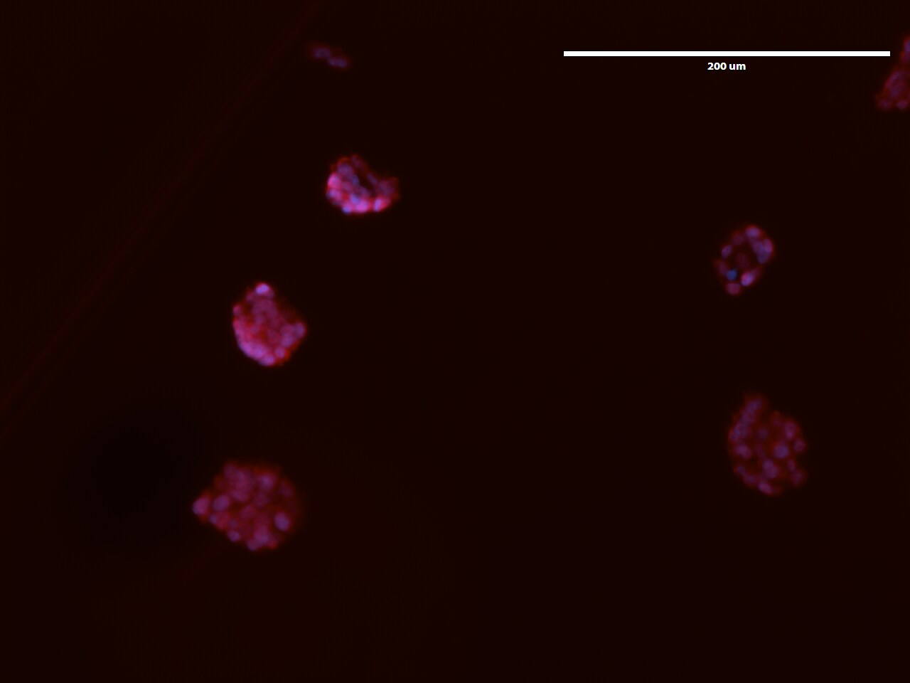
Immunocytochemistry/ Immunofluorescence: MAT2A Antibody - BSA Free [NB110-94158]
Immunocytochemistry/Immunofluorescence: MAT2A Antibody [NB110-94158] - Mouse embryonic stem cells (mESCs) were fixed with paraformaldehyde, permeabilized with Triton, blocked with 4% BSA and 1% normal goat serum, and incubated with 1:200 dilution of MAT2A antibody. Then, samples were stained with anti-rabbit Alexa Fluor 594 (1:1000) and NucBlue. ICC/IF image submitted by a verified customer review.![Simple Western: MAT2A AntibodyBSA Free [NB110-94158]](https://resources.rndsystems.com/images/products/MAT2A-Antibody-Simple-Western-NB110-94158-img0007.jpg "Simple Western: MAT2A AntibodyBSA Free [NB110-94158]")
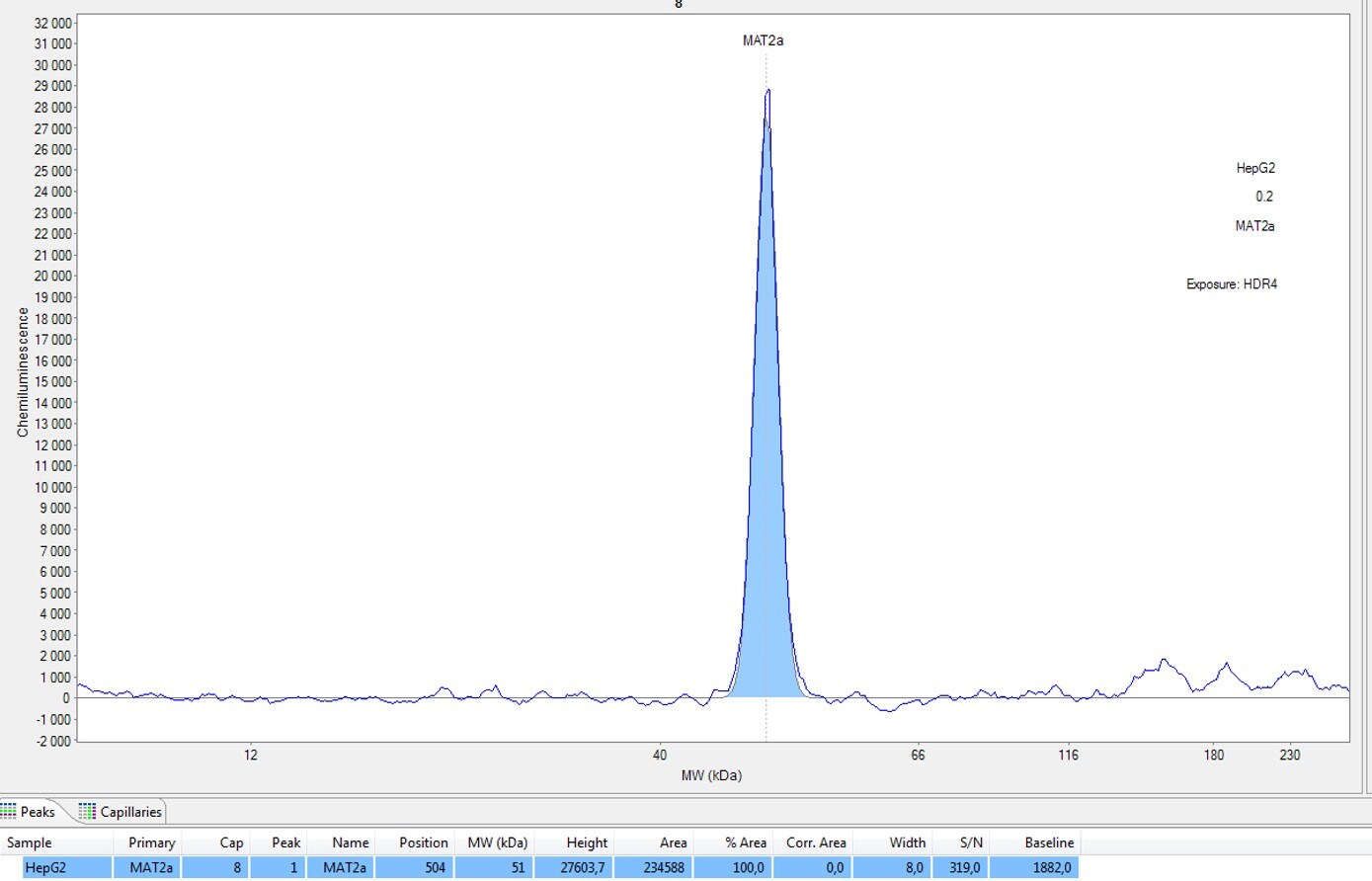
Simple Western: MAT2A AntibodyBSA Free [NB110-94158]
Simple Western: MAT2A Antibody [NB110-94158] - Human HepG2 cell lysate (0.2 ug/uL total protein). Antibody at 1:250. Simple Western image submitted by a verified customer review.Applications for MAT2A Antibody - BSA Free
Immunocytochemistry/ Immunofluorescence
Immunohistochemistry
Immunohistochemistry-Paraffin
Immunoprecipitation
Western Blot
Reviewed Applications
Read 3 reviews rated 4.3 using NB110-94158 in the following applications:
Formulation, Preparation, and Storage
Purification
Formulation
Format
Preservative
Concentration
Shipping
Stability & Storage
Background: MAT2A
Long Name
Alternate Names
Gene Symbol
Additional MAT2A Products
Product Documents for MAT2A Antibody - BSA Free
Certificate of Analysis
To download a Certificate of Analysis, please enter a lot or batch number in the search box below.
Product Specific Notices for MAT2A Antibody - BSA Free
This product is for research use only and is not approved for use in humans or in clinical diagnosis. Primary Antibodies are guaranteed for 1 year from date of receipt.
Citations for MAT2A Antibody - BSA Free
 Powered by Bioz
Customer Reviews for MAT2A Antibody - BSA Free (3)
Have you used MAT2A Antibody - BSA Free?
Submit a review and receive an Amazon gift card!
$25/€18/£15/$25CAN/¥2500 Yen for a review with an image
$10/€7/£6/$10CAN/¥1110 Yen for a review without an image
Submit a review
Customer Images
-
Application: Simple WesternSample Tested: HepG2 CELLSSpecies: HumanVerified Customer | Posted 02/03/2020Matériel: 1x25caps 12-230kDa échantillon : 0.2µg/µL anticorps : 1/250
 Bio-Techne ResponseThis review was submitted through the legacy Novus Innovators Program, reflecting a new species or application tested on a primary antibody.
 -
Application: ImmunocytochemistrySample Tested: J1 embryonic stem cells and Mouse Embryonic Stem CellsSpecies: Mus musculus and MouseVerified Customer | Posted 02/18/2019mESCs were fixed with paraformaldehyde, permeabilized with Triton, blocked with 4% BSA and 1% normal goat serum, and incubated with 1:200 dilution of Mat2a antibody. Then, samples were stained with anti-rabbit Alexa Fluor 594 (1:1000) and NucBlue.Antibody behaved as expected in IF (different conditions gave different levels of fluorescence that matched qPCR and Western blot results very well).
-
Application: Western BlotSample Tested: Whole cell lysate prepared from HEK 293 cellsSpecies: HumanVerified Customer | Posted 11/26/2014Western blot for MAT1A in HEK293 cells
There are no reviews that match your criteria.
Protocols
View specific protocols for MAT2A Antibody - BSA Free (NB110-94158):
Immunocytochemistry Protocol
Culture cells to appropriate density in 35 mm culture dishes or 6-well plates.
1. Remove culture medium and add 10% formalin to the dish. Fix at room temperature for 30 minutes.
2. Remove the formalin and add ice cold methanol. Incubate for 5-10 minutes.
3. Remove methanol and add washing solution (i.e. PBS). Be sure to not let the specimen dry out. Wash three times for 10 minutes.
4. To block nonspecific antibody binding incubate in 10% normal goat serum from 1 hour to overnight at room temperature.
5. Add primary antibody at appropriate dilution and incubate at room temperature from 2 hours to overnight at room temperature.
6. Remove primary antibody and replace with washing solution. Wash three times for 10 minutes.
7. Add secondary antibody at appropriate dilution. Incubate for 1 hour at room temperature.
8. Remove antibody and replace with wash solution, then wash for 10 minutes. Add Hoechst 33258 to wash solution at 1:25,0000 and incubate for 10 minutes. Wash a third time for 10 minutes.
9. Cells can be viewed directly after washing. The plates can also be stored in PBS containing Azide covered in Parafilm (TM). Cells can also be cover-slipped using Fluoromount, with appropriate sealing.
*The above information is only intended as a guide. The researcher should determine what protocol best meets their needs. Please follow safe laboratory procedures.
Antigen Unmasking:
Bring slides to a boil in 10 mM sodium citrate buffer (pH 6.0) then maintain at a sub-boiling temperature for 10 minutes. Cool slides on bench-top for 30 minutes (keep slides in the sodium citrate buffer all the time).
Staining:
1. Wash sections in deionized water three times for 5 minutes each.
2. Wash sections in PBS for 5 minutes.
3. Block each section with 100-400 ul blocking solution (1% BSA in PBS) for 1 hour at room temperature.
4. Remove blocking solution and add 100-400 ul diluted primary antibody. Incubate overnight at 4 C.
5. Remove antibody solution and wash sections in wash buffer three times for 5 minutes each.
6. Add 100-400 ul HRP polymer conjugated secondary antibody. Incubate 30 minutes at room temperature.
7. Wash sections three times in wash buffer for 5 minutes each.
8. Add 100-400 ul DAB substrate to each section and monitor staining closely.
9. As soon as the sections develop, immerse slides in deionized water.
10. Counterstain sections in hematoxylin.
11. Wash sections in deionized water two times for 5 minutes each.
12. Dehydrate sections.
13. Mount coverslips.
Western Blot Protocol
1. Perform SDS-PAGE (4-12% MOPS) on samples to be analyzed, loading 40 ug of total protein per lane.
2. Transfer proteins to Nitrocellulose according to the instructions provided by the manufacturer of the transfer
apparatus.
3. Rinse membrane with dH2O and then stain the blot using Ponceau S for 1-2 minutes to access the transfer of proteins onto the nitrocellulose membrane. Rinse the blot in water to remove excess stain and mark the lane locations and locations of molecular weight markers using a pencil.
4. Rinse the blot in TBS for approximately 5 minutes.
5. Block the membrane using 5% NFDM + 1% BSA in TBS + Tween, 1 hour at RT.
6. Rinse the membrane in dH2O and then wash the membrane in wash buffer [TBS + 0.1% Tween] 3 times for 10 minutes each.
7. Dilute the rabbit anti-MAT2a primary antibody (NB 110-94158) in blocking buffer and incubate 1 hour at room temperature.
8. Rinse the membrane in dH2O and then wash the membrane in wash buffer [TBS + 0.1% Tween] 3 times for 10 minutes each.
9. Apply the diluted rabbit-IgG HRP-conjugated secondary antibody in blocking buffer (as per manufacturers
instructions) and incubate 1 hour at room temperature.
10. Wash the blot in wash buffer [TBS + 0.1% Tween] 3 times for 10 minutes each (this step can be repeated as required to reduce background).
11. Apply the detection reagent of choice in accordance with the manufacturers instructions (Pierce ECL).
Note: Tween-20 can be added to the blocking or antibody dilution buffer at a final concentration of 0.05-0.2%, provided it does not interfere with antibody-antigen binding.
Find general support by application which include: protocols, troubleshooting, illustrated assays, videos and webinars.
- Antigen Retrieval Protocol (PIER)
- Antigen Retrieval for Frozen Sections Protocol
- Appropriate Fixation of IHC/ICC Samples
- Cellular Response to Hypoxia Protocols
- Chromogenic IHC Staining of Formalin-Fixed Paraffin-Embedded (FFPE) Tissue Protocol
- Chromogenic Immunohistochemistry Staining of Frozen Tissue
- ClariTSA™ Fluorophore Kits
- Detection & Visualization of Antibody Binding
- Fluorescent IHC Staining of Frozen Tissue Protocol
- Graphic Protocol for Heat-induced Epitope Retrieval
- Graphic Protocol for the Preparation and Fluorescent IHC Staining of Frozen Tissue Sections
- Graphic Protocol for the Preparation and Fluorescent IHC Staining of Paraffin-embedded Tissue Sections
- Graphic Protocol for the Preparation of Gelatin-coated Slides for Histological Tissue Sections
- ICC Cell Smear Protocol for Suspension Cells
- ICC Immunocytochemistry Protocol Videos
- ICC for Adherent Cells
- IHC Sample Preparation (Frozen sections vs Paraffin)
- Immunocytochemistry (ICC) Protocol
- Immunocytochemistry Troubleshooting
- Immunofluorescence of Organoids Embedded in Cultrex Basement Membrane Extract
- Immunofluorescent IHC Staining of Formalin-Fixed Paraffin-Embedded (FFPE) Tissue Protocol
- Immunohistochemistry (IHC) and Immunocytochemistry (ICC) Protocols
- Immunohistochemistry Frozen Troubleshooting
- Immunohistochemistry Paraffin Troubleshooting
- Immunoprecipitation Protocol
- Preparing Samples for IHC/ICC Experiments
- Preventing Non-Specific Staining (Non-Specific Binding)
- Primary Antibody Selection & Optimization
- Protocol for Heat-Induced Epitope Retrieval (HIER)
- Protocol for Making a 4% Formaldehyde Solution in PBS
- Protocol for VisUCyte™ HRP Polymer Detection Reagent
- Protocol for the Fluorescent ICC Staining of Cell Smears - Graphic
- Protocol for the Fluorescent ICC Staining of Cultured Cells on Coverslips - Graphic
- Protocol for the Preparation & Fixation of Cells on Coverslips
- Protocol for the Preparation and Chromogenic IHC Staining of Frozen Tissue Sections
- Protocol for the Preparation and Chromogenic IHC Staining of Frozen Tissue Sections - Graphic
- Protocol for the Preparation and Chromogenic IHC Staining of Paraffin-embedded Tissue Sections
- Protocol for the Preparation and Chromogenic IHC Staining of Paraffin-embedded Tissue Sections - Graphic
- Protocol for the Preparation and Fluorescent ICC Staining of Cells on Coverslips
- Protocol for the Preparation and Fluorescent ICC Staining of Non-adherent Cells
- Protocol for the Preparation and Fluorescent ICC Staining of Stem Cells on Coverslips
- Protocol for the Preparation and Fluorescent IHC Staining of Frozen Tissue Sections
- Protocol for the Preparation and Fluorescent IHC Staining of Paraffin-embedded Tissue Sections
- Protocol for the Preparation of Gelatin-coated Slides for Histological Tissue Sections
- Protocol for the Preparation of a Cell Smear for Non-adherent Cell ICC - Graphic
- R&D Systems Quality Control Western Blot Protocol
- TUNEL and Active Caspase-3 Detection by IHC/ICC Protocol
- The Importance of IHC/ICC Controls
- Troubleshooting Guide: Immunohistochemistry
- Troubleshooting Guide: Western Blot Figures
- Western Blot Conditions
- Western Blot Protocol
- Western Blot Protocol for Cell Lysates
- Western Blot Troubleshooting
- Western Blot Troubleshooting Guide
- View all Protocols, Troubleshooting, Illustrated assays and Webinars
FAQs for MAT2A Antibody - BSA Free
-
Q: We received an enquiry about the following antibody: NB110-94158. Since the protein sequence between MAT1A and MAT2A shares high homology, the customer would like to know how specific NB110-94158 is for MAT2A. I checked the alignment between the antibody immunogen and the MAT1A protein and this is actually high at 76%. Therefore, it is likely that the antibody would cross-react with the MAT1A protein especially as it is a polyclonal antibody. I would appreciate if you can provide any available data about cross-reactivity testing on the NB110-94158 specificity for MAT2A. Any information you can provide is highly appreciated.
A: The immunogen for NB110-94158 shares under 45% similarity with MAT1A. It is extremely unlikely that this antibody would cross-react with MAT1A, given the large difference in homology. Customer feedback suggests that this antibody is absolutely specific for MAT2A.