MCAM/CD146 Antibody (MUC18/1130)
Novus Biologicals | Catalog # NBP2-44512
Key Product Details
Species Reactivity
Validated:
Human
Cited:
Human
Applications
Validated:
Immunohistochemistry, Immunohistochemistry-Paraffin, Flow Cytometry, Immunofluorescence, Immunocytochemistry/ Immunofluorescence
Cited:
Flow Cytometry, Immunoprecipitation
Label
Unconjugated
Antibody Source
Monoclonal Mouse IgG1 kappa Clone # MUC18/1130
Loading...
Product Specifications
Immunogen
Recombinant full-length human MCAM/CD146 protein (Uniprot: P43121)
Localization
Cell surface
Clonality
Monoclonal
Host
Mouse
Isotype
IgG1 kappa
Theoretical MW
130 kDa.
Disclaimer note: The observed molecular weight of the protein may vary from the listed predicted molecular weight due to post translational modifications, post translation cleavages, relative charges, and other experimental factors.
Disclaimer note: The observed molecular weight of the protein may vary from the listed predicted molecular weight due to post translational modifications, post translation cleavages, relative charges, and other experimental factors.
Description
200ug/ml of antibody purified from Bioreactor Concentrate by Protein A or G. Prepared in 10 mM PBS with 0.05% BSA & 0.05% azide. Also available WITHOUT BSA & azide at 1.0 mg/ml. (NBP2-47777)
Antibody with azide - store at 2 to 8C. Antibody without azide - store at -20 to -80C.
Antibody with azide - store at 2 to 8C. Antibody without azide - store at -20 to -80C.
Scientific Data Images for MCAM/CD146 Antibody (MUC18/1130)
![Immunohistochemistry-Paraffin: MCAM/CD146 Antibody (MUC18/1130) [NBP2-44512]](https://resources.rndsystems.com/images/products/MCAM-CD146-Antibody-MUC18-1130-Immunohistochemistry-Paraffin-NBP2-44512-img0003.jpg "Immunohistochemistry-Paraffin: MCAM/CD146 Antibody (MUC18/1130) [NBP2-44512]")
Immunohistochemistry-Paraffin: MCAM/CD146 Antibody (MUC18/1130) [NBP2-44512]
Immunohistochemistry-Paraffin: MCAM/CD146 Antibody (MUC18/1130) [NBP2-44512] - Human Tonsil stained with MUC18 Monoclonal Antibody (MUC18/1130)![Immunohistochemistry-Paraffin: MCAM/CD146 Antibody (MUC18/1130) [NBP2-44512]](https://resources.rndsystems.com/images/products/MCAM-CD146-Antibody-MUC18-1130-Immunohistochemistry-Paraffin-NBP2-44512-img0001.jpg "Immunohistochemistry-Paraffin: MCAM/CD146 Antibody (MUC18/1130) [NBP2-44512]")
Immunohistochemistry-Paraffin: MCAM/CD146 Antibody (MUC18/1130) [NBP2-44512]
Immunohistochemistry-Paraffin: MCAM/CD146 Antibody (MUC18/1130) [NBP2-44512] - Human Melanoma stained with MUC18 Monoclonal Antibody (MUC18/1130)![Immunohistochemistry-Paraffin: MCAM/CD146 Antibody (MUC18/1130) [NBP2-44512]](https://resources.rndsystems.com/images/products/MCAM-CD146-Antibody-MUC18-1130-Immunohistochemistry-Paraffin-NBP2-44512-img0002.jpg "Immunohistochemistry-Paraffin: MCAM/CD146 Antibody (MUC18/1130) [NBP2-44512]")
Immunohistochemistry-Paraffin: MCAM/CD146 Antibody (MUC18/1130) [NBP2-44512]
Immunohistochemistry-Paraffin: MCAM/CD146 Antibody (MUC18/1130) [NBP2-44512] - Human Melanoma stained with MUC18 Monoclonal Antibody (MUC18/1130)![Immunohistochemistry-Paraffin: MCAM/CD146 Antibody (MUC18/1130) [NBP2-44512]](https://resources.rndsystems.com/images/products/MCAM-CD146-Antibody-MUC18-1130-Immunohistochemistry-Paraffin-NBP2-44512-img0006.jpg "Immunohistochemistry-Paraffin: MCAM/CD146 Antibody (MUC18/1130) [NBP2-44512]")
Immunohistochemistry-Paraffin: MCAM/CD146 Antibody (MUC18/1130) [NBP2-44512]
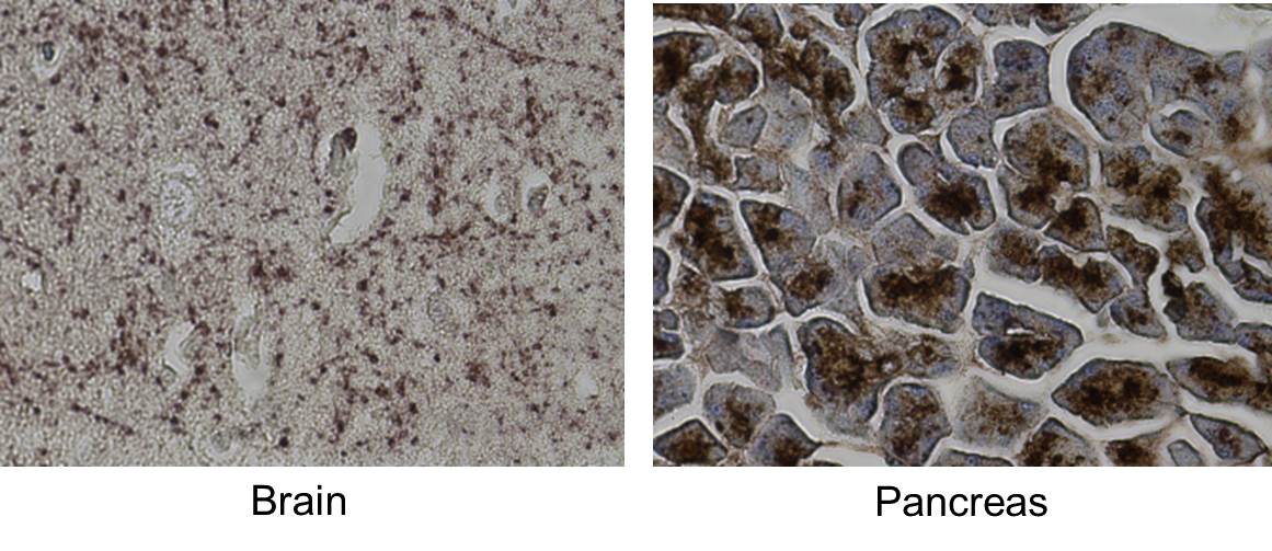
Immunohistochemistry-Paraffin: MCAM/CD146 Antibody (MUC18/1130) [NBP2-44512] - CD146 was stained in both human brain and pancreatic tissues. CD146 expression was high in pancreatic tissue, yet low in brain tissue as shown by the brown coloration. Image from a verified customer review.![Immunohistochemistry-Paraffin: MCAM/CD146 Antibody (MUC18/1130) [NBP2-44512]](https://resources.rndsystems.com/images/products/MCAM-CD146-Antibody-MUC18-1130-Immunohistochemistry-Paraffin-NBP2-44512-img0007.jpg "Immunohistochemistry-Paraffin: MCAM/CD146 Antibody (MUC18/1130) [NBP2-44512]")
Immunohistochemistry-Paraffin: MCAM/CD146 Antibody (MUC18/1130) [NBP2-44512]
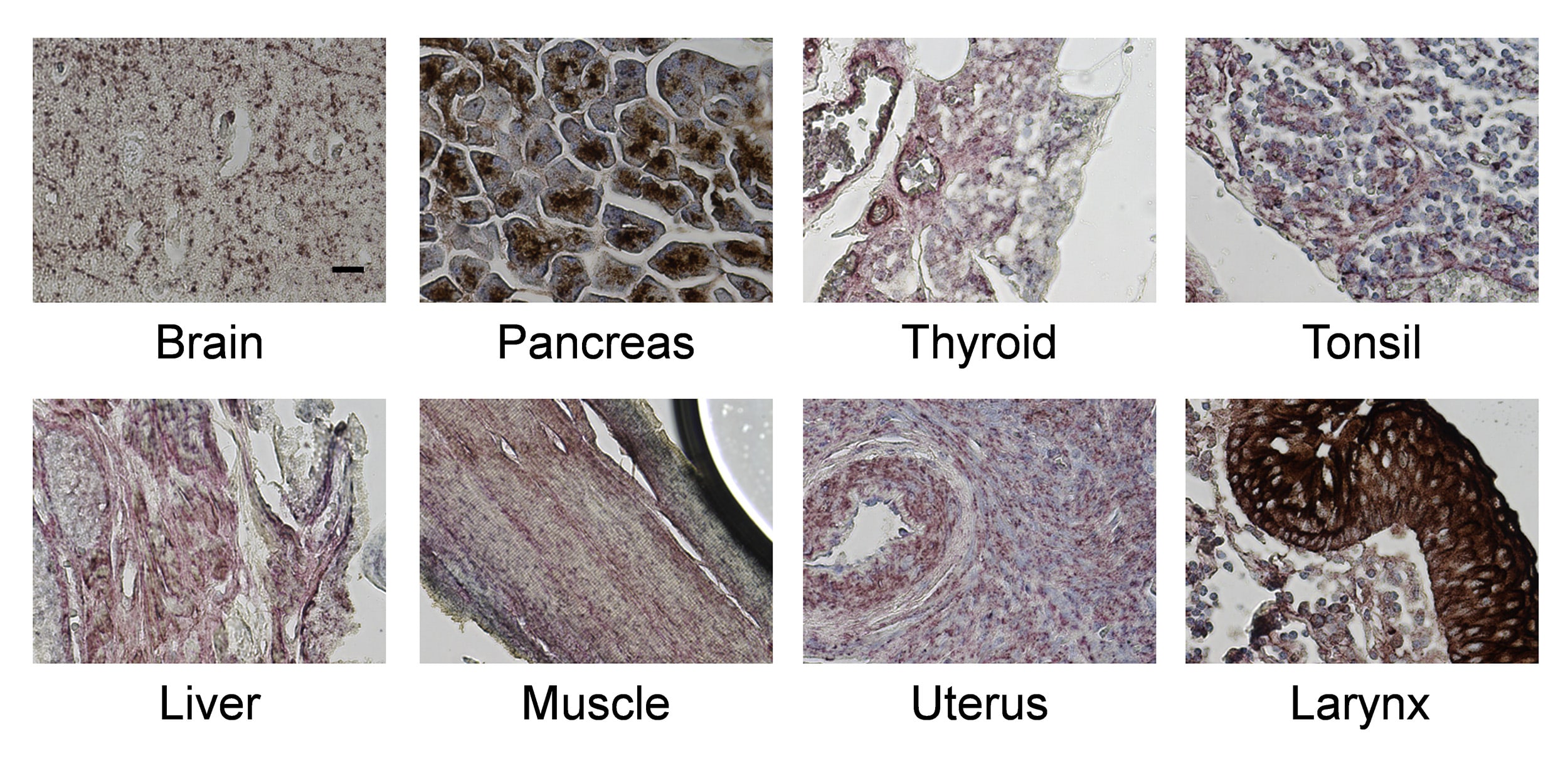
Immunohistochemistry-Paraffin: MCAM/CD146 Antibody (MUC18/1130) [NBP2-44512] - CD146 Staining of Normal Human Tissues. Image from a verified customer review.Applications for MCAM/CD146 Antibody (MUC18/1130)
Application
Recommended Usage
Flow Cytometry
1-2 ug/million cells
Immunocytochemistry/ Immunofluorescence
1-2 ug/ml
Immunofluorescence
0.5 - 1.0 ug/ml
Immunohistochemistry-Paraffin
1-2 ug/ml
Application Notes
Immunohistochemistry (Formalin-fixed): 1-2ug/ml for 30 minutes at RT. Staining of formalin-fixed tissues requires heating tissue sections in 1mM EDTA, pH 8.0, for 45 min at 95C followed by cooling at RT for 20 minutes.
Optimal dilution for a specific application should be determined.
Optimal dilution for a specific application should be determined.
Reviewed Applications
Read 2 reviews rated 5 using NBP2-44512 in the following applications:
Flow Cytometry Panel Builder
Bio-Techne Knows Flow Cytometry
Save time and reduce costly mistakes by quickly finding compatible reagents using the Panel Builder Tool.
Advanced Features
- Spectra Viewer - Custom analysis of spectra from multiple fluorochromes
- Spillover Popups - Visualize the spectra of individual fluorochromes
- Antigen Density Selector - Match fluorochrome brightness with antigen density

Formulation, Preparation, and Storage
Purification
Protein A or G purified
Formulation
10 mM PBS with 0.05% BSA
Preservative
0.05% Sodium Azide
Concentration
0.2 mg/ml
Shipping
The product is shipped with polar packs. Upon receipt, store it immediately at the temperature recommended below.
Stability & Storage
Store at 4C.
Background: MCAM/CD146
Long Name
Melanoma Cell Adhesion Molecule
Alternate Names
CD146, L-Gicerin, MUC18
Gene Symbol
MCAM
UniProt
Additional MCAM/CD146 Products
Product Documents for MCAM/CD146 Antibody (MUC18/1130)
Certificate of Analysis
To download a Certificate of Analysis, please enter a lot or batch number in the search box below.
Product Specific Notices for MCAM/CD146 Antibody (MUC18/1130)
This product is for research use only and is not approved for use in humans or in clinical diagnosis. Primary Antibodies are guaranteed for 1 year from date of receipt.
Citations for MCAM/CD146 Antibody (MUC18/1130)
 Powered by Bioz
Customer Reviews for MCAM/CD146 Antibody (MUC18/1130) (2)
5 out of 5
2 Customer Ratings
Have you used MCAM/CD146 Antibody (MUC18/1130)?
Submit a review and receive an Amazon gift card!
$25/€18/£15/$25CAN/¥2500 Yen for a review with an image
$10/€7/£6/$10CAN/¥1110 Yen for a review without an image
Submit a review
Customer Images
Showing
1
-
2 of
2 reviews
Showing All
Filter By:
-
Application: Immunohistochemistry-ParaffinSample Tested: brain and pancreatic tissueSpecies: uman tissueVerified Customer | Posted 12/05/2016CD146 was stained in both human brain and pancreatic tissues. CD146 expression was high in pancreatic tissue, yet low in brain tissue as shown by the brown coloration.The antibody was used at a ratio of 1:200.

-
Application: Immunohistochemistry-ParaffinSample Tested: Human Normal Tissue MicroarraySpecies: HumanVerified Customer | Posted 06/04/2016CD146 Staining of Normal Human Tissues
There are no reviews that match your criteria.
Protocols
Find general support by application which include: protocols, troubleshooting, illustrated assays, videos and webinars.
- 7-Amino Actinomycin D (7-AAD) Cell Viability Flow Cytometry Protocol
- Antigen Retrieval Protocol (PIER)
- Antigen Retrieval for Frozen Sections Protocol
- Appropriate Fixation of IHC/ICC Samples
- Cellular Response to Hypoxia Protocols
- Chromogenic IHC Staining of Formalin-Fixed Paraffin-Embedded (FFPE) Tissue Protocol
- Chromogenic Immunohistochemistry Staining of Frozen Tissue
- ClariTSA™ Fluorophore Kits
- Detection & Visualization of Antibody Binding
- Extracellular Membrane Flow Cytometry Protocol
- Flow Cytometry Protocol for Cell Surface Markers
- Flow Cytometry Protocol for Staining Membrane Associated Proteins
- Flow Cytometry Staining Protocols
- Flow Cytometry Troubleshooting Guide
- Fluorescent IHC Staining of Frozen Tissue Protocol
- Graphic Protocol for Heat-induced Epitope Retrieval
- Graphic Protocol for the Preparation and Fluorescent IHC Staining of Frozen Tissue Sections
- Graphic Protocol for the Preparation and Fluorescent IHC Staining of Paraffin-embedded Tissue Sections
- Graphic Protocol for the Preparation of Gelatin-coated Slides for Histological Tissue Sections
- ICC Cell Smear Protocol for Suspension Cells
- ICC Immunocytochemistry Protocol Videos
- ICC for Adherent Cells
- IHC Sample Preparation (Frozen sections vs Paraffin)
- Immunocytochemistry (ICC) Protocol
- Immunocytochemistry Troubleshooting
- Immunofluorescence of Organoids Embedded in Cultrex Basement Membrane Extract
- Immunofluorescent IHC Staining of Formalin-Fixed Paraffin-Embedded (FFPE) Tissue Protocol
- Immunohistochemistry (IHC) and Immunocytochemistry (ICC) Protocols
- Immunohistochemistry Frozen Troubleshooting
- Immunohistochemistry Paraffin Troubleshooting
- Intracellular Flow Cytometry Protocol Using Alcohol (Methanol)
- Intracellular Flow Cytometry Protocol Using Detergents
- Intracellular Nuclear Staining Flow Cytometry Protocol Using Detergents
- Intracellular Staining Flow Cytometry Protocol Using Alcohol Permeabilization
- Intracellular Staining Flow Cytometry Protocol Using Detergents to Permeabilize Cells
- Preparing Samples for IHC/ICC Experiments
- Preventing Non-Specific Staining (Non-Specific Binding)
- Primary Antibody Selection & Optimization
- Propidium Iodide Cell Viability Flow Cytometry Protocol
- Protocol for Heat-Induced Epitope Retrieval (HIER)
- Protocol for Liperfluo
- Protocol for Making a 4% Formaldehyde Solution in PBS
- Protocol for VisUCyte™ HRP Polymer Detection Reagent
- Protocol for the Characterization of Human Th22 Cells
- Protocol for the Characterization of Human Th9 Cells
- Protocol for the Fluorescent ICC Staining of Cell Smears - Graphic
- Protocol for the Fluorescent ICC Staining of Cultured Cells on Coverslips - Graphic
- Protocol for the Preparation & Fixation of Cells on Coverslips
- Protocol for the Preparation and Chromogenic IHC Staining of Frozen Tissue Sections
- Protocol for the Preparation and Chromogenic IHC Staining of Frozen Tissue Sections - Graphic
- Protocol for the Preparation and Chromogenic IHC Staining of Paraffin-embedded Tissue Sections
- Protocol for the Preparation and Chromogenic IHC Staining of Paraffin-embedded Tissue Sections - Graphic
- Protocol for the Preparation and Fluorescent ICC Staining of Cells on Coverslips
- Protocol for the Preparation and Fluorescent ICC Staining of Non-adherent Cells
- Protocol for the Preparation and Fluorescent ICC Staining of Stem Cells on Coverslips
- Protocol for the Preparation and Fluorescent IHC Staining of Frozen Tissue Sections
- Protocol for the Preparation and Fluorescent IHC Staining of Paraffin-embedded Tissue Sections
- Protocol for the Preparation of Gelatin-coated Slides for Histological Tissue Sections
- Protocol for the Preparation of a Cell Smear for Non-adherent Cell ICC - Graphic
- Protocol: Annexin V and PI Staining by Flow Cytometry
- Protocol: Annexin V and PI Staining for Apoptosis by Flow Cytometry
- TUNEL and Active Caspase-3 Detection by IHC/ICC Protocol
- The Importance of IHC/ICC Controls
- Troubleshooting Guide: Fluorokine Flow Cytometry Kits
- Troubleshooting Guide: Immunohistochemistry
- View all Protocols, Troubleshooting, Illustrated assays and Webinars
Loading...
Associated Pathways
