MCM3 Antibody - BSA Free
Novus Biologicals | Catalog # NB100-289
![Western Blot: MCM3 Antibody [NB100-289]](https://resources.rndsystems.com/images/products/MCM3-Antibody-Western-Blot-NB100-289-img0012.jpg "Western Blot: MCM3 Antibody [NB100-289]")
Key Product Details
Validated by
Biological Validation
Species Reactivity
Human, Mouse
Applications
Immunohistochemistry, Immunohistochemistry-Paraffin, Western Blot, Immunoprecipitation
Label
Unconjugated
Antibody Source
Polyclonal Rabbit IgG
Format
BSA Free
Loading...
Product Specifications
Immunogen
A 38-amino acid synthetic peptide, which represented a portion of human Minichromosome Maintenance Deficient 3 encoded in part by exons 10 and 11 (LocusLink ID 4172).
Clonality
Polyclonal
Host
Rabbit
Isotype
IgG
Scientific Data Images for MCM3 Antibody - BSA Free
Western Blot: MCM3 Antibody [NB100-289]
Western Blot: MCM3 Antibody [NB100-289] - Detection of Human MCM3 by Western Blot. Samples: Whole cell lysate (50 ug) from HeLa and 293T cells prepared using NETN lysis buffer. Antibody: Affinity purified rabbit anti-MCM3 antibody NB100-289 used for WB at 0.1 ug/ml. Detection: Chemiluminescence with an exposure time of 30 seconds.![Immunohistochemistry: MCM3 Antibody [NB100-289]](https://resources.rndsystems.com/images/products/MCM3-Antibody-Immunohistochemistry-NB100-289-img0010.jpg "Immunohistochemistry: MCM3 Antibody [NB100-289]")
Immunohistochemistry: MCM3 Antibody [NB100-289]
Immunohistochemistry: MCM3 Antibody [NB100-289] - Sample: FFPE section of human colon carcinoma. Antibody: Affinity purified rabbit anti-MCM3 used at a dilution of 1:1,000 (1ug/ml). Detection: DAB![Western Blot: MCM3 Antibody [NB100-289]](https://resources.rndsystems.com/images/products/MCM3-Antibody-Western-Blot-NB100-289-img0002.jpg "Western Blot: MCM3 Antibody [NB100-289]")
Western Blot: MCM3 Antibody [NB100-289]
Western Blot: MCM3 Antibody [NB100-289] - RIPA extract (50 ug) from HeLa cells. Antibody used at 0.2 ug/ml.
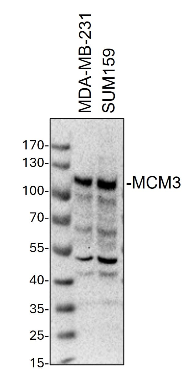
Western Blot: Rabbit Polyclonal MCM3 Antibody [NB100-289]
Western Blot: Rabbit Polyclonal MCM3 Antibody [NB100-289] - Whole cell lysates from MDA-MB-231 and SUM159 cells were loaded with 50 ug/lane. 10% SDS-PAGE. MCM3 Antibody (NB100-289) was used for primary antibody: 1:1000, 4℃, overnight. Image from a verified customer review.Applications for MCM3 Antibody - BSA Free
Application
Recommended Usage
Immunohistochemistry
1:1000 to 1:5000
Immunohistochemistry-Paraffin
1:1000 to 1:5000
Immunoprecipitation
2-5 ug//mg lysate
Western Blot
1:2000-1:10000
Application Notes
Western blot and immunoprecipitation. Suggested working dilutions: Western Blot - 1: 2,000-1:10,000 Immunohistochemistry/Immunocytochemistry - ND Immunoprecipitation - 2 to 5 ug/mg lysate** *The investigator should determine the optimal working dilution for a specific application. **Use NB 100-288 (MCM2 antibody) to IP the MCM complex
Reviewed Applications
Read 1 review rated 5 using NB100-289 in the following applications:
Formulation, Preparation, and Storage
Purification
Immunogen affinity purified
Formulation
Tris-Citrate/Phosphate (pH 7.0 - 8.0)
Format
BSA Free
Preservative
0.09% Sodium Azide
Concentration
1.0 mg/ml
Shipping
The product is shipped with polar packs. Upon receipt, store it immediately at the temperature recommended below.
Stability & Storage
Store at 4C. Do not freeze.
Background: MCM3
Alternate Names
cervical cancer proto-oncogene 5, DNA polymerase alpha holoenzyme-associated protein P1, DNA replication factor MCM3, DNA replication licensing factor MCM3, EC 3.6.4.12, HCC5, hRlf beta subunit, MCM3 minichromosome maintenance deficient 3, MCM3 minichromosome maintenance deficient 3 (S. cerevisiae), MGC1157, minichromosome maintenance complex component 3, minichromosome maintenance deficient (S. cerevisiae) 3, minichromosome maintenance deficient 3, P1.h, p102, P1-MCM3, replication licensing factor, beta subunit, RLF subunit beta, RLFB
Entrez Gene IDs
4172 (Human)
Gene Symbol
MCM3
Additional MCM3 Products
Product Documents for MCM3 Antibody - BSA Free
Certificate of Analysis
To download a Certificate of Analysis, please enter a lot or batch number in the search box below.
Product Specific Notices for MCM3 Antibody - BSA Free
This product is for research use only and is not approved for use in humans or in clinical diagnosis. Primary Antibodies are guaranteed for 1 year from date of receipt.
Citations for MCM3 Antibody - BSA Free
 Powered by Bioz
Customer Reviews for MCM3 Antibody - BSA Free (1)
5 out of 5
1 Customer Rating
Have you used MCM3 Antibody - BSA Free?
Submit a review and receive an Amazon gift card!
$25/€18/£15/$25CAN/¥2500 Yen for a review with an image
$10/€7/£6/$10CAN/¥1110 Yen for a review without an image
Submit a review
Customer Images
Showing
1
-
1 of
1 review
Showing All
Filter By:
-
Application: Western BlotSample Tested: Human MDA-MB-231 cells and SUM-159PTSpecies: HumanVerified Customer | Posted 11/28/2024Western Blot: whole cell lysates from MDA-MB-231 and SUM159 cells were loaded with 50 ug/lane. 10% SDS-PAGE. MCM3 Antibody (NB100-289) was used for primary antibody: 1:1000, 4℃, overnight.

There are no reviews that match your criteria.
Protocols
Find general support by application which include: protocols, troubleshooting, illustrated assays, videos and webinars.
- Antigen Retrieval Protocol (PIER)
- Antigen Retrieval for Frozen Sections Protocol
- Appropriate Fixation of IHC/ICC Samples
- Cellular Response to Hypoxia Protocols
- Chromogenic IHC Staining of Formalin-Fixed Paraffin-Embedded (FFPE) Tissue Protocol
- Chromogenic Immunohistochemistry Staining of Frozen Tissue
- ClariTSA™ Fluorophore Kits
- Detection & Visualization of Antibody Binding
- Fluorescent IHC Staining of Frozen Tissue Protocol
- Graphic Protocol for Heat-induced Epitope Retrieval
- Graphic Protocol for the Preparation and Fluorescent IHC Staining of Frozen Tissue Sections
- Graphic Protocol for the Preparation and Fluorescent IHC Staining of Paraffin-embedded Tissue Sections
- Graphic Protocol for the Preparation of Gelatin-coated Slides for Histological Tissue Sections
- IHC Sample Preparation (Frozen sections vs Paraffin)
- Immunofluorescent IHC Staining of Formalin-Fixed Paraffin-Embedded (FFPE) Tissue Protocol
- Immunohistochemistry (IHC) and Immunocytochemistry (ICC) Protocols
- Immunohistochemistry Frozen Troubleshooting
- Immunohistochemistry Paraffin Troubleshooting
- Immunoprecipitation Protocol
- Preparing Samples for IHC/ICC Experiments
- Preventing Non-Specific Staining (Non-Specific Binding)
- Primary Antibody Selection & Optimization
- Protocol for Heat-Induced Epitope Retrieval (HIER)
- Protocol for Making a 4% Formaldehyde Solution in PBS
- Protocol for VisUCyte™ HRP Polymer Detection Reagent
- Protocol for the Preparation & Fixation of Cells on Coverslips
- Protocol for the Preparation and Chromogenic IHC Staining of Frozen Tissue Sections
- Protocol for the Preparation and Chromogenic IHC Staining of Frozen Tissue Sections - Graphic
- Protocol for the Preparation and Chromogenic IHC Staining of Paraffin-embedded Tissue Sections
- Protocol for the Preparation and Chromogenic IHC Staining of Paraffin-embedded Tissue Sections - Graphic
- Protocol for the Preparation and Fluorescent IHC Staining of Frozen Tissue Sections
- Protocol for the Preparation and Fluorescent IHC Staining of Paraffin-embedded Tissue Sections
- Protocol for the Preparation of Gelatin-coated Slides for Histological Tissue Sections
- R&D Systems Quality Control Western Blot Protocol
- TUNEL and Active Caspase-3 Detection by IHC/ICC Protocol
- The Importance of IHC/ICC Controls
- Troubleshooting Guide: Immunohistochemistry
- Troubleshooting Guide: Western Blot Figures
- Western Blot Conditions
- Western Blot Protocol
- Western Blot Protocol for Cell Lysates
- Western Blot Troubleshooting
- Western Blot Troubleshooting Guide
- View all Protocols, Troubleshooting, Illustrated assays and Webinars
Loading...