MCM7 Antibody - BSA Free
Novus Biologicals | Catalog # NBP1-31350
![Immunocytochemistry/ Immunofluorescence: MCM7 Antibody [NBP1-31350]](https://resources.rndsystems.com/images/products/MCM7-Antibody-Immunocytochemistry-Immunofluorescence-NBP1-31350-img0004.jpg "Immunocytochemistry/ Immunofluorescence: MCM7 Antibody [NBP1-31350]")
Loading...
Key Product Details
Species Reactivity
Validated:
Human
Cited:
Human
Predicted:
Bovine (98%), Mouse (92%), Rat (93%). Backed by our 100% Guarantee.
Applications
Validated:
Immunohistochemistry, Immunohistochemistry-Paraffin, Western Blot, Immunocytochemistry/ Immunofluorescence, Immunoprecipitation, Chromatin Immunoprecipitation (ChIP)
Cited:
Western Blot
Label
Unconjugated
Antibody Source
Polyclonal Rabbit IgG
Format
BSA Free
Loading...
Product Specifications
Immunogen
Recombinant protein encompassing a sequence within the center region of human MCM7. The exact sequence is proprietary.
Reactivity Notes
Immunogen displays the following percentage of sequence identity for non-tested species: Zebrafish (85%).
Localization
Nucleus
Clonality
Polyclonal
Host
Rabbit
Isotype
IgG
Theoretical MW
81 kDa.
Disclaimer note: The observed molecular weight of the protein may vary from the listed predicted molecular weight due to post translational modifications, post translation cleavages, relative charges, and other experimental factors.
Disclaimer note: The observed molecular weight of the protein may vary from the listed predicted molecular weight due to post translational modifications, post translation cleavages, relative charges, and other experimental factors.
Scientific Data Images for MCM7 Antibody - BSA Free
Immunocytochemistry/ Immunofluorescence: MCM7 Antibody [NBP1-31350]
Immunocytochemistry/Immunofluorescence: MCM7 Antibody [NBP1-31350] - Paraformaldehyde-fixed HeLa, using antibody at 1:200 dilution.![Immunohistochemistry-Paraffin: MCM7 Antibody [NBP1-31350]](https://resources.rndsystems.com/images/products/MCM7-Antibody-Immunohistochemistry-Paraffin-NBP1-31350-img0005.jpg "Immunohistochemistry-Paraffin: MCM7 Antibody [NBP1-31350]")
Immunohistochemistry-Paraffin: MCM7 Antibody [NBP1-31350]
Immunohistochemistry-Paraffin: MCM7 Antibody [NBP1-31350] - Paraffin-embedded Cal27 Xenograft, using antibody at 1:100 dilution.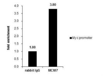
Chromatin Immunoprecipitation (ChIP): MCM7 Antibody [NBP1-31350] - Cross-linked ChIP was performed with HeLa chromatin extract and 5 ug of either control rabbit igG or anti-MCM7 antibody. The precipitated DNA was detected by PCR with primer set targeting to Myc promoter.
![Immunoprecipitation: MCM7 Antibody [NBP1-31350]](https://resources.rndsystems.com/images/products/MCM7-Antibody-Immunoprecipitation-NBP1-31350-img0007.jpg "Immunoprecipitation: MCM7 Antibody [NBP1-31350]")
Immunoprecipitation: MCM7 Antibody [NBP1-31350]
Immunoprecipitation: MCM7 Antibody [NBP1-31350] - MCM7 antibody [N2C2], Internal immunoprecipitates MCM7 protein in IP experiments. IP samples: HeLa whole cell extract. A. 40 ug HeLa whole cell extract. B. Control with 4 ug of preimmune Rabbit IgG. C. Immunoprecipitation of MCM7 protein by 4 ug MCM7 antibody [N2C2], Internal. 7.5 % SDS-PAGE. The immunoprecipitated MCM7 protein was detected by MCM7 antibody [N2C2], Internal diluted at 1:1000. [EasyBlot anti-rabbit IgG was used as a secondary reagent]
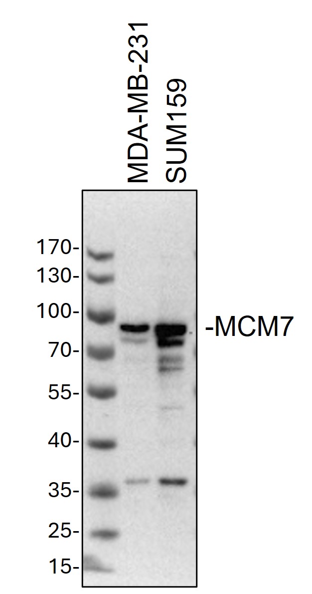
Western Blot: Rabbit Polyclonal MCM7 Antibody [NBP1-31350]
Western Blot: Rabbit Polyclonal MCM7 Antibody [NBP1-31350] - Whole cell lysates from MDA-MB-231 and SUM159 cells were loaded with 50 ug/lane.10% SDS-PAGE. MCM7 Antibody (NBP1-31350) was used for primary antibody: 1:1000, 4℃, overnight. Image from a verified customer review.
Western Blot: MCM7 Antibody [NBP1-31350] -
Various whole cell extracts (30 ug) were separated by 7.5% SDS-PAGE, and the membrane was blotted with MCM7 antibody [N2C2], Internal (NBP1-31350) diluted at 1:1000. The HRP-conjugated anti-rabbit IgG antibody was used to detect the primary antibody.
Western Blot: MCM7 Antibody [NBP1-31350] -
Various whole cell extracts (30 ug) were separated by 7.5% SDS-PAGE, and the membrane was blotted with MCM7 antibody [N2C2], Internal (NBP1-31350) diluted at 1:1000. The HRP-conjugated anti-rabbit IgG antibody was used to detect the primary antibody.
Western Blot: MCM7 Antibody [NBP1-31350] -
Various whole cell extracts (30 ug) were separated by 7.5% SDS-PAGE, and the membrane was blotted with MCM7 antibody [N2C2], Internal (NBP1-31350) diluted at 1:1000. The HRP-conjugated anti-rabbit IgG antibody was used to detect the primary antibody.
Western Blot: MCM7 Antibody [NBP1-31350] -
Various whole cell extracts (30 ug) were separated by 7.5% SDS-PAGE, and the membrane was blotted with MCM7 antibody [N2C2], Internal (NBP1-31350) diluted at 1:1000. The HRP-conjugated anti-rabbit IgG antibody was used to detect the primary antibody.Applications for MCM7 Antibody - BSA Free
Application
Recommended Usage
Chromatin Immunoprecipitation (ChIP)
Assay dependent
Immunocytochemistry/ Immunofluorescence
1:100-1:1000
Immunohistochemistry
1:100-1:1000
Immunohistochemistry-Paraffin
1:100-1:1000
Immunoprecipitation
1:100-1:500
Western Blot
1:500-1:3000
Reviewed Applications
Read 1 review rated 5 using NBP1-31350 in the following applications:
Formulation, Preparation, and Storage
Purification
Antigen Affinity-purified
Formulation
0.1M Tris, 0.1M Glycine, 10% Glycerol
Format
BSA Free
Preservative
0.01% Thimerosal
Concentration
Concentrations vary lot to lot. See vial label for concentration. If unlisted please contact technical services.
Shipping
The product is shipped with polar packs. Upon receipt, store it immediately at the temperature recommended below.
Stability & Storage
Aliquot and store at -20C or -80C. Avoid freeze-thaw cycles.
Background: MCM7
Long Name
Minichromosome Maintenance Protein 7
Alternate Names
CDC47, MCM2, P1CDC47, P85MCM, PNAS146, PPP1R104, Protein Phosphatase 1
Gene Symbol
MCM7
Additional MCM7 Products
Product Documents for MCM7 Antibody - BSA Free
Certificate of Analysis
To download a Certificate of Analysis, please enter a lot or batch number in the search box below.
Product Specific Notices for MCM7 Antibody - BSA Free
This product is for research use only and is not approved for use in humans or in clinical diagnosis. Primary Antibodies are guaranteed for 1 year from date of receipt.
⚠ WARNING: This product can expose you to chemicals including mercury, which is known to the State of California to cause reproductive toxicity with developmental effects. For more information go to www.P65Warnings.ca.gov.Related Research Areas
Citations for MCM7 Antibody - BSA Free
 Powered by Bioz
Customer Reviews for MCM7 Antibody - BSA Free (1)
5 out of 5
1 Customer Rating
Have you used MCM7 Antibody - BSA Free?
Submit a review and receive an Amazon gift card!
$25/€18/£15/$25CAN/¥2500 Yen for a review with an image
$10/€7/£6/$10CAN/¥1110 Yen for a review without an image
Submit a review
Customer Images
Showing
1
-
1 of
1 review
Showing All
Filter By:
-
Application: Western BlotSample Tested: Human MDA-MB-231 cells and SUM-159PTSpecies: HumanVerified Customer | Posted 11/28/2024Western Blot: whole cell lysates from MDA-MB-231 and SUM159 cells were loaded with 50 ug/lane. 10% SDS-PAGE. MCM7 Antibody (NBP1-31350) was used for primary antibody: 1:1000, 4℃, overnight.

There are no reviews that match your criteria.
Protocols
Find general support by application which include: protocols, troubleshooting, illustrated assays, videos and webinars.
- Antigen Retrieval Protocol (PIER)
- Antigen Retrieval for Frozen Sections Protocol
- Appropriate Fixation of IHC/ICC Samples
- Cellular Response to Hypoxia Protocols
- ChIP Protocol Video
- Chromatin Immunoprecipitation (ChIP) Protocol
- Chromatin Immunoprecipitation Protocol
- Chromogenic IHC Staining of Formalin-Fixed Paraffin-Embedded (FFPE) Tissue Protocol
- Chromogenic Immunohistochemistry Staining of Frozen Tissue
- ClariTSA™ Fluorophore Kits
- Detection & Visualization of Antibody Binding
- Fluorescent IHC Staining of Frozen Tissue Protocol
- Graphic Protocol for Heat-induced Epitope Retrieval
- Graphic Protocol for the Preparation and Fluorescent IHC Staining of Frozen Tissue Sections
- Graphic Protocol for the Preparation and Fluorescent IHC Staining of Paraffin-embedded Tissue Sections
- Graphic Protocol for the Preparation of Gelatin-coated Slides for Histological Tissue Sections
- ICC Cell Smear Protocol for Suspension Cells
- ICC Immunocytochemistry Protocol Videos
- ICC for Adherent Cells
- IHC Sample Preparation (Frozen sections vs Paraffin)
- Immunocytochemistry (ICC) Protocol
- Immunocytochemistry Troubleshooting
- Immunofluorescence of Organoids Embedded in Cultrex Basement Membrane Extract
- Immunofluorescent IHC Staining of Formalin-Fixed Paraffin-Embedded (FFPE) Tissue Protocol
- Immunohistochemistry (IHC) and Immunocytochemistry (ICC) Protocols
- Immunohistochemistry Frozen Troubleshooting
- Immunohistochemistry Paraffin Troubleshooting
- Immunoprecipitation Protocol
- Preparing Samples for IHC/ICC Experiments
- Preventing Non-Specific Staining (Non-Specific Binding)
- Primary Antibody Selection & Optimization
- Protocol for Heat-Induced Epitope Retrieval (HIER)
- Protocol for Making a 4% Formaldehyde Solution in PBS
- Protocol for VisUCyte™ HRP Polymer Detection Reagent
- Protocol for the Fluorescent ICC Staining of Cell Smears - Graphic
- Protocol for the Fluorescent ICC Staining of Cultured Cells on Coverslips - Graphic
- Protocol for the Preparation & Fixation of Cells on Coverslips
- Protocol for the Preparation and Chromogenic IHC Staining of Frozen Tissue Sections
- Protocol for the Preparation and Chromogenic IHC Staining of Frozen Tissue Sections - Graphic
- Protocol for the Preparation and Chromogenic IHC Staining of Paraffin-embedded Tissue Sections
- Protocol for the Preparation and Chromogenic IHC Staining of Paraffin-embedded Tissue Sections - Graphic
- Protocol for the Preparation and Fluorescent ICC Staining of Cells on Coverslips
- Protocol for the Preparation and Fluorescent ICC Staining of Non-adherent Cells
- Protocol for the Preparation and Fluorescent ICC Staining of Stem Cells on Coverslips
- Protocol for the Preparation and Fluorescent IHC Staining of Frozen Tissue Sections
- Protocol for the Preparation and Fluorescent IHC Staining of Paraffin-embedded Tissue Sections
- Protocol for the Preparation of Gelatin-coated Slides for Histological Tissue Sections
- Protocol for the Preparation of a Cell Smear for Non-adherent Cell ICC - Graphic
- R&D Systems Quality Control Western Blot Protocol
- TUNEL and Active Caspase-3 Detection by IHC/ICC Protocol
- The Importance of IHC/ICC Controls
- Troubleshooting Guide: Immunohistochemistry
- Troubleshooting Guide: Western Blot Figures
- Western Blot Conditions
- Western Blot Protocol
- Western Blot Protocol for Cell Lysates
- Western Blot Troubleshooting
- Western Blot Troubleshooting Guide
- View all Protocols, Troubleshooting, Illustrated assays and Webinars
Loading...