Mitofilin Antibody - BSA Free
Novus Biologicals | Catalog # NB100-1919
![Western Blot: Mitofilin Antibody [NB100-1919]](https://resources.rndsystems.com/images/products/Mitofilin-Antibody-Western-Blot-NB100-1919-img0006.jpg "Western Blot: Mitofilin Antibody [NB100-1919]")
Key Product Details
Species Reactivity
Validated:
Cited:
Applications
Validated:
Cited:
Label
Antibody Source
Format
Product Specifications
Immunogen
Reactivity Notes
Localization
Marker
Clonality
Host
Isotype
Scientific Data Images for Mitofilin Antibody - BSA Free
Western Blot: Mitofilin Antibody [NB100-1919]
Western Blot: Mitofilin Antibody [NB100-1919] - Western blot of dog testis. Total protein extract 1:2000 dilution of antibody.![Immunocytochemistry/ Immunofluorescence: Mitofilin Antibody [NB100-1919]](https://resources.rndsystems.com/images/products/Mitofilin-Antibody-Immunocytochemistry-Immunofluorescence-NB100-1919-img0004.jpg "Immunocytochemistry/ Immunofluorescence: Mitofilin Antibody [NB100-1919]")
Immunocytochemistry/ Immunofluorescence: Mitofilin Antibody [NB100-1919]
Immunocytochemistry/Immunofluorescence: Mitofilin Antibody [NB100-1919] - HeLa cells with antibody 1:500 dilution labeling mitochondria.![Immunohistochemistry-Paraffin: Mitofilin Antibody [NB100-1919]](https://resources.rndsystems.com/images/products/Mitofilin-Antibody-Immunohistochemistry-Paraffin-NB100-1919-img0009.jpg "Immunohistochemistry-Paraffin: Mitofilin Antibody [NB100-1919]")
Immunohistochemistry-Paraffin: Mitofilin Antibody [NB100-1919]
Immunohistochemistry-Paraffin: Mitofilin Antibody [NB100-1919] - IHC-P detection of Mitofilin/Mitochondrial inner membrane protein in a section of normal human skin using Mitofilin antibody at 1:200 dilution. The representative image shows a specific and expected granular staining of Mitofilin in the keratinocytes as well as the other cells of epidermal layer with no staining in basal cells. [60X Magnification]![Western Blot: Mitofilin Antibody [NB100-1919]](https://resources.rndsystems.com/images/products/Mitofilin-Antibody-Western-Blot-NB100-1919-img0012.jpg "Western Blot: Mitofilin Antibody [NB100-1919]")
Western Blot: Mitofilin Antibody [NB100-1919]
Mitofilin-Antibody-Western-Blot-NB100-1919-img0012.jpg![Immunohistochemistry-Paraffin: Mitofilin Antibody [NB100-1919]](https://resources.rndsystems.com/images/products/Mitofilin-Antibody-Immunohistochemistry-Paraffin-NB100-1919-img0011.jpg "Immunohistochemistry-Paraffin: Mitofilin Antibody [NB100-1919]")
Immunohistochemistry-Paraffin: Mitofilin Antibody [NB100-1919]
Immunohistochemistry-Paraffin: Mitofilin Antibody [NB100-1919] - IHC-P analysis of Mitofilin/Mitochondrial inner membrane protein in a transverse section of normal human skeletal muscle using Mitofilin antibody at 1:200 dilution. The myocytes showed an expected and very specific granular staining in the cytoplasm. [10X Magnification]![Immunohistochemistry-Paraffin: Mitofilin Antibody [NB100-1919]](https://resources.rndsystems.com/images/products/Mitofilin-Antibody-Immunohistochemistry-Paraffin-NB100-1919-img0010.jpg "Immunohistochemistry-Paraffin: Mitofilin Antibody [NB100-1919]")
Immunohistochemistry-Paraffin: Mitofilin Antibody [NB100-1919]
Immunohistochemistry-Paraffin: Mitofilin Antibody [NB100-1919] - IHC-P detection of Mitofilin/Mitochondrial inner membrane protein in a section of normal human skin using Mitofilin antibody at 1:200 dilution. Specific cytoplasmic positivity of Mitofilin was observed in the keratinocytes as well as other cells of the epidermal layer except for basal cells and dermal connective tissue. [10X Magnification]
Western Blot: Mitofilin Antibody [NB100-1919] -
Western Blot: Mitofilin Antibody [NB100-1919] - COA7 pathological variants are import‐defectiveASchematic representation of wild‐type & mutant COA7.BCellular protein extracts were isolated from HEK293 cells that were transfected with a plasmid that encoded wild‐type or mutant COA7. The samples were analyzed by reducing SDS–PAGE & Western blot.CCellular fractions were prepared from HEK293 cells that were transfected with a plasmid that encoded wild‐type or mutant COA7. The fractions were analyzed by reducing SDS–PAGE & Western blot. T, total; C, cytosol; M, mitochondria.DHEK293 cells that transiently expressed wild‐type or mutant COA7HIS were solubilized, & the affinity purification of COA7HIS was performed. The samples were analyzed by reducing & non‐reducing SDS–PAGE & Western blot. Load: 3%. Eluate: 100%.EEqual amounts of wild‐type & mutant radiolabeled [35S]COA7 precursors were imported into mitochondria isolated from HEK293 cells. The samples were analyzed by reducing SDS–PAGE & autoradiography. The results of three biological replicates were analyzed, quantified, & normalized to wild‐type COA7 at 45 min. The data are expressed as a mean ± SEM (n = 3). IAA, iodoacetamide.Source data are available online for this figure. Image collected & cropped by CiteAb from the following publication (https://pubmed.ncbi.nlm.nih.gov/30885959), licensed under a CC-BY license. Not internally tested by Novus Biologicals.Applications for Mitofilin Antibody - BSA Free
Immunocytochemistry/ Immunofluorescence
Immunohistochemistry
Immunohistochemistry-Paraffin
Western Blot
Reviewed Applications
Read 3 reviews rated 4.3 using NB100-1919 in the following applications:
Formulation, Preparation, and Storage
Purification
Formulation
Format
Preservative
Concentration
Shipping
Stability & Storage
Background: Mitofilin
Alternate Names
Gene Symbol
UniProt
Additional Mitofilin Products
Product Documents for Mitofilin Antibody - BSA Free
Certificate of Analysis
To download a Certificate of Analysis, please enter a lot or batch number in the search box below.
Product Specific Notices for Mitofilin Antibody - BSA Free
This product is for research use only and is not approved for use in humans or in clinical diagnosis. Primary Antibodies are guaranteed for 1 year from date of receipt.
Citations for Mitofilin Antibody - BSA Free
 Powered by Bioz
Customer Reviews for Mitofilin Antibody - BSA Free (3)
Have you used Mitofilin Antibody - BSA Free?
Submit a review and receive an Amazon gift card!
$25/€18/£15/$25CAN/¥2500 Yen for a review with an image
$10/€7/£6/$10CAN/¥1110 Yen for a review without an image
Submit a review
Customer Images
-
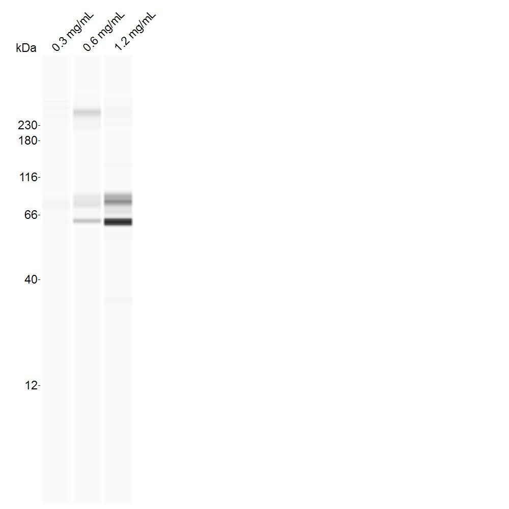
Application: Simple WesternSample Tested: cell line, whole cell lysateSpecies: RatVerified Customer | Posted 08/18/2017B103 whole-cell lysates (concentrations indicated), probed with 1:25 dilution of antibody on the standard separation matrix (12-230 kDa).

-
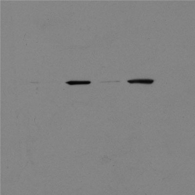
Application: Western BlotSample Tested: Protein lysate from mitochondria isolated from SH-SY5Y cellsSpecies: HumanVerified Customer | Posted 06/14/2016Neuroblastoma SH-SY5Y mitochondrial protein lysate probed for mitofillin: Each lane run at 12ug of protein per lane
-
Application: ImmunoprecipitationSample Tested: Mouse heartSpecies: MouseVerified Customer | Posted 08/27/2013
There are no reviews that match your criteria.
Protocols
Find general support by application which include: protocols, troubleshooting, illustrated assays, videos and webinars.
- Antigen Retrieval Protocol (PIER)
- Antigen Retrieval for Frozen Sections Protocol
- Appropriate Fixation of IHC/ICC Samples
- Cellular Response to Hypoxia Protocols
- Chromogenic IHC Staining of Formalin-Fixed Paraffin-Embedded (FFPE) Tissue Protocol
- Chromogenic Immunohistochemistry Staining of Frozen Tissue
- ClariTSA™ Fluorophore Kits
- Detection & Visualization of Antibody Binding
- Fluorescent IHC Staining of Frozen Tissue Protocol
- Graphic Protocol for Heat-induced Epitope Retrieval
- Graphic Protocol for the Preparation and Fluorescent IHC Staining of Frozen Tissue Sections
- Graphic Protocol for the Preparation and Fluorescent IHC Staining of Paraffin-embedded Tissue Sections
- Graphic Protocol for the Preparation of Gelatin-coated Slides for Histological Tissue Sections
- ICC Cell Smear Protocol for Suspension Cells
- ICC Immunocytochemistry Protocol Videos
- ICC for Adherent Cells
- IHC Sample Preparation (Frozen sections vs Paraffin)
- Immunocytochemistry (ICC) Protocol
- Immunocytochemistry Troubleshooting
- Immunofluorescence of Organoids Embedded in Cultrex Basement Membrane Extract
- Immunofluorescent IHC Staining of Formalin-Fixed Paraffin-Embedded (FFPE) Tissue Protocol
- Immunohistochemistry (IHC) and Immunocytochemistry (ICC) Protocols
- Immunohistochemistry Frozen Troubleshooting
- Immunohistochemistry Paraffin Troubleshooting
- Immunoprecipitation Protocol
- Preparing Samples for IHC/ICC Experiments
- Preventing Non-Specific Staining (Non-Specific Binding)
- Primary Antibody Selection & Optimization
- Protocol for Heat-Induced Epitope Retrieval (HIER)
- Protocol for Making a 4% Formaldehyde Solution in PBS
- Protocol for VisUCyte™ HRP Polymer Detection Reagent
- Protocol for the Fluorescent ICC Staining of Cell Smears - Graphic
- Protocol for the Fluorescent ICC Staining of Cultured Cells on Coverslips - Graphic
- Protocol for the Preparation & Fixation of Cells on Coverslips
- Protocol for the Preparation and Chromogenic IHC Staining of Frozen Tissue Sections
- Protocol for the Preparation and Chromogenic IHC Staining of Frozen Tissue Sections - Graphic
- Protocol for the Preparation and Chromogenic IHC Staining of Paraffin-embedded Tissue Sections
- Protocol for the Preparation and Chromogenic IHC Staining of Paraffin-embedded Tissue Sections - Graphic
- Protocol for the Preparation and Fluorescent ICC Staining of Cells on Coverslips
- Protocol for the Preparation and Fluorescent ICC Staining of Non-adherent Cells
- Protocol for the Preparation and Fluorescent ICC Staining of Stem Cells on Coverslips
- Protocol for the Preparation and Fluorescent IHC Staining of Frozen Tissue Sections
- Protocol for the Preparation and Fluorescent IHC Staining of Paraffin-embedded Tissue Sections
- Protocol for the Preparation of Gelatin-coated Slides for Histological Tissue Sections
- Protocol for the Preparation of a Cell Smear for Non-adherent Cell ICC - Graphic
- R&D Systems Quality Control Western Blot Protocol
- TUNEL and Active Caspase-3 Detection by IHC/ICC Protocol
- The Importance of IHC/ICC Controls
- Troubleshooting Guide: Immunohistochemistry
- Troubleshooting Guide: Western Blot Figures
- Western Blot Conditions
- Western Blot Protocol
- Western Blot Protocol for Cell Lysates
- Western Blot Troubleshooting
- Western Blot Troubleshooting Guide
- View all Protocols, Troubleshooting, Illustrated assays and Webinars
FAQs for Mitofilin Antibody - BSA Free
-
Q: One of our customers is interested in testing your Anti-Mitofilin antibody (NB100-1919) with IHC. Do you know how likely this Ab will be in succeeding to this attempt?
A: This Ab has been validated for use in IHC-P in Canine, Feline, Mouse, Rat and Human.