![Western Blot: MKP-1/DUSP1 Antibody [NB100-834]](https://resources.rndsystems.com/images/products/MKP-1-DUSP1-Antibody-Western-Blot-NB100-834-img0004.jpg "Western Blot: MKP-1/DUSP1 Antibody [NB100-834]")
Loading...
Key Product Details
Species Reactivity
Validated:
Human, Rat
Predicted:
Bovine (100%), Canine (100%), Mouse (100%). Backed by our 100% Guarantee.
Applications
Immunohistochemistry, Immunohistochemistry-Paraffin, Western Blot, Peptide ELISA
Label
Unconjugated
Antibody Source
Polyclonal Goat IgG
Loading...
Product Specifications
Immunogen
Peptide with sequence SYLQSPITTSPSC corresponding to C-Terminus according to NP_004408.1.
Reactivity Notes
Rat reactivity reported from a verified customer review.
Clonality
Polyclonal
Host
Goat
Isotype
IgG
Scientific Data Images for MKP-1/DUSP1 Antibody
Western Blot: MKP-1/DUSP1 Antibody [NB100-834]
Western Blot: MKP-1/DUSP1 Antibody [NB100-834] - Staining of HeLa cell lysate (35 ug protein in RIPA buffer). Antibody at 1 ug/mL. Detected by chemiluminescence.![Immunohistochemistry-Paraffin: MKP-1/DUSP1 Antibody [NB100-834]](https://resources.rndsystems.com/images/products/MKP-1-DUSP1-Antibody-Immunohistochemistry-Paraffin-NB100-834-img0006.jpg "Immunohistochemistry-Paraffin: MKP-1/DUSP1 Antibody [NB100-834]")
Immunohistochemistry-Paraffin: MKP-1/DUSP1 Antibody [NB100-834]
Immunohistochemistry-Paraffin: MKP-1/DUSP1 Antibody [NB100-834] - Negative Control showing staining of paraffin embedded Human Prostate, with no primary antibody.![Immunohistochemistry-Paraffin: MKP-1/DUSP1 Antibody [NB100-834]](https://resources.rndsystems.com/images/products/MKP-1-DUSP1-Antibody-Immunohistochemistry-Paraffin-NB100-834-img0005.jpg "Immunohistochemistry-Paraffin: MKP-1/DUSP1 Antibody [NB100-834]")
Immunohistochemistry-Paraffin: MKP-1/DUSP1 Antibody [NB100-834]
Immunohistochemistry-Paraffin: MKP-1/DUSP1 Antibody [NB100-834] - Staining of paraffin embedded Human Prostate. Antibody at 8 ug/mL. Heat induced antigen retrieval with citrate buffer pH 6, HRP-staining.Applications for MKP-1/DUSP1 Antibody
Application
Recommended Usage
Immunohistochemistry-Paraffin
8 ug/mL
Peptide ELISA
Detection limit 1:64000
Western Blot
1 - 3 ug/mL
Application Notes
WB: Approx. 40 kDa band observed in human lysates of HeLa (calculated MW of 39.3 kDa band according to NP_004408.1). Customers have reported positive results on mouse microglia (BV2 cells), rat primary microglia, and rat spinal cord homogenate.
Reviewed Applications
Read 1 review rated 4 using NB100-834 in the following applications:
Formulation, Preparation, and Storage
Purification
Immunogen affinity purified
Formulation
Tris saline (20 mM Tris pH 7.3, 150 mM NaCl), 0.5% BSA
Preservative
0.02% Sodium Azide
Concentration
0.5 mg/ml
Shipping
The product is shipped with polar packs. Upon receipt, store it immediately at the temperature recommended below.
Stability & Storage
Store at -20C. Avoid freeze-thaw cycles.
Background: MKP-1
Long Name
MAP Kinase Phosphatase 1
Alternate Names
CL100, DUSP1, HVH1, MKP1, PTPN10
Gene Symbol
DUSP1
UniProt
Additional MKP-1 Products
Product Documents for MKP-1/DUSP1 Antibody
Certificate of Analysis
To download a Certificate of Analysis, please enter a lot or batch number in the search box below.
Product Specific Notices for MKP-1/DUSP1 Antibody
This product is for research use only and is not approved for use in humans or in clinical diagnosis. Primary Antibodies are guaranteed for 1 year from date of receipt.
Related Research Areas
Customer Reviews for MKP-1/DUSP1 Antibody (1)
4 out of 5
1 Customer Rating
Have you used MKP-1/DUSP1 Antibody?
Submit a review and receive an Amazon gift card!
$25/€18/£15/$25CAN/¥2500 Yen for a review with an image
$10/€7/£6/$10CAN/¥1110 Yen for a review without an image
Submit a review
Customer Images
Showing
1
-
1 of
1 review
Showing All
Filter By:
-
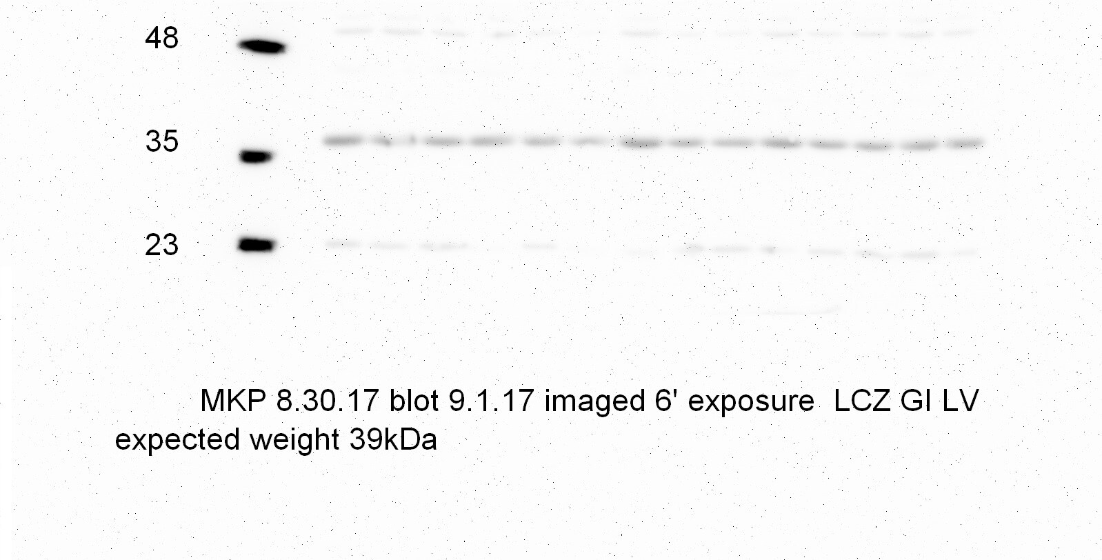
Application: Western BlotSample Tested: Heart lysatesSpecies: RatVerified Customer | Posted 03/19/2018Probing MKP-1 with LV Heart tissue from RatsProteins were separated on SDS-PAGE and transferred onto PVDF membrane. Membrane was blocked with 5% milk in TBS 0.01% tween and then incubated with primary antibody overnight 1:1000. Secondary was used at 1:5000 and then membrane was ECL and imaged with GE Imagequant imager.

There are no reviews that match your criteria.
Protocols
Find general support by application which include: protocols, troubleshooting, illustrated assays, videos and webinars.
- Antigen Retrieval Protocol (PIER)
- Antigen Retrieval for Frozen Sections Protocol
- Appropriate Fixation of IHC/ICC Samples
- Cellular Response to Hypoxia Protocols
- Chromogenic IHC Staining of Formalin-Fixed Paraffin-Embedded (FFPE) Tissue Protocol
- Chromogenic Immunohistochemistry Staining of Frozen Tissue
- ClariTSA™ Fluorophore Kits
- Detection & Visualization of Antibody Binding
- ELISA Sample Preparation & Collection Guide
- ELISA Troubleshooting Guide
- Fluorescent IHC Staining of Frozen Tissue Protocol
- Graphic Protocol for Heat-induced Epitope Retrieval
- Graphic Protocol for the Preparation and Fluorescent IHC Staining of Frozen Tissue Sections
- Graphic Protocol for the Preparation and Fluorescent IHC Staining of Paraffin-embedded Tissue Sections
- Graphic Protocol for the Preparation of Gelatin-coated Slides for Histological Tissue Sections
- How to Run an R&D Systems DuoSet ELISA
- How to Run an R&D Systems Quantikine ELISA
- How to Run an R&D Systems Quantikine™ QuicKit™ ELISA
- IHC Sample Preparation (Frozen sections vs Paraffin)
- Immunofluorescent IHC Staining of Formalin-Fixed Paraffin-Embedded (FFPE) Tissue Protocol
- Immunohistochemistry (IHC) and Immunocytochemistry (ICC) Protocols
- Immunohistochemistry Frozen Troubleshooting
- Immunohistochemistry Paraffin Troubleshooting
- Preparing Samples for IHC/ICC Experiments
- Preventing Non-Specific Staining (Non-Specific Binding)
- Primary Antibody Selection & Optimization
- Protocol for Heat-Induced Epitope Retrieval (HIER)
- Protocol for Making a 4% Formaldehyde Solution in PBS
- Protocol for VisUCyte™ HRP Polymer Detection Reagent
- Protocol for the Preparation & Fixation of Cells on Coverslips
- Protocol for the Preparation and Chromogenic IHC Staining of Frozen Tissue Sections
- Protocol for the Preparation and Chromogenic IHC Staining of Frozen Tissue Sections - Graphic
- Protocol for the Preparation and Chromogenic IHC Staining of Paraffin-embedded Tissue Sections
- Protocol for the Preparation and Chromogenic IHC Staining of Paraffin-embedded Tissue Sections - Graphic
- Protocol for the Preparation and Fluorescent IHC Staining of Frozen Tissue Sections
- Protocol for the Preparation and Fluorescent IHC Staining of Paraffin-embedded Tissue Sections
- Protocol for the Preparation of Gelatin-coated Slides for Histological Tissue Sections
- Quantikine HS ELISA Kit Assay Principle, Alkaline Phosphatase
- Quantikine HS ELISA Kit Principle, Streptavidin-HRP Polymer
- R&D Systems Quality Control Western Blot Protocol
- Sandwich ELISA (Colorimetric) – Biotin/Streptavidin Detection Protocol
- Sandwich ELISA (Colorimetric) – Direct Detection Protocol
- TUNEL and Active Caspase-3 Detection by IHC/ICC Protocol
- The Importance of IHC/ICC Controls
- Troubleshooting Guide: ELISA
- Troubleshooting Guide: Immunohistochemistry
- Troubleshooting Guide: Western Blot Figures
- Western Blot Conditions
- Western Blot Protocol
- Western Blot Protocol for Cell Lysates
- Western Blot Troubleshooting
- Western Blot Troubleshooting Guide
- View all Protocols, Troubleshooting, Illustrated assays and Webinars
Loading...